Introduction
Nematodes of the superfamily Aproctoidea are oviparous parasites in the air sac, and nasal and orbicular cavities of birds. Its type genus Aprocta von Linstow, Reference von Linstow1883 was erected for Aprocta cylindrica von Linstow, Reference von Linstow1883 collected from Monticola solitarius (L.) (Passeriformes: Muscicapidae) in the Turkestan Region of Central Asia by von Linstow (Reference von Linstow1883). Later, von Linstow (Reference von Linstow1903) established another oviparous genus, Lissonema, for Lissonema rotundatum von Linstow, Reference von Linstow1903 collected from Centropus sinensis (Stephens) (Cuculiformes: Cuculidae) in Thailand. The two genera were treated as synonymous by some authors (e.g. Anderson and Bain Reference Anderson and Bain1976; Anderson and Chabaud Reference Anderson and Chabaud1958; Chabaud et al. Reference Chabaud, Anderson and Brygoo1959; Yorke and Maplestone Reference Yorke and Maplestone1926), while others recognised Lissonema as valid (e.g. Sandground Reference Sandground1933; Skrjabin et al. Reference Skrjabin, Shikhobalova, Sobolev and Skrjabin1949; Sonin Reference Sonin1966). Eventually, Bain and Mawson (Reference Bain and Mawson1981) justified the reinstatement of Lissonema and provided a clear morphological differentiation between the two genera.
Nematodes corresponding to the genus Aprocta, in the morphology of the ovejector, the appearance of the male caudal papillae, the shape of the spicules and the size of the eggs, were found in the orbits of a flammulated owl, Psiloscops flammeolus (Kaup) (Strigiformes: Strigidae) in Montana, USA. These nematodes, morphologically distinct from all known Aprocta spp., with the combination of their body length and spicule size, are here described as a new species based on light and scanning electron microscopies. Additionally, fragments of 18S and 12S rRNA genes of the new species are provided.
Material and methods
Sample collection and morphological examination
Ten nematodes were collected from the orbits of a flammulated owl, Psiloscops flammeolus (Kaup) (Strigiformes: Strigidae) found dead in March 2021 in Helena, Lewis and Clark County, Montana, and donated to the Philip L. Wright Museum of the University of Montana (UMZM), Missoula (coll. number UMZM:Bird:22418) by the Montana Department of Fish, Wildlife and Parks. From these nematodes, three males and five females were studied morphologically; two specimens, that were not studied morphologically, were deposited in the UMZM under accession UMZM:Para:15.
Nematodes were cleared with lactophenol and examined and measured under a compound microscope as temporary mounts. Light photomicrographs were taken using a compound microscope Carl Zeiss Axio Imager M2 equipped with a digital camera (ProgRes C7) and software CapturePro. The material used for scanning electron microscopy (SEM) was dehydrated through a graded ethanol series immersed in hexamethyldisilazane for 20 min, air-dried and coated with gold-palladium in an Emitech K500X sputter coater, and examined using a Tescan LYRA 3 XMU FEG/SEMxFIB microscope at an accelerating voltage of 10 kV.
All measurements are in micrometres unless otherwise indicated and are given as the range followed by the mean in parentheses.
The type and voucher materials of the new species were deposited in the Helminthological Collection of the Institute of Biodiversity and Ecosystem Research, Bulgarian Academy of Sciences (IBER-BAS).
DNA extraction, polymerase chain reaction (PCR) amplification and sequencing
Total DNA was isolated from a fragment of the genital system containing eggs of a female preserved in 100% ethanol, using genomic DNA extraction kit (‘NZY Tissue gDNA Isolation kit’, Cat. No. MB13502, NZYtech, Portugal).
For amplification of a region of 18S rRNA nuclear gene a universal nematode primer pair was chosen – forward primer – For_18S_Nema 5′-CAAGTCTGGTGCCAGCAG-3′ and reverse primer – Rev_18S_NemaSec 5′-CGGAAACCTTGTTACGACT-3′, amplifying a region of approximately 1250 nucleotides. The PCR mixtures contained 25 μL of NZYTaq II 2× Green Master Mix (Cat. No. MB358; Nzytech, Portugal), 1 μM of each primer (FOR/REV), 10 ng template DNA and PCR-grade water to a total volume of 50 μL. PCR reactions were carried out under the following conditions: initial denaturation at 94°C for 5 min, 30 cycles (denaturation at 94°C for 30 s; primer annealing at 50°C for 30 s; extension at 72°C for 90s) and final extension at 72°C for 10 min. PCR products were visualised on 1% agarose gel with ‘GreenSafe Premium’ staining (Cat. No. MB13201 NZYTech, Portugal) under ultraviolet light. Fragment size was determined using ‘Perfect 100 bp DNA Ladder’ (Cat. No. E3134-01, EURx, Poland). PCR purification and Sanger sequencing in both directions were carried out by Macrogen Europe (Netherlands) using the PCR primers and two additional internal sequencing primers – forward primer – For_18S_NemaSec 5′-TGCGGCTTAATTTGACTCAAC-3′ and reverse primer – Rev_18S_NemaB 5′-TCTAAGGGCATCACAGACCT-3′.
For amplification of a region of 12S rRNA mitochondrial gene, the following primer pair was chosen – forward primer – 12SF 5′-GTTCCAGAATAATCGGCTA-3′ (as suggested by Casiraghi et al. Reference Casiraghi, Bain, Guerrero, Martin, Pocacqua, Gardner, Franceschi and Bandi2004) and a custom reverse primer – 12SRev_Spiru 5′-CTACCATACTACAACTTACGC-3′. The PCR reactions were performed under the same conditions as the reactions for 18S, with the only difference being the elongation phase of each cycle being 60 s; PCR product visualisation was carried in a similar method with the same reagents as described above for 18S rDNA. DNA sequencing for this gene was performed as described above, with only the PCR primers used for sequencing, no internal primers were used due the shorter product length.
Both genes were manually edited and deposited in the GenBank database under the accession numbers: PV661993 (18S rRNA gene) and PV697359 (12S rRNA gene).
Phylogenetic analysis
The 18S rRNA sequence was aligned with the sequences used for phylogenetic analyses by Neidringhaus et al. (2023) using MEGA software, version 7.0 (Kumar et al. Reference Kumar, Stecher and Tamura2016) and version 11 (Tamura et al. Reference Tamura, Stecher and Kumar2021). Two sequences used by Neidringhaus et al. (2023) – LM006781 ‘Wuchereria bancrofti genome assembly, scaffold: WBA_contig0005517’ and LK982445 ‘Thelazia callipaeda genome assembly, scaffold: TCLT_contig0001594’ – were not used in the phylogenetic reconstruction, because they were retracted from GenBank by their authors. Instead AY843436 ‘Wuchereria bancrofti isolate Wardha 18S small subunit ribosomal RNA gene, partial sequence’ and OZ239431 ‘Thelazia callipaeda genome assembly, chromosome: 2’ were used in the analysis. Phylogenetic analysis was performed using Bayesian inference (BI) with MrBayes (Ronquist et al. Reference Ronquist, Teslenko, Van Der Mark, Ayres, Darling, Höhna, Larget, Liu, Suchard and Huelsenbeck2012). Prior to analysis, the best model of nucleotide substitution was selected using MrModeltest2 (Nylander Reference Nylander2004); this was the general time reversible model, with estimates of invariant sites and gamma distributed among-site rate variation (GTR+G+I). The analysis was run for 1 × 10⁷ generations, two separate runs, each with four chains, discarding 25% of resulting trees as burn-in. Average standard deviation of split frequencies below 0.01 was observed and served as an indication of the Markov chains reaching convergence. Branches persisting in less than 50% of post burn-in samples were treated as polytomies. Nodal support was expressed as posterior probabilities (PP). All positions with less than 95% site coverage were eliminated – that is, fewer than 5% alignment gaps, missing data, and ambiguous bases were allowed at any position. There were a total of 1690 positions in the final dataset.
Results
Morphological description
Aprocta bainae sp. nov. Mutafchiev & Kinsella (Figures 1–3)
Aprocta bainae sp. nov. A, Anterior region, female, lateral view; B, Anterior extremity, male, lateral view (B1), dorsoventral view (B2) and apical view (B3); C, posterior extremity, male, lateral view; note the caudal papillae (arrows) and the phasmid (arrowhead); D, spicules of three different males, right spicules dextral view (R) and left spicules sinistral view (L); E, gubernaculum, lateral view; F, posterior, extremity, female, lateral view; note the phasmids (arrows); G, eggs, note the anterior extremity (G1) and posterior extremity (G2) of the first stage larva inside.

General description. Medium-sized aproctid nematodes with marked sexual dimorphism, males about half length of females. Body long, cylindrical, tapering anteriorly, anterior extremity round in lateral view (Figure 1A, B). Body cuticle thinner anteriorly and thicker posteriorly, with fine transversely orientated and anastomosing striae (Figure 3C). Mouth opening laterally compressed by two rounded, inconspicuous pseudolabia each bearing two external and two internal sublateral cephalic papillae and single amphid (Figures 1B, 2A, 3A, B). Somatic musculature well-developed. Buccal cavity with thick walls, dorso-ventraly elongated, in lateral view, slightly longer than wide. Deirids and excretory pore not observed. Nerve ring not distinct (Figure 2B). Oesophagus divided at level of nerve ring into anterior muscular and posterior glandular part (Figure 2B). Tails round with subterminal phasmids (Figure 1C, F).
Aprocta bainae sp. nov., male, light photomicrographs. A, Anterior extremity, apical view; note the external cephalic papillae (black arrowheads), the internal cephalic papillae (white arrowheads) and the amphids (arrows); B, anterior extremity, lateral view; note the border between the muscular and glandular portion of the oesophagus (arrowhead) and the nerve ring (asterisk); C, posterior extremity, lateral view; D, left precloacal caudal papillae (arrows); E, left postcloacal papillae (arrows).

Aprocta bainae sp. nov., male, scanning electron microscopy. A, Anterior extremity, apical view; note an external cephalic papilla (black arrowhead) and an internal cephalic papilla (white arrowhead) situated on a common cuticular elevation; B, detail of the pair of cephalic papillae indicated on Figure 3A; note an external cephalic papilla (black arrowhead) and an internal cephalic papilla (white arrowhead); C, detail of the body surface; D, tail, dextral view.

Male (based on three individuals). Total length 19.6–23.0 mm (21.7 mm), posterior body end ventrally curved into two tight spirals. Cuticle up to 15 μm thick at posterior end. Maximum body width 540–660 (613), body width at oesophago-intestinal junction 340–447 (377). Cloacal region swollen, body width at cloaca 143–183 (160). Buccal cavity 32–45 (41) long and 25–35 (31) wide, measured in lateral view. Nerve ring at 130–174 (149) from anterior extremity. Oesophagus 720–1050 (847) long with maximum width of 75–106 (94) measured at its posterior end; oesophageal width to oesophageal length ratio 1:7.5–9.9 (1:9.0). Caudal alae and area rugosa absent. Tail 108–135 (122) long (Figures 1C, 2C). Caudal papillae total four pairs (Figures 1C, 2D, E): two pairs precloacal and two pairs postcloacal. Spicules dissimilar with simple tip (Figures 1D, 2C); left spicule 233–308 (280) long; right spicule 203–275 (242), gubernaculum longer than wide, with irregular shape about 40–50 long (Figure 1E)
Female (based on five measured individuals, unless otherwise indicated). Total length 32.6–41.8 mm (37.4 mm); maximum body width 790–900 (828); body width at vulva 320–390 (355, n = 4), body width at oesophago-intestinal junction 415–470 (435, n = 4), anal body width 285–380 (334). Cuticle up to 8 μm thick. Buccal cavity 30–48 (44) long and 33–38 (35, n = 4) wide. Nerve ring at 163–185 (171) from anterior extremity. Oesophagus 816–980 (893) long and with maximum width 105–123 (112) measured at its posterior end. Oesophageal width to oesophageal length ratio 1:7.6–8.9 (1:8.2). Vulval lips with slight swelling, vulval orifice situated at 640–770 (713, n = 4) from anterior extremity (Figure 1A); Ovejector 375–575 (459, n = 4) long; didelphic. Tail 100–210 (170) long (Figure 1F). Eggs 51–56 × 30–32 (54 × 31, n = 10) with shell c. 3 μm thick, containing developed first-stage larva 177–198 (186, n = 6) in length with larval hook and rasp on its anterior end and armed tail (Figure 1G).
Taxonomic summary
Type-host: Psiloscops flammeolus (Kaup) (Strigiformes: Strigidae).
Site of infection: orbital cavity.
Type-locality: Helena, Montana, USA.
Type-material: Holotype, male preserved in 70% ethanol (IBER-BAS N001.467); Paratypes preserved in 70% ethanol – IBER-BAS N001.468 (1 male and 4 females) and IBER-BAS N001.469 (one female from which tissue was used for generation of DNA sequences); SEM stub IBER-BAS N001.470 (one male).
Other material: five permanent slides with eggs (IBER-BAS N001.462–N001.466).
Etymology: The new species is named for the late Dr Odile Bain in recognition of her contribution to the taxonomy of this nematode group.
ZooBank Life Science Identifier: urn:lsid:zoobank.org:act:A6D922FB-1D0F-437A-B6ED-7F064F45A831
Molecular characterisation
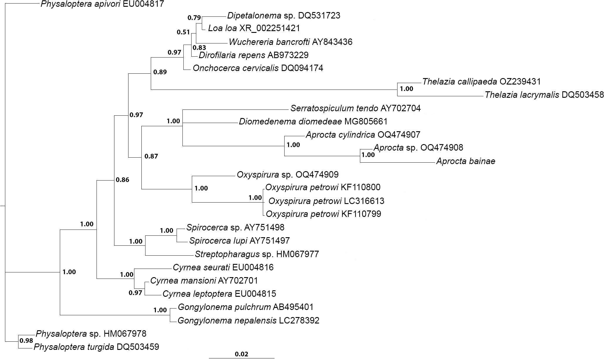
Partial 12S rDNA (429 bp in length) and 18S rDNA (1285 bp in length) sequences were generated from a single female (deposited under accession number IBER-BS N001.469). The phylogenetic tree inferred from Bayesian inference of the partial 18S rDNA (Figure 4) revealed the three representatives of Aprocta as forming a strongly supported monophyletic group (PP 1.00). Aprocta bainae sp. nov. was grouped with ‘Aprocta sp.’ (PP 1.00) collected from Strix varia Barton (Strigiformes) in the USA. The analysis grouped (PP 1.00) Aprocta together with the member of the genus Diomedenema Johnston & Mawson, 1952 (Desmidocercidae), a representative of another family within the superfamily Aproctoidea, and the genus Serratospiculum Skrjabiin, 1915 (Diplotriaenidae), member of the superfamily Diplotriaenoidae. However, the relationships between the three genera were not resolved by the phylogenetic reconstruction. This is the first publication of a 12S rRNA gene sequence for a member of the superfamily Aproctoidea.
Phylogram from Bayesian inference analysis of the 18S rDNA sequences of Aprocta bainae sp. nov. and selected spirurid nematodes with their respective GenBank accession numbers. Posterior probabilities are shown at the nodes. The scale bar represents the expected number of substitutions per site.

Discussion
Based on a few morphologically well-studied species from both Aprocta and Lissonema, Bain and Mawson (Reference Bain and Mawson1981) concluded that Lissonema differs from Aprocta by their relatively long ovejector (1500–5000 μm vs. 200–600 μm long), at least one large papilla or one or more large pairs (vs. small caudal papillae in males), equal spicules with wide and blunt distal end (vs. spicules subequal with pointed distal end), eggs over 65 μm long (vs. eggs less than 55 μm long), deirids present (vs. absent), first stage larva more than 400 μm long, with long tail without terminal spines (vs. larva 200 μm long with short tail with terminal spines). While Bain and Mawson (Reference Bain and Mawson1981) listed the species that, according to them, belong to Aprocta and Lissonema, they did not justify each treatment and also failed to provide the synonyms recognised by them.
However, reviewing the previous descriptions of Aprocta spp., we found that many of these are superficial, and in several instances, we have noticed significant discrepancies between the textual descriptions and the illustrations. In addition, there are a few species for which the host and the type locality was not given; the site of infection of a few species is also unknown. All these issues inevitably affect our knowledge on the diversity and host specificity of the genus Aprocta.
Aprocta bainae sp. nov. described here corresponds to the diagnoses of the genus Aprocta as coined by Bain and Mawson (Reference Bain and Mawson1981). The new species can be differentiated from the known species of Aprocta by the combination of body length and spicule size. Other morphological characters that could be potentially used for differentiation of the species within the genus could be the shape of the two spicules, the position of the vulvar opening in relation to the oesophago-intestinal junction, and the size of the eggs.
Body lengths of both male and females of A. bainae sp. nov. considerably exceed those of the following species:
-
• Aprocta cylindrica and the several species recognised as its junior synonyms by Sonin (Reference Sonin1966), including: Aprocta microanalis Skrjabin, Reference Skrjabin1917 from the orbital cavity of Luscinia svecica (L.) (Muscicapidae) in Semirechyenskaya Province (Russian Turkestan, Russian Empire), now a territory located mostly in southeastern Kazakhstan and northeastern Kyrgyzstan; Aprocta striata Sandground, Reference Sandground1933 from the orbital cavity and nasal sinuses of Ficedula hyperythra (Blyth) (Muscicapidae) from French Indo-China (today’s Cambodia, Laos, and Vietnam); Aprocta circumcularis Hartwich, Reference Hartwich1951 from Luscinia megarhynchos Brehm (Muscicapidae) in Germany; Aprocta skrjabini Makarenko, Reference Makarenko1959 from the orbital cavity of Phoenicurus phoenicurus (L.) (Muscicapidae) in European Russia. The body lengths, as provided in the original descriptions of these samples, vary from 13.3–16.4 mm for the males and 24.0–33.5 mm for the females (summarised from Hartwich Reference Hartwich1951; Makarenko Reference Makarenko1959; Sandground Reference Sandground1933; Skrjabin Reference Skrjabin1917; von Linstow Reference von Linstow1883). The above species, considered synonyms of A. cylindrica by Sonin (Reference Sonin1966), were not listed in Bain and Mawson (Reference Bain and Mawson1981).
-
• Aprocta pillersi (Yorke & Maplestone, Reference Yorke and Maplestone1926) Bain & Mawson, Reference Bain and Mawson1981 was described from a ‘blue warbler flycatcher’ by Yorke and Maplestone (Reference Yorke and Maplestone1926). The scientific name of the host and the locality were not provided in the original publication; Sonin (Reference Sonin1966) suggested that its type host is possibly ‘Dendroica caerulescens (Parulidae)’ now Setophaga caerulescens (Gmelin) from North America. However, the host could be a species of the genus Cyornis Blyth (Muscicapidae) known as ‘blue flycatchers’ with South-east Asia distribution. Yorke and Maplestone (Reference Yorke and Maplestone1926) described the male and the female of Aprocta pillersi as 16 mm and 32 mm long, respectively.
-
• Aprocta textori Vuylsteke, Reference Vuylsteke1953, a parasite of Textor melanocephalus duboisi Hartlaub (Ploceidae) in the DR Congo, was characterised with a male 9.5 mm long and a female 15 mm long (Vuylsteke Reference Vuylsteke1953).
-
• Aprocta angolica (Vuylsteke, Reference Vuylsteke1953) from Prionops plumatus poliocephalus (Stanley) (=Prionops poliocephala angolica) (Vangidae) in the DR Congo was described with a male 13 mm long and a female 21 mm long (Vuylsteke Reference Vuylsteke1953).
-
• Aprocta cercomelae Vuylsteke, Reference Vuylsteke1953, a taxon originally proposed as a variety of A. cylindrica, and later elevated to a full species rank by Sonin (Reference Sonin1966), was described from Oenanthe familiaris falkensteini (Cabanis) (=Cercomela familiaris modesta Shelley) (Muscicapidae) in the DR Congo. This species was characterised with a male 8.7 mm long and a female 21 mm long (Vuylsteke Reference Vuylsteke1953).
-
• Aprocta laevicutis Sandground, Reference Sandground1933 collected from the conjunctival sac of Cyornis whitei whitei Harington (Muscicapidae) (=Cyornis banyumas whitei) from French Indo-China has males and females measuring 8.0–9.0 mm long and 15.0–17.0 mm long, respectively (Sandground Reference Sandground1933).
-
• Aprocta vestibulata (Johnston and Mawson Reference Johnston and Mawson1940) from Aphelocephala nigricincta (North) (Acanthizidae) in South Australia has males 8.5–9.5 mm long and females 20.0–25.0 mm long (Johnston and Mawson Reference Johnston and Mawson1940).
-
• Aprocta sudarikowi Sobolev, Reference Sobolev1947, collected from the orbital cavity of Charadrius dubius Scopoli (Charadriidae) in European Russia, was characterised with males 11.6–12.0 mm long and females 18.9–19.3 mm long (Sobolev Reference Sobolev1947). Aprocta grallaria Serkowa, 1948, described from the same host in Siberia, was recognised as a junior synonym of A. sudarikowi; the body lengths of males and females were measured as 12–18 mm and 24–30 mm, respectively (Sonin Reference Sonin1963). Aprocta grallaria was not included in the list of Aprocta spp. by Bain and Mawson (Reference Bain and Mawson1981).
-
• Aprocta brevipenis Rodrigues & Rodrigues, Reference Rodrigues and Rodrigues1980, collected from the orbital cavity of Guira guira (Gmelin) (Cuculidae) in Brazil, was described from a male measuring 8.53 mm long and females 25.32 mm (Rodrigues and Rodrigues Reference Rodrigues and Rodrigues1980). This species was omitted by Bain and Mawson (Reference Bain and Mawson1981), likely because they were not aware of its description whose publication almost coincided with their work. Aprocta brevipenis was characterised with morphology corresponding to the diagnosis of Aprocta – that is, subequal spicules with pointed distal ends, short ovejector, and eggs 44–48 × 26-29 μm in size.
-
• Aprocta intraorbitalis Hernandez-Rodriguez, Gutierrez-Palomino & Martinez-Gomez, 1986, a parasite from the orbital cavity of Cyanopica cooki Bonaparte (reported as Cyanopica cyanus (Pallas)) (Corvidae) in Spain, has males 11.4–16.6 mm long and females 19.0–33.8 mm long (Hernandez-Rodriguez et al. Reference Hernandez-Rodriguez, Gutiérrez-Palomino and Martinez-Gomez1986).
Aprocta bainae sp. nov. has body length close to or smaller than the remaining Aprocta spp., but it can be distinguished from them as follows:
-
• Aprocta orbitalis von Linstow, 1901 was described from the orbits of Milvus migrans (Boddaert) (Accipitridae) (= Falco fuscoater) by von Linstow (Reference von Linstow1901a) without a reference to the locality. The male was characterised as 21 mm long with spicules 400 and 470 μm long, the female was 38 mm long. Sonin (Reference Sonin1966) considered Falco fuscoater a synonym of Buteo buteo vulpinus, however Falco fuscoater Meyer & Wolf was a name given to the black kite (see Meyer and Wolf Reference Meyer and Wolf1810). The males of A. bainae sp. nov. have shorter spicules than A. orbitalis.
-
• Aprocta narium von Linstow, 1901 was collected from the nasal cavity of a Buteo sp. from Ukraine and described with a male measuring 21 mm in length having spicules 350 μm and 370 μm long and a female 33 mm long (von Linstow Reference von Linstow1901b). The oesophageal lengths were originally provided as ratios to the body length, their calculated lengths of 1190 μm in the male and 2000 μm in the females exceed considerably that of the other known Aprocta spp., including the new species; the position of the vulva at 180 μm from anterior body extremity, also provided as a ratio of the body length, is more anterior to that of the congeners. It is possible that these ratios were mistaken. The males of A. bainae sp. nov. differ from A. narium also by their shorter spicules.
-
• Aprocta turgida Stossich, Reference Stossich1902, from the nasal cavity of Larus argentatus (Laridae) from Italy, was briefly described, based on a few females measuring 20–27 mm long by Stossich (Reference Stossich1902). Aprocta milinskii Leonov, Reference Leonov1958, described from L. argentatus in Ukraine (Leonov Reference Leonov1958), was recognised as its junior synonym by Sonin (Reference Sonin1966), who further considered A. turgida as a specific parasite of gulls and sterns. Aprocta milinskii was not mentioned by Bain and Mawson (Reference Bain and Mawson1981). Chabaud and Choquet (Reference Chabaud and Choquet1955) delivered a more detailed morphological description based on material collected from Larus michahellis Naumann in France; the studied male was 26 mm long, with spicules 380 μm long, whereas the female was 33 mm long. The males of A. bainae sp. nov. can be differentiated from Aprocta turgida by their shorter spicules.
-
• Aprocta matronensis Railliet & Henry, Reference Railliet and Henry1910 was collected from the infraorbital canal of Corvus cornix L. (Corvidae) in France; Its males were 21–24 mm long, with spicules 310–370 μm long, the females were damaged but their eggs were described as being 40–45 × 23–27 μm in size (Railliet and Henry Reference Railliet and Henry1910). The description of A. matronensis was not complemented with any illustrations. Aprocta bainae sp. nov. differs from this species by its shorter spicules and larger eggs.
-
• Aprocta crassa Railliet & Henry, Reference Railliet and Henry1910 was collected from Otis tarda (Otidae) in Ethiopia by Railliet and Henry (Reference Railliet and Henry1910). As originally described, this species has males 20–30 mm long, with spicules 335 and 385 μm long with roundish distal end and females 35–50 mm long with eggs 55–60 × 28–30 μm in size; illustrations were not provided. The sample initially identified as A. orbitalis from Otis tarda (Otidae) in Spain by Yeh (Reference Yeh1957), but later recognised as conspecific with A. crassa by Chabaud et al. (Reference Chabaud, Anderson and Brygoo1959) and Sonin (Reference Sonin1966), was characterised by males 28–30 mm long, with left and right spicules, 400–440 μm and 420–470 μm long, respectively, and females 38–50 mm long with eggs measured 50–56 × 34–36 μm. Aprocta bainae sp. nov. can be distinguished from A. crassa, as originally described, by its shorter spicules and smaller eggs. The spicules of the new species are even shorter than those of the sample from Spain, but the two samples have eggs similar in size.
-
• Aprocta corvicola Johnston & Mawson, Reference Johnston and Mawson1940 was collected behind the eye of Corvus coronoides Vigors & Horsfield (Corvidae) in Australia. The species was characterised by Johnston and Mawson (Reference Johnston and Mawson1940) with males 12–20 mm long, with equal spicules 280–320 μm long, and females 50–60 mm long. Sonin (Reference Sonin1966) treated this species a junior synonym of A. matronensis. According to Sonin (Reference Sonin1966) this conclusion was based on a sample from Corvus corone L. from Kyrgyzstan studied by Sonin and Borgarenko (Reference Sonin and Borgarenko1965) and considered conspecific with A. matronensis. The comparative table in Sonin (Reference Sonin1966) contains more detailed morphometrical data of the sample from C. corone than those provided by Sonin and Borgarenko (Reference Sonin and Borgarenko1965), and males were described as 16–31 mm long, with spicules 250–310 μm and 280–330 μm long, thus overlapping the males of the original description of A. matronensis and partly overlapping these of A. corvicola. However, Aprocta corvicola and A. matronensis, as originally described, differ considerably from one another in their body dimensions, spicule lengths, as well as the size of their eggs, which were measured 40–45 × 23–27 μm in Aprocta matronensis from Australia, 45–50 × 30 μm in Aprocta corvicola from France. The eggs of the sample from Kyrgyzstan were 50–60 × 30–40 μm. Therefore, we consider Sonin’s conclusion that the three samples are conspecific as premature, Aprocta corvicola was not mentioned by Bain and Mawson (Reference Bain and Mawson1981). Aprocta bainae sp. nov. can be distinguished from A. corvicola by its longer males, subequal spicules, and larger eggs.
-
• Aprocta caballeroi Ybarra, 1969 was described from Buteo jamaicensis borealis (Gmelin) in Mexico as having a male 18.1 mm long, with left and right spicule 395 μm and 270 μm long, respectively, and an oesophagus 769 μm long and 83 μm wide, and female 30.0 mm long with an oesophagus 900 μm long and 83 μm wide (Ybarra Reference Ybarra1949). Aprocta bainae sp. nov. can be distinguished from A. caballeroi by its longer body and slightly shorter spicules. Also, the new species has a broader oesophagus than A. caballeroi. The oesophageal width to oesophageal length ratio of Aprocta bainae sp. nov. is in the range of 1:6.9–8.8 in the males and 1:6.5–8.5 in the females, whereas according to provided metrics in A. caballeroi the ratio is 1:9.3 in the male and 1:10.8 in the female. However, there are considerable discrepancies between the textual description and illustrations provided by Ybarra (Reference Ybarra1949); thus in Figure 1 the vulva is located at the mid-length of the oesophagus, whereas according to the textual description the vulva is located at 691 μm from the anterior end and the oesophagus is 900 μm long; based on Figure 1 the calculated oesophageal width to length ratio is 1:12.9; also all scale bars to the figures do not correspond to the morphometrics given in the textual description.
-
• Aprocta ptiloscelidis Schuurmans-Stekhoven, Reference Schuurmans-Stekhoven1951 was described from the nasal cavity of Vanellus resplendens (Tschudi) (Charadriidae) (=Ptiloscelis resplendens (Tschudi)) in Argentina (Schuurmans-Stekhoven Reference Schuurmans-Stekhoven1951). The males were described as 2 cm long, whereas the females were 80 mm long; eggs were illustrated in the vagina, but there is no information about their size, as well as about the length of the vagina. The lengths of the spicules were not provided but their shape is reminiscent of the other Aprocta spp. Aprocta bainae sp. nov. can be distinguished from Aprocta ptiloscelidis by its considerably shorter females. In addition, the females of A. ptiloscelidis were described with shorter oesophagus than that of A. bainae sp. nov. (285 μm vs. 825–1,100 μm) and subterminal anus.
-
• Aprocta boulengeri Mawson & Bain, 1981, a parasite from Strepera graculina Shaw (Cracticidae) in Australia, was described by Bain and Mawson (Reference Bain and Mawson1981) by one intact male 25.6 mm long and right and left spicules 400 μm and 440 μm long, respectively. A female was selected as the holotype and her body length was given as ‘50 μm’ which is an apparent typographical error, whereas the other female collected was 37 mm long. This species differs from A. bainae sp. nov. by the presence of a median subterminal papilla on the male tail and its spicules exceeding 400 μm in length characterised further with tricuspid distal extremities.
-
• Aprocta bakeri Mawson & Bain, 1981, from Corvus orru Bonaparte (Corvidae) in Australia, was described based on a single male 29 mm long with left and right spicules 390 μm and 330 μm long, respectively (Bain and Mawson Reference Bain and Mawson1981). This species, as in A. bainae sp. nov. has a cloacal protuberance, but the new species differs from it by its shorter spicules and shorter body length.
The following two species are considered by us as species inquirendae:
-
• Aprocta ophthalmophaga Stossich, Reference Stossich1902 was described from the ocular cavity of a ‘un grosso’ Falco sp. collected in Ethiopia (Stossich Reference Stossich1902). The original description was only textual, and the only measurements provided were those of the body lengths of the males ranging 20–24 mm and the females measured 40 mm. Bain and Mawson (Reference Bain and Mawson1981) treated A. ophthalmophaga as an Aprocta, however, taking into account the superficial morphological description and unknown type host we treat this species a species inquirenda.
-
• Aprocta colaptidis Schuurmans-Stekhoven, Reference Schuurmans-Stekhoven1950 was described from the abdominal cavity of Xiphocolaptes sp. (Furnariidae) in Paraguay by Schuurmans-Stekhoven (Reference Schuurmans-Stekhoven1950). The material was composed of five females; the morphological description was superficial and not accompanied with illustrations, the only morphometric provided was that the females measured 40 cm long. Thus, the females of Aprocta colaptidis differ from all other Aprocta spp. by their terminal anus and the body length that is 6–7 times as long as that of the other females (unless the body length was incorrectly provided in centimetres instead of millimetres). We consider Aprocta colaptidis Schuurmans-Stekhoven, Reference Schuurmans-Stekhoven1950 as a species inquirenda.
The following species is considered by us a species incertae sedis:
-
• Aprocta colaptidis Schuurmans-Stekhoven, Reference Schuurmans-Stekhoven1951 was indicated as a new species in Schuurmans-Stekhoven (Reference Schuurmans-Stekhoven1951). The name was attributed to two samples composed by males and females collected under the skin and in the muscles of the neck of Colaptes campestroides (Malherbe) (Picidae) in Argentina. Schuurmans-Stekhoven (Reference Schuurmans-Stekhoven1951) described the males as having two subequal spicules, the longer one being thin and the shorter one being stouter, their lengths were not provided; the females were described as having a terminal anus. The description does not contain information about the length of the vagina and whether there were eggs in the vagina and uteri. The terminal position of the anus and the site of infection of the material studied by Schuurmans-Stekhoven (Reference Schuurmans-Stekhoven1951) differ from those of the other known Aprocta spp. Therefore, we consider A. colaptidis Schuurmans-Stekhoven, Reference Schuurmans-Stekhoven1951 a species incertae sedis. Schuurmans-Stekhoven (Reference Schuurmans-Stekhoven1951) did not refer to the material described as Aprocta colaptidis in his earlier work, thus Aprocta colaptidis Schuurmans-Stekhoven, Reference Schuurmans-Stekhoven1951 appears to be a homonym of Aprocta colaptidis Schuurmans-Stekhoven, Reference Schuurmans-Stekhoven1950.
Besides A. bainae sp. nov., there are no males of Aprocta described with four pairs of caudal papillae. However, we refrain from using caudal papillae as a distinguishing character because the posterior pair of papillae of A. bainae sp. nov. was difficult to observe as it was not associated with any particular structure, also the left papillae of the first and the second pair were not observed on the holotype (the dextral caudal papillae of this specimen were not studied due to the densely coiled posterior end). Therefore, it cannot be excluded that in the other Aprocta spp. some of caudal papillae might have been overlooked.
A bifurcation of the testes was reported for A. laevicutis, A. bakeri and A. intraorbitalis (Bain and Mawson Reference Bain and Mawson1981; Hernandez-Rodriguez et al. Reference Hernandez-Rodriguez, Gutiérrez-Palomino and Martinez-Gomez1986; Sandground Reference Sandground1933). We did not observe a bifurcation of the testes in A. bainae sp. nov., however, the bifurcation could be missed as the detailed internal morphological observations were obstructed by thick somatic musculature.
The sample, identified as Aprocta sp. from the orbits of three barred owls in California by Niedringhaus et al. (Reference Niedringhaus, Dumbacher, Dunker, Medina, Lawson, Fenton, Higley, Haynes and Yabsley2023), was characterised with a morphology corresponding to that of Aprocta. The measured male was 17.3 mm in length, with left and rights spicule, 320 μm and 308 μm long, respectively, the three females were 16.0–21.0 mm in length, containing eggs 45–47 × 25–32 μm in size, thus distinct morphologically from A. bainae sp. nov. with their smaller body length, longer spicules, and smaller eggs.
Bain and Mawson (Reference Bain and Mawson1981) and Bain (Reference Bain1982) concluded that the two genera have mutually exclusive range of hosts, Aprocta being adapted to Charadriiformes, Gruiformes, Falconiformes, and Passeriformes, whereas Lissonema spp. appears to parasitic in Strigiformes, Cuculiformes, Piciformes, Trogoniformes, Caprimulgiformes, and Coraciiformes. However, A. brevipenis, A. bainae sp. nov., and ‘Aprocta sp.’ of Niedringhaus et al. (Reference Niedringhaus, Dumbacher, Dunker, Medina, Lawson, Fenton, Higley, Haynes and Yabsley2023) were collected from hosts that were considered typical for the genus Lissonema.
Our phylogenetic analysis similar to that performed, based on maximum likelihood, by Niedringhaus et al. (Reference Niedringhaus, Dumbacher, Dunker, Medina, Lawson, Fenton, Higley, Haynes and Yabsley2023) revealed the genera Aprocta, Diomedenema, and Serratospiculum (Diplotriaenoidea) as forming together a monophyletic clade. However, in our consensus tree the genera Aprocta, Diomedenema, and Serratospiculum formed a monophyletic group with the members of the genus Oxyspirura (Thelaziidae, Thelazioidae), whereas in Niedringhaus et al. (Reference Niedringhaus, Dumbacher, Dunker, Medina, Lawson, Fenton, Higley, Haynes and Yabsley2023) these were sister to a clade formed by the members of the family Onchocercidae included in the analysis and the genus Thelazia (Thelaziidae, Thelazioidae). The differences in the two phylogenetic hypotheses could be due to the shorter matrix used in our analysis – we targeted 463 bp shorter fragment of A. bainae than that obtained by Niedringhaus et al. (Reference Niedringhaus, Dumbacher, Dunker, Medina, Lawson, Fenton, Higley, Haynes and Yabsley2023) – or due to the difference of the statistical inference methods used (BI vs. maximum likelihood). Both analyses revealed Thelaziidae as a polyphyletic group.
12S rRNA gene has been shown to be easy to amplify and convenient for both DNA barcoding and phylogenetic studies at intra-phyla relationships genus level (Chan et al. Reference Chan, Chaisiri, Morand, Saralamba and Thaenkham2020; Ferri et al. Reference Ferri, Barbuto, Bain, Galimberti, Uni, Guerrero, Ferté, Bandi, Martin and Casiraghi2009). Thus, the generated 12S rDNA sequence can facilitate future studies on the taxonomy and systematics of the group.
Author contribution
YM Conceptualisation, Visualisation, Resources, Investigation, Writing – original draft, Writing – review and editing. JMK Investigation, Writing – review and editing. SCG Sample collection; Writing – review and editing. BN Writing – original draft; Analyses, data curation, Writing – review and editing.
Financial support
Molecular studies were covered by the Bulgarian National Science Fund (Grant КП-06-Н71/11). This study used facilities of the Institute of Biodiversity and Ecosystem Research – Bulgarian Academy of Sciences upgraded in the frames of the project DiSSCo-BG (Upgrade of the Research Infrastructure ‘Distributed System of Scientific Collections — Bulgaria’) funded by the National Roadmap for Research Infrastructures, Ministry of Education and Science of the Republic of Bulgaria. SEM observations were covered by the Bulgarian National Endowment Fund ‘13 Centuries Bulgaria’.
Competing interests
The authors declare none.
Ethical standard
Not applicable.