Introduction
Calcium hydroxylapatite (Radiesse, Bioform, San Mateo, California, USA) is used extensively for soft tissue augmentation in the correction of cosmetic defects and facial lipoatrophy. In 2004, its utility in correcting vocal fold insufficiency was first reported.Reference Chhetri, Jahan-Parwar, Hart, Bhuta and Berke1–Reference Belafsky and Postma3 Since that time, it has become increasingly popular in the treatment of unilateral vocal fold paralysis and vocal fold bowing.Reference Rosen, Garner-Schmidt, Casiano, Anderson, Johnson and Reussner4 It has also been used to treat glottic insufficiency and aspiration following partial laryngectomy.Reference Lee and Woo5 While the functional outcome following injection is the subject of intense ongoing study, the ultimate fate of the injected paste is less well characterised.
We report the computed tomography (CT) and positron emission tomography (PET) findings which characterise injected calcium hydroxylapatite, findings which may be mistaken for malignancy.
Case report
Near-total laryngectomy is a form of laryngectomy sparing a single arytenoid, which is used to form a one-way, myomucosal, tracheopharyngeal shunt. Via this shunt, a patient can develop tracheopharyngeal speech without the need for a prosthesis. Unfortunately, incompetence of the shunt is a common complication of the procedure and can lead to intractable aspiration.Reference Suits, Cohen and Everts6 Several methods have been designed to correct this, including the injection of collagen and Teflon® into the shunt to close it.Reference Coniglio, Winkle, Bennett, Martin and Pacella7, Reference Brasnu, Strome, Laccourreye, Weinstein and Menard8
A 69-year-old man had undergone near-total laryngectomy, left modified radical neck dissection and post-operative radiation therapy for a T4 N0 M0 squamous cell carcinoma of the left pyriform sinus.
For six years, the patient had remained free of disease, but then began to develop incompetence of the myomucosal shunt, gross extravasation of swallowed liquids and consequent aspiration.
Computed tomography scanning, laryngoscopy, oesophagoscopy and bronchoscopy showed no sign of tumour recurrence.
Under general anaesthesia, calcium hydroxylapatite was injected transorally and trans-stomally to seal the pharyngeal and tracheal openings of the shunt, respectively, resolving the aspiration.
The patient did well for 17 months following injection, before developing a mild recurrence of his aspiration. A second injection was performed, and the patient was symptom free for 15 months.
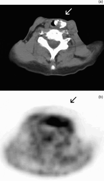
As part of routine follow up, the patient underwent several subsequent CT and PET scans, which afforded incidental opportunities to monitor the status of the implanted calcium hydroxylapatite. A PET CT scan using 18F-deoxyglucose was performed within three days of the second injection. The injected calcium hydroxylapatite appeared bright white on CT (Figure 1a), while PET showed intense 18F-deoxyglucose uptake at the site of the injection (Figure 1b).
(a) Axial computed tomography scan showing calcium hydroxylapatite injected into the pharyngeal aspect of a myomucosal shunt; calcium particles appear bright white (arrow). (b) Corresponding positron emission tomography image, showing intense 18F-deoxyglucose uptake at injection site (arrow).

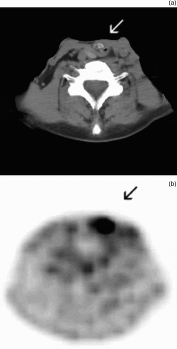
One year later, the patient underwent a second whole body PET CT using 18F-deoxyglucose. On the CT, much of the injected calcium particles appeared to have been reabsorbed (Figure 2a). However, PET scanning still showed intense 18F-deoxyglucose uptake at the site of the injection (Figure 2b). While the PET findings aroused suspicion of a recurrence of disease, correlation with the CT images and the patient's physical examination showed the origin of the signal to be the site of injection, not a new tumour. Despite apparent reabsorption of the calcium particle component, the augmentative effect of the implant persisted, and the patient remained symptom free at the time of writing. Ongoing physical examinations showed no evidence of cancer recurrence, either at the injection site or elsewhere.
Imaging scans taken one year after injection. (a) Axial computed tomography scan showing that much of the injected calcium hydroxylapatite has been reabsorbed (arrow). (b) Corresponding positron emission tomography image, showing intense 18F-deoxyglucose uptake persisting at the injection site (arrow).

Discussion
Malignant lesions and their metastases readily concentrate 18F-deoxyglucose, and it is this property that renders PET a useful tool in the staging and monitoring of patients with squamous cell carcinoma of the head and neck. However, various forms of laryngeal pathology can and do produce false positive findings on PET. Unilateral vocal fold paralysis produces intense uptake in the contralateral mobile vocal fold.Reference Heller, Meltzer, Fukui, Rosen, Chander and Martinelli9 Teflon granuloma has also been shown to produce an intense signal on PET.Reference Yeretsian, Blodgett, Branstetter, Roberts and Meltzer10–Reference Chadwick, Khalid, Wagner and Stack13
18F-deoxyglucose concentrates within tissues in which glucose uptake is increased, corresponding to elevated metabolic activity, compared with surrounding tissue. In the presented patient, the intense uptake at the injection site, as seen on PET, suggested that a robust host–implant interaction was occurring. Cancer patients with unilateral vocal fold paralysis or those who have undergone prior laryngeal surgery may require treatment with calcium hydroxylapatite. It is therefore important to recognise that this implant material will produce intense uptake within the injection site region on PET scanning, which may persist for at least a year following injection. As both injectable calcium hydroxylapatite and PET become more widely used, physicians caring for patients with head and neck cancer need to be cognisant of this potential for a false positive finding on PET.
When injected into tissue, the calcium hydroxylapatite implant undergoes several changes. The aqueous carrier gel is slowly reabsorbed over three months. Multinucleated giant cells and lymphocytes surround the calcium particles, in a manner histologically consistent with a chronic foreign body reaction.Reference Chhetri, Jahan-Parwar, Hart, Bhuta and Berke1 After one year, fibrovascular tissue may infiltrate around the particles, and it is believed that this ingrowth may afford long-lasting augmentation of the injection site.Reference Chhetri, Jahan-Parwar, Hart, Bhuta and Berke1–Reference Belafsky and Postma3
• Use of injectable calcium hydroxylapatite is increasing, particularly in the treatment of vocal fold paralysis or glottic incompetence following partial laryngectomy
• Injection sites demonstrate high 18F-deoxyglucose uptake on positron emission tomography (PET) scanning, which can be mistaken for malignancy in patients with a history of head and neck carcinoma
• Injected calcium hydroxylapatite appears bright white on computed tomography scanning, a finding which dissipates over time; however, the corresponding PET findings remain positive for at least one year following injection
• Clinicians should be aware of this potentially misleading false positive PET finding
• The augmentative properties of injected calcium hydroxylapatite persist even after the calcium particles have apparently been reabsorbed
In the present case, calcium hydroxylapatite paste proved very effective in stopping aspiration through an incompetent myomucosal shunt. The efficacy persisted despite apparent reabsorption of the calcium particles over time. Whether this was due to tissue ingrowth and/or other factors was not clear, since tissue was not taken from the patient for histological analysis. Ongoing research is currently being conducted to assess how long calcium hydroxylapatite persists, and the degree and duration of tissue augmentation.
Conclusion
Injectable calcium hydroxylapatite is being increasingly used in the treatment of vocal fold paralysis and glottic incompetence following partial laryngectomy. These conditions may be readily seen in patients with head and neck carcinoma. Many of these patients will undergo PET scanning as a means of staging initial disease or monitoring response to treatment. Injected calcium hydroxylapatite produces intense uptake on PET scanning, which may be mistaken for malignancy, and this effect will persist for at least a year following injection. Clinicians should be aware of this potential diagnostic pitfall when conducting PET scanning on patients who have undergone calcium hydroxylapatite injection.
Following injection, the implant itself appears bright white on CT, but this finding will fade over time. Despite apparent reabsorption of the calcium particles, the augmentative effect of the implant persists, implying an impact on the surrounding tissues independent of persistence of the calcium particles.