INTRODUCTION
The genus Leishmania constitutes dimorphic protozoa in the order Kinetoplastida, that alternate between a flagellated forms found in invertebrate hosts (sand flies) and a non-flagellated form in vertebrate hosts. Several orders of mammals, including Edentata, Carnivora, Hyracoidea, Rodentia, Primates, Marsupialia and Perissodactyla, are natural vertebrate hosts of Leishmania species (Gramiccia and Gradoni, Reference Gramiccia and Gradoni2005). Human infections by over 20 species of Leishmania have been described, with clinical signs ranging from localized cutaneous lesions to fatal systemic visceral disease (Bañuls et al. Reference Bañuls, Hide and Tibayrenc1999).
At least four major clinical forms of leishmaniasis are recognized: cutaneous leishmaniasis (CL), either diffuse or localized; mucocutaneous leishmaniasis (ML); visceral leishmaniasis (VL); and PKDL (post-kala-azar dermal leishmaniasis) (Babiker et al. Reference Babiker, Ravagnan, Fusaro, Hassan, Bakheit, Mukhtar, Cattoli and Capelli2014). Whereas CL has a tendency to spontaneously resolve, VL is fatal when left untreated. The clinical form is dependent on both the parasite species and the host immune response. For instance, Leishmania (Leishmania) infantum is the principal agent of VL in South America, Europe and Africa (Gramiccia, Reference Gramiccia2011), whereas L. (L.) amazonensis is the etiological agent of human tegumentary leishmaniasis, which has been reported across a considerable portion of the South American continent. However, L. amazonensis can cause a wide spectrum of diseases in humans, resulting in cutaneous, mucosal, diffuse and even visceral leishmaniasis (Barral et al. Reference Barral, Badaró, Barral-Netto, Grimaldi, Momem and Carvalho1986, Reference Barral, Pedral-Sampaio, Grimaldi, Momen, McMahon-Pratt, de Jesus, Almeida, Badaro, Barral-Netto, Carvalho and Johnson1991). Because L. infantum and L. amazonensis have a wide area of overlap, mixed infections may explain differences in the clinical courses of these infections. In fact, human co-infections with Leishmania species have been observed in the Old World (Mebrahtu et al. Reference Mebrahtu, Lawyer, Hendricks, Muigai, Oster, Perkins, Koech, Pamba and Roberts1991; Ibrahim et al. Reference Ibrahim, Smyth, Ali, Barker and Kharazmi1994; al-Diwany et al. Reference al-Diwany, al-Awkati, Atia and Rassam1995; Shirian et al. Reference Shirian, Oryan, Hatam and Daneshbod2012; Babiker et al. Reference Babiker, Ravagnan, Fusaro, Hassan, Bakheit, Mukhtar, Cattoli and Capelli2014), as well in the New World (Silveira et al. Reference Silveira, Lainson, Shaw and Ribeiro1984; Oliveira Neto et al. Reference Oliveira Neto, Marzochi, Grimaldi, Pacheco, Toledo and Momen1986; Martinez et al. Reference Martinez, Mollinedo, Torrez, Muñoz and Bañuls2002; Monroy-Ostria et al. Reference Monroy-Ostria, Nasereddin, Monteon, Guzmán-Bracho and Jaffe2014). However, only one of these reports is on L. amazonensis and L. infantum (Martinez et al. Reference Martinez, Mollinedo, Torrez, Muñoz and Bañuls2002).
Thus, in South America, VL is frequently caused by L. infantum and, at an unknown frequency, by L. amazonensis, and mixed infections with these organisms are possible. Mixed infection might affect the clinical course, diagnosis, treatment and epidemiology of the disease. Despite this, there are no reports of experiments in animal models with mixed infections of these species.
The use of animal models has been very important to our understanding of the immunology and cell biology of host-Leishmania spp. interactions. Although murine models, according to lineage, are limited in their use with some Leishmania species, the golden hamster (Mesocricetus auratus) has been used for both species that cause dermotropic leish maniasis (L. amazonensis, L. Braziliensis, L. mexicana and others) as well as species that causes visceral forms of the disease (L. infantum and L. donovani) (Campos-Neto and Bunn-Moreno, Reference Campos-Neto and Bunn-Moreno1982).
In the present study, we describe the clinical course of mixed infections with L. amazonensis and L. infantum in a hamster model using two routes of inoculation, we used several methods to detect parasites in skin, spleen and liver samples, and we also describe histopathological changes and humoral immune responses to these infections.
MATERIALS AND METHODS
Ethics statement
Experiments were performed in accordance with guidelines of the Institutional Animal Care (Comissão de Ética no Uso de Animais, CEUA) from the Universidade Federal de Minas Gerais, protocol no. 129/2013.
Parasite culture
Promastigotes of L. infantum (MCAN/BR/2002/BH401) and L. amazonensis (IFLA/BR/1967/PH8) were isolated from the spleen and skin, respectively, of hamsters that were previously infected, and inoculated into a biphasic NNN (Novy, McNeal and Nicolle) culture medium enriched with Schneider's medium (Sigma, St. Louis, MO, USA) supplemented with 10% (v/v) heat-inactivated fetal bovine serum (Cultilab, Campinas, Brazil), 100 U mL−1 penicillin, 100 µg mL−1 streptomycin, 1% vitamin solution (BME Vitamins 100×, Sigma, St. Louis, MO, EUA), and 1% L-glutamine (200 mm), at pH 7·4 and 23 ± 1 °C. After 4 weeks, the promastigotes were grown only in the Schneider's medium supplemented as described above. The species were cultivated under identical conditions (exponential growth phase, temperature, parasite concentration and medium) until the fifth passage, and then were used for experimental infections.
Hamster infection
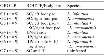
Sixty-three 4–6-week-old male golden hamsters (M. auratus) that weighed 40–60 g were held in appropriate cages with water and food ad libitum throughout the experiment. The animals were divided into groups according to the infection route (subcutaneous = SC or intraperitoneal = IP) and parasite species (L. amazonensis or L. infantum or both). Each animal was inoculated with 107 promastigotes from each species in a volume of 20 µL (SC) or 500 µL (IP). For mixed infections, each animal was inoculated with L. infantum on the left and L. amazonensis on the right side. Uninfected animals were used as a control group. The experiment was performed with seven groups and nine hamsters per group. Table 1 shows the groups, infection route/body site and species of Leishmania.
Experimental groups and respective route and site of infection and Leishmania species inoculated

Clinical follow-up
The animals were followed for 90 days post-infection (d.p.i.) and evaluated at intervals of 30 days. For each time point (0, 30, 60 and 90 d.p.i.), the animals were clinically evaluated for weight, behaviour, irritability, movement, breathing, changes in hair, dermatitis, skin lesion, ascites, spleen enlargement, liver enlargement or any other relevant change. The inoculated footpads were measured using a digital caliber, data expressed as mean ± standard deviation (s.d.). Then, three animals per group were euthanized, livers and spleens were harvested, weighed and measured, and skin and blood samples were collected. Skin, spleens and livers were used for parasite detection and histopathology, and blood was used to obtain serum for immunoglobulin G (IgG) detection by enzyme-linked immunosorbent assay (ELISA) and Western blot.
Parasite detection
To parasite detection in skin, samples were obtained from site of parasite inoculation, i.e. from the footpad in infection via SC, or from the abdomen in infection via IP. The parasite detection was done using microscopy, culture isolation and polymerase chain reaction (PCR).
Microscopy
One fragment from each tissue (skin, spleens and liver) were dried in paper filter, then used to prepare imprint in glass slides. The glass slides were prepared in triplicate for each tissue. The slides were fixed with methanol and Giemsa stained for routine optical microscopy (OM) examination for amastigote forms of the parasite.
Culture isolation
Samples of skin, spleen and liver were collected and immediately seeded into a biphasic NNN culture medium enriched with Schneider's medium, as described above. In order to detect promastigote forms, cultures were maintained at 23 ± 1 °C and examined every 7 days for 30 days. When promastigote forms were observed, the parasites were cultivated just in Schneider's medium, until the exponential growth phase, and then washed [4000 g, 10 min in phosphate buffered saline (PBS) at 20°C]. The pellet was used for DNA extraction and real-time PCR (RT–PCR) and PCR–restriction fragment length polymorphism (PCR–RFLP) assays.
RT–PCR
To quantify parasite burdens in spleen and liver at 90 d.p.i., we used primers (Bretagne et al. Reference Bretagne, Durand, Olivi, Garin, Sulahian, Rivollet, Vidaud and Deniau2001) to amplify a 90-bp fragment of the DNA polymerase gene. This is a single copy-number gene of Leishmania (GenBank accession number AF009147) that was amplified with the following primers: forward: 5′-TGTCGCTTGCAGACCAGATG-3′ and reverse: 5′-GCATCGCAGGTGTGAGCAC-3′. To construction of the calibration curves, serial dilutions (10×) of a fragment containing 109–102 copies were employed. PCR was carried out in a final volume of 10 µL containing 1·25 pm of forward and reverse primers, 1× SYBR GREEN reaction master mix1 (Applied Biosystems, Foster City, CA, USA) and 1 µL (50 ng) of template DNA. The PCR conditions were as follows: an initial denaturation step at 95 °C for 10 min followed by 40 cycles of denaturation at 95 °C for 15 s and annealing/extension at 60 °C for 1 min. To quantify parasite burdens, CT values that were obtained for each test sample were compared with those that were obtained for the corresponding standard curve (Bruna-Romero et al. Reference Bruna-Romero, Hafalla, González-Aseguinolaza, Sano, Tsuji and Zavala2001). The reactions were processed and analysed in a Step One Sequence Detection System device (SDS; Applied Biosystems). Data are expressed as mean ± s.d.
PCR–RFLP
DNA was obtained from Leishmania promastigotes observed in the culture medium from skin, spleen and liver samples, and directly in these tissues. DNA from all samples, including from cultured reference strains of Leishmania held in the cryobank at our laboratory, was extracted using a Wizard kit (Promega, Madison, WI, USA) according to manufacturer's recommendations. In the PCR reaction, the Leishmania DNA target was the internal transcribed spacer 1 (ITS1), and for the RFLP assay, PCR products were digested with the HaeIII enzyme (Promega), according to the method of Schönian et al. (Reference Schönian, Nasereddin, Dinse, Schweynoch, Schallig, Presber and Jaffe2003). Briefly, the samples were analysed for ITS1 using 400 nm primers: LITSR: 5′-CTTGGATCATTTTCCGATG-3′ and L5·8S 5′-TGATACCACTTATCGCATT-3′. The reaction was carried out in 25 µL of total volume. PCR products (10 µL) were digested with the HaeIII enzyme. The amplicons of approximately 300–350 bp were analysed on 1·5% agarose gels visualized by UV light after staining with ethidium bromide (0·3 µg mL−1). The restriction fragments, together with a 50-bp DNA ladder were analysed by 6% polyacrylamide gel electrophoresis (PAGE) and stained with silver, for a comparison of the patterns obtained when the PCR products were digested with the reference strains of L. (L.) amazonensis (IFLA/BR/1967/PH8) or L. (L.) infantum (MHOM/BR/2002/BH401).
Histopathology and immunohistochemistry
Samples of skin, spleen and liver were fixed in 10% neutral buffered formalin. Then, the samples were dehydrated, cleared, embedded in paraffin, cut into 3–4-μm sections, and stained with haematoxylin and eosin (H&E). Immunohistochemistry was performed according to Tafuri et al. (Reference Tafuri, Santos, Arantes, Gonçalves, De Melo, Michalick and Tafuri2004) to detect Leishmania amastigotes in tissue.
Under microscopic analysis, the major histological alterations were as follow: (1) In the skin, epithelial alterations such as acantosis, hypercheratosis, parakeratosis, vacuolar degeneration and intercellular oedema were observed. Chronic inflammatory reactions were observed in the dermis, whereas focal mononuclear exudates were found around vessels and glands or diffuse exudates were found in the upper or deep dermis. (2) In the spleen, the thickness of the capsule and trabeculae system associated with chronic inflammatory exudates was evaluated. In addition, classical depletion of the white pulp (T-dependent area) and hypertrophy and hyperplasia of macrophages of the red pulp were observed. (3) In the liver, we observed a chronic inflammatory reaction characterized by mononuclear exudate localized to the hepatic portal area or the lobules, organized as intralobular granulomas. Classical hypertrophy and hyperplasia of Kupffer cells, as well as regression of hepatocyte lesions as hydropic or steatosis degeneration and necrosis, were also evaluated. All these changes were evaluated semi-quantitatively, with a determination of the extent of changes throughout the histological section, and they were classified as follows: 1, no changes; 2, discrete changes (20–30%); 3, moderate changes (30–60%); and 4, severe changes (>60%).
Humoral immune response
ELISA
Anti-Leishmania IgG titers in serum were determined by ELISA using soluble promastigotes antigen (SLA) from L. infantum (MHOM/BR/2002/BH401) and L. amazonensis (IFLA/BR/1967/PH8). We performed a conventional ELISA (McLaren et al. Reference McLaren, Lillywhite and Sirr1980). Ninety-six well microplates (Eppendorf, Hamburg, Germany) were used with SLA at a concentration of 2 µg well−1. Previously, the optimal serum dilution was determined to be 1:100, and the optimal anti-hamster IgG horseradish peroxidase-labelled (Santa Cruz Biotechnology, Dallas, TX, USA) dilution was determined to be 1:2500. The reactions were developed with SIGMAFAST OPD (Sigma, St. Louis, MO, EUA), and plates were incubated for 30 min in the dark. Reactions were stopped with 4 N H2SO4, and the plates (optical density = OD) were read at 492 nm in a Multiskan Go Reader (Thermo Scientific, Waltham, MA, USA). Statistical analysis was performed using Prism 5·0 software (GraphPad, San Diego, CA, USA).
Western blot
Sodium dodecyl sulphate (SDS)–PAGE was performed in a vertical gel apparatus (Bio-Rad, Hercules, CA, USA). The antigen [SLA from L. infantum (MHOM/BR/2002/BH401) and L. amazonensis (IFLA/BR/1967/PH8)] was boiled for 5 min in sample buffer and immediately subjected to electrophoresis (40 µg well−1) in a 12% PAGE containing 0·1% SDS, as described by Laemmli (Reference Laemmli1970). The gels were run at 70 V for the stacking gel and 100 V for the resolving gel until the bromophenol blue dye migrated to 1 cm from the bottom of the gel in Tris–glycine–SDS buffer (pH 8·3). A Western blot was carried out as described previously (Cuquerella et al. Reference Cuquerella, Gómez-Muñoz and Alunda1991) with few adaptations. Proteins were transferred onto nitrocellulose membranes (Amersham Biosciences, Piscataway, NJ, USA) in a trans-blot semidry transfer unit (GE Healthcare, Chicago, IL, USA) by applying a current of 1·6 mA cm−2 for 45 min. Blots were blocked overnight with blocking buffer (6% low fat milk powder in PBS/0·1% Tween 20), and then washed with PBS containing 0·5% Tween 20. After blocking, blots were incubated for 2 h at 37 °C with pooled hamster sera at a dilution of 1:100 in PBS. After washing, the blots were incubated with a horseradish peroxidase-labelled IgG (Santa Cruz Biotechnology) at a dilution of 1:2500. Plates were incubated at 37 °C for 1 h, and then washed three times at 24°C for 15 min. The immunoreaction was allowed to develop with 3,3′-diaminobenzidine plus 4-chloro-1 naphthol system for 5 min, and then stopped with deionized water. Molecular masses of bands were obtained using Gel Analyzer 2010a software (http://www.gelanalyzer.com) and molecular weight markers (120, 85, 50, 35, 25 and 20 kDa- (Promega).
Statistical analysis
Data analyses were performed using GraphPad Prism 5 software (Version 5.00 for Windows, GraphPad Software, San Diego, CA, USA) and the significance was defined as P < 0·05. Mann–Whitney test was used in RT–PCR, while comparisons of multiple groups were done by the analysis of variance (two-way ANOVA), followed by pairwise comparisons using Student's t-test with a Bonferroni correction.
RESULTS
Clinical assessments
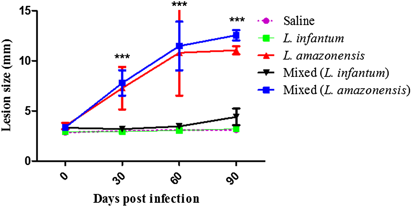
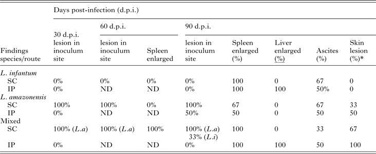
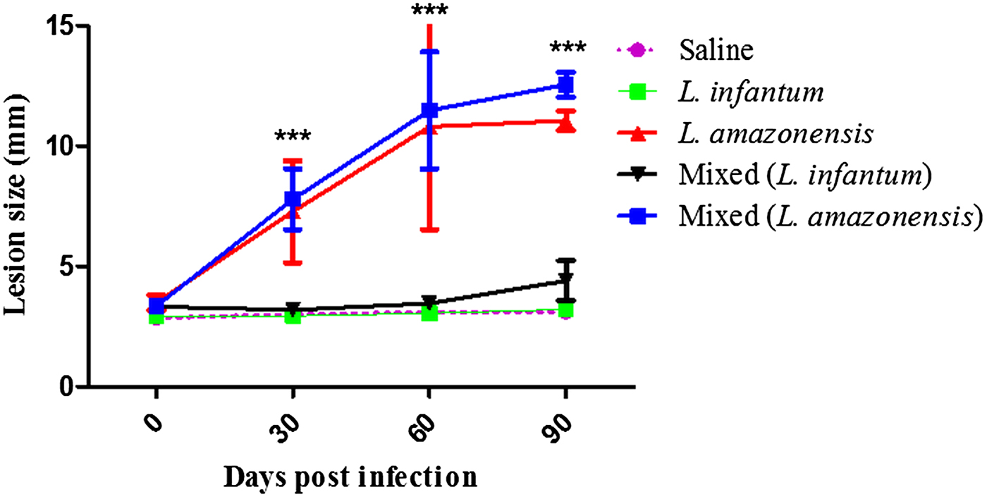
During follow-up of the animals experimentally infected with L. amazonensis, L. infantum and both (mixed infection), only SC infection with L. amazonensis on the footpad resulted in leishmanial lesions; these lesions increased in site were L. amazonensis was inoculated at 30, 60 and 90 d.p.i., either in single infection or in mixed infection (Fig. 1). The animals with mixed infections developed more severe clinical disease. At 90 d.p.i., skin lesions were presented in 100% of the scrota when hamsters were mixed inoculated by the IP route. Infection with L. infantum did not cause skin lesions, even in mixed infections. Enlargement of the spleen was observed in all groups, but was earliest in mixed infections (60 d.p.i.). In contrast, enlargement of the liver was only observed at 90 d.p.i. in animals inoculated IP with L. infantum and with mixed infection. Table 2 shows the percentage of animals with clinical changes during the follow-up period, according to the species of Leishmania and route of infection.
Kinetics of footpad lesion formation during Leishmania amazonensis and L. infantum infection. The Leishmania species were inoculated as single infections or mixed infection. In mixed infection, the species were inoculated in different footpads. Data from each footpad according to inoculated species. Data were expressed as mean ± s.d. Groups were compared using ANOVA, followed by pairwise comparisons using Student's t-test with a Bonferroni correction. Significance was defined as ***P < 0·001.
Percentage of animals with clinical alterations during follow up according to Leishmania species and infection route

(L.a) = Site inoculated with L. amazonensis. (L.i) = Site inoculated with L. infantum. *Skin lesion in uninoculated site. ND, Not done.
Parasite detection
Leishmania was detected in skin, spleen and liver samples by OM on glasses slides, culture isolation and PCR. The samples were collected during the follow-up period, at time points of 30, 60 and 90 d.p.i. In mixed infections, identification of the Leishmania species was made by PCR–RFLP assay directly on tissue samples and on DNA from culture of each tissue (skin, spleen and liver).
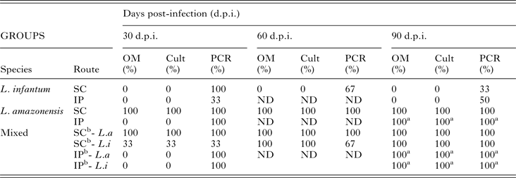
The results of OM and culture were 100% concordant in all tissues. In the skin from hamsters infected with L. amazonensis via SC inoculation, including mixed infections, Leishmania was found beginning at 30 d.p.i. by all methods, with 100% positivity. Infection with L. infantum via the SC and IP routes was only detected in skin by PCR. As expected, PCR presented a higher positivity rate in all groups during the follow-up period. The percentage of samples positive for the parasites in skin by OM, culture and PCR during follow up of the infected groups is shown in Table 3.
Percentage of positive parasite detection in SKIN by optical microscopy (OM), Culture (Cult) and PCR during follow up of groups infected with L. infantum, L. amazonensis and mixed infection

ND, Not done.
a Sample of lesion skin from uninoculum site.
b Each species was inoculated in a different site. L.a = L. amazonensis. L.i = L. infantum.
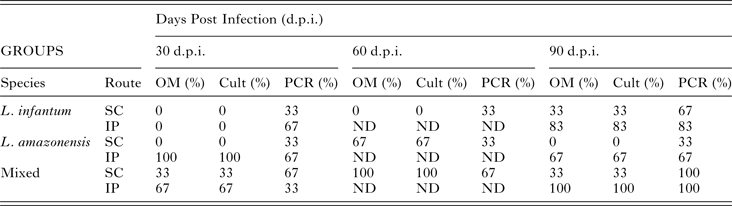
Table 4 shows the parasite detection rates in spleens by OM, culture and PCR during follow up of the infected groups. Hamsters infected via the IP route with L. amazonensis showed 100% positivity by OM and culture at 30 d.p.i. Surprisingly, L. amazonensis, SC route, was detected in only 67% of samples by PCR, and at 90 d.p.i., the positivity rate by OM and culture decreased to 67%, so that the rate was equal to that obtained by PCR. Likewise, lower rates of PCR positivity were observed in mixed infections via the IP route at 30 d.p.i. and via the SC route at 60 d.p.i. It also interesting to observe that, although L. amazonensis was found in spleens from hamsters beginning at 30 d.p.i., in spleens from hamsters infected with L. infantum, the parasite was found only at 90 d.p.i., except by PCR.
Percentage of positive parasite detection in SPLEEN by optical microscopy (OM), Culture (Cult) and PCR during follow up of groups infected with L. infantum, L. amazonensis and mixed infection

ND, Not done.
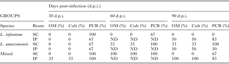
The liver was the tissue that presented the lowest positivity. At 30 d.p.i., 33% of sample from mixed infections via the IP route were positive by OM and culture, whereas 100% were positive by PCR. At this time point, the livers from the others groups were positive only by PCR. Hamsters infected with L. infantum via the SC route and mixed infections (SC and IP) were 100% PCR positive, whereas L. infantum infected via the IP route presented 67% PCR positivity. In animals without mixed infections, similar to the spleen, L. amazonensis was detected in livers earlier than L. infantum. In addition, when L. infantum was IP inoculated, the parasites were more easily detected in the spleen and liver than those infected by the SC route, mainly at 90 d.p.i. The parasite detection rates in livers by OM, culture and PCR during follow up of the infected groups are shown in Table 5.
Percentage of positive parasite detection in LIVER by optical microscopy (OM), Culture (Cult) and PCR during follow up of groups infected with L. infantum, L. amazonensis and mixed infection

ND, Not done.
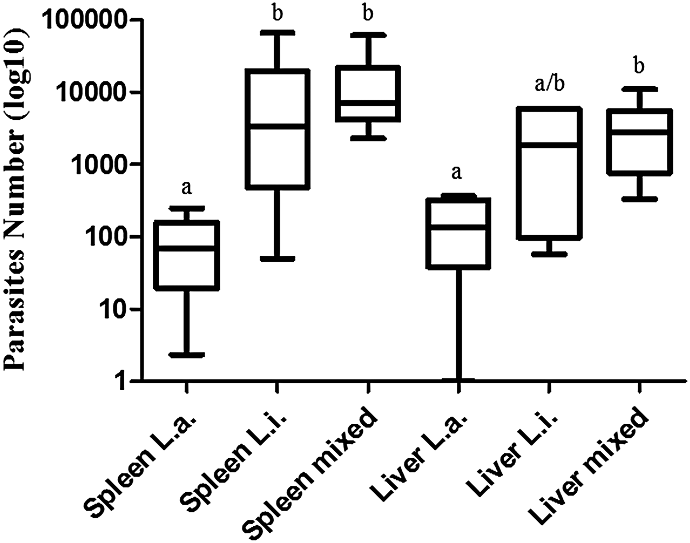
To quantify parasite burdens in spleen and liver at 90 d.p.i. RT–PCR was made. In liver and spleen samples from animals with mixed infection via IP lower dispersion of data was observed, i.e. the upper and lower quartiles are closer to the median than in the other groups, and higher parasitic load when compared mixed infection vs L. amazonensis infection, P < 0·05 (Fig. 2). Likewise, in spleen as well as in liver samples from animals with mixed infection via SC lower dispersion of data was observed, however, without statistical significance (data not shown).
Box and whisker plots of the parasite quantification in hamsters infected with L. amazonensis (L.a.), L. infantum (L.i.) and mixed infection. Amastigote number determined by real-time PCR in spleen and liver samples. The horizontal line within the box indicates the median, boundaries of the box indicate the 25th and 75th percentile, and the whiskers indicate the highest and lowest values of the results. Groups were compared using the Mann–Whitney test. Significance was defined as P < 0·05. Different letters indicated significant differences.

As described above, PCR–RFLP was performed directly on tissue samples and DNA from culture of each tissue (skin, spleen and liver). In groups infected with L. infantum or L. amazonensis, the PCR–RFLP assay was performed, and the results agreed with the species inoculated, it means L. infantum bands with approximately 184, 72 and 55 bp (base pairs) and L. amazonensis bands with approximately 186 and 142 bp (data not shown). The results of the PCR–RFLP assay were very interesting in mixed infections (bands with approximately 186, 142, 72 and 55 bp) via both the SC and IP routes. In these groups, three profiles were observed: L. amazonensis (L.a.), L. infantum (L.i.) and both species (mixed).
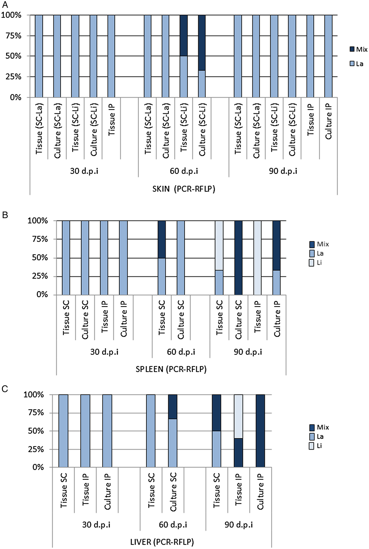
PCR–RFLP results showed that in all skin samples, even in the skin where L. infantum was inoculated and in both tissue and culture samples, only L. amazonensis were detected at 30 and 90 d.p.i. Nonetheless, a mixed profile was observed at 60 d.p.i. (Fig. 3A). At the beginning of infection (30 d.p.i.), L. amazonensis was predominant in the spleen and liver. However, it was very interesting to observe that at 60 and 90 d.p.i., L. infantum was found in tissue and culture, but with a higher frequency in tissue than in culture, mainly in infections via the IP route. In spleens at 60 d.p.i., 100% of the cultures were positive for L. amazonensis, whereas, in tissue, 50% of cultures indicated L. amazonensis and 50% showed a mixed profile. From spleens in the group inoculated via SC route, culture of samples at 90 d.p.i. showed a 100% mixed profile, but in tissues, the mixed profile changed to L. infantum (67%) or L. amazonensis (33%). In the group inoculated via IP route, tissue was found 100% positive for L. infantum at 90 d.p.i., whereas, in culture, 33% of samples were found positive for L. amazonensis and 67% showed a mixed profile (Fig. 3B). In liver samples (Fig. 3C), 100% of the tissue at 60 d.p.i. were positive for L. amazonensis, whereas, in culture, 67% of the samples were positive for L. amazonensis and 33% showed a mixed profile. At 90 d.p.i., the tissue profile changed to 50% L. amazonensis and 50% mixed. In the group inoculated via IP route, 67% of tissues were found positive for L. infantum and 33% were mixed, and in culture, 100% of samples were mixed.
Percentage of each Leishmania species identified in skin (A), spleen (B) and liver (C) from animals with mixed infection of L. infantum and L. amazonensis at 30, 60 and 90 d.p.i.. The hamsters were inoculated via subcutaneous (SC) or intraperitoneal (IP) routes, in different sites of the skin for each species. PCR–RFLP was performed on tissue or culture. La = L. amazonensis; Li = L. infantum; or mix (both species).
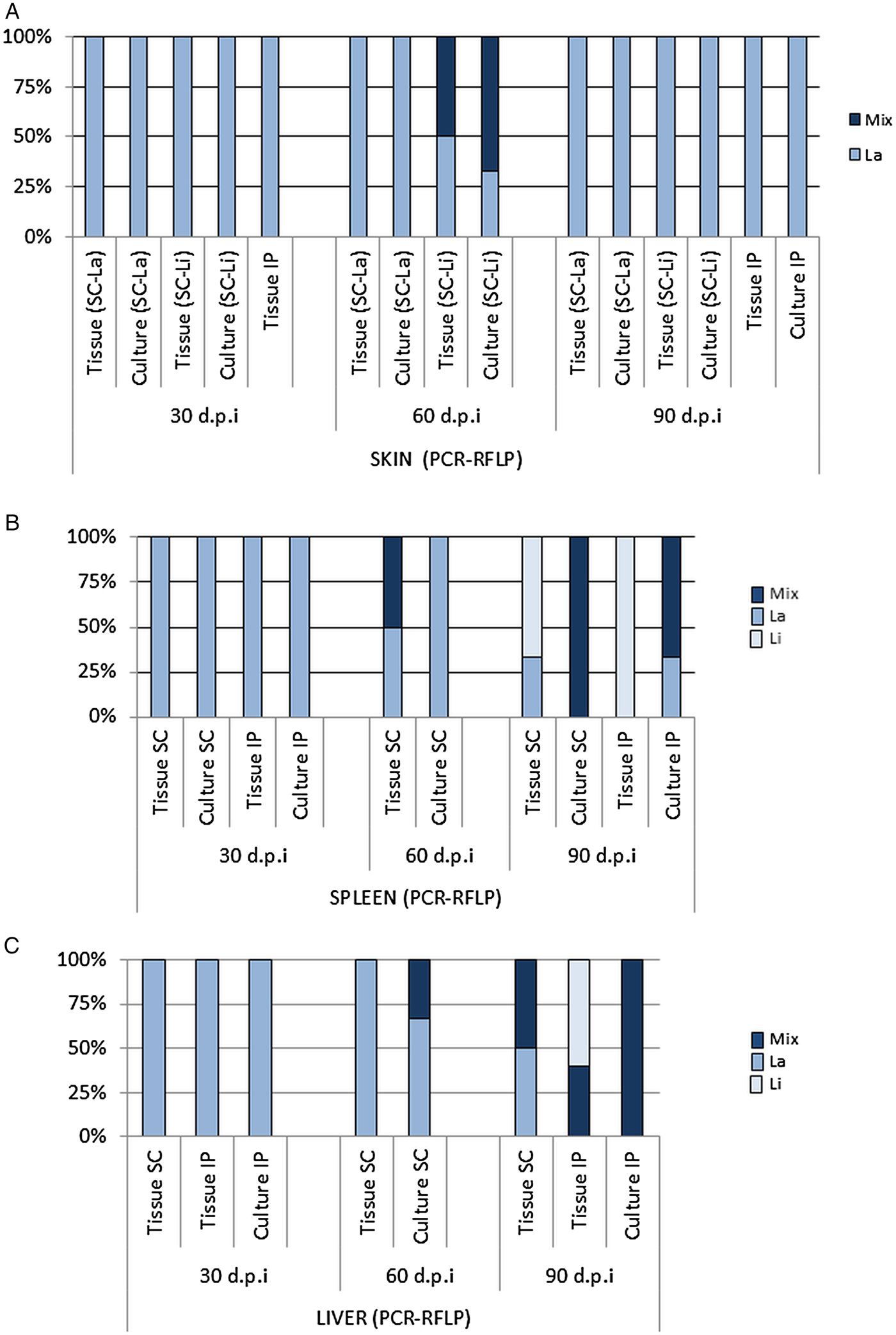
Thus, these results suggest that, in mixed infections with L. amazonensis and L. infantum, L. amazonensis spreads in the skin, appears earlier in the spleen and liver than L. infantum, and grows better in the culture medium than L. infantum. On the other hand, L. infantum is concentrated in the spleen and liver, mainly when inoculated via the IP route, whereas L. amazonensis decreases in these tissues.
Histopathology
Skin
Microscopic analysis of the group of hamsters infected with L. amazonensis showed intense epidermal or dermal alterations. In fact, all animals infected with L. amazonensis showed intense acantosis, hypercheratosis, parakeratosis, vacuolar degeneration and necrosis with the formation of ulcers. An intense and diffuse chronic inflammatory reaction was observed until 90 d.p.i. (30, 60 and 90 d.p.i.), which was associated with numerous Leishmania amastigotes in macrophages in the upper or deep dermis. Interestingly, this inflammation only appeared in the IP-infected group at 90 d.p.i., different from the SC-infected group, in which appeared at 30 and 60 d.p.i.
Spleen
Upon microscopic examination, in all infected groups, spleens showed morphological signs of T-area (white pulp) depletion, whereas the control group did not. Moreover, throughout the experiment, we observed a capsular and trabecular chronic inflammatory reaction, as well as hypertrophy and hyperplasia of macrophages in the red pulp. In general, macrophages were parasitized and vacuolized with the presence of intracellular amastigotes forms of Leishmania. In some areas, macrophages were arranged in nodules (granulomas) in the red pulp. However, these alterations where mainly observed in the mixed infection group at 60 and 90 d.p.i. In addition, parasitism was more evident in this group at 60 and 90 d.p.i. in the IP- and SC-inoculated groups.
Liver
Upon microscopic examination, in all infected groups, livers showed a chronic inflammatory reaction that was absent in controls. At 30 d.p.i., especially in the IP mixed group, we observe a moderate amount of mononuclear exudate of plasma cells, lymphocytes, and macrophages in hepatic portal space. At 60 and 90 d.p.i., this chronic exudate was also noted inside the sinusoid vessels. The exudate formed nodules characterizing intralobular granulomas. Granulomas were easily observed in the mixed infection group. These were constituted mainly of macrophages (epithelioid cells), plasma cells, and lymphocytes. In general, the parasitism was less intense in livers throughout the course of infection than that observed in spleens, and it was always more intense in the mixed infection group.
Humoral immune response
ELISA
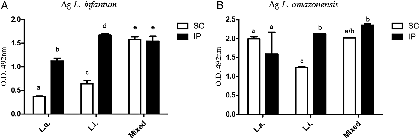
For ELISA, we used two antigens, one from L. amazonensis and one from L. infantum, and serum from all animals infected with L. infantum, L. amazonensis and mixed infection. The O.D. values were slightly higher against antigens from L. amazonensis than antigens from L. infantum in hamsters infected with single species as well as in mixed infection. It was observed that in time point 90 d.p.i. there were significant differences in OD within the groups when compared with 30 and 60 d.p.i. (Supplementary Fig. 1). Results show that in mixed infection IgG levels are high, but not the highest, and not influenced by infection route or antigen used in ELISA. On the other hand, infection with L. infantum induced the highest IgG levels when infection route was IP, but not in SC, and L. infantum antigen was used in ELISA. In addition, IgG levels in L. amazonensis infection are high and not influenced by infection route only when L.a. antigen was used in ELISA (Fig. 4A, B).
ELISA to detected IgG in hamsters experimentally infected with Leishmania amazonensis (L.a.), L. infantum (L.i.), and both species (mixed) by different routes of inoculation. (A) Antigen from L. infantum; (B) Antigen from L. amazonensis. The animals were inoculated via subcutaneous (SC) or intraperitoneal (IP) routes and serum samples were obtained at 90 d.p.i. Two-way ANOVA was performed to analyse the OD values. For all comparisons, P < 0·05 was considered statistically significant. Data are presented as mean ± standard deviation (s.d.). Different letters indicated significant differences.

Western blot
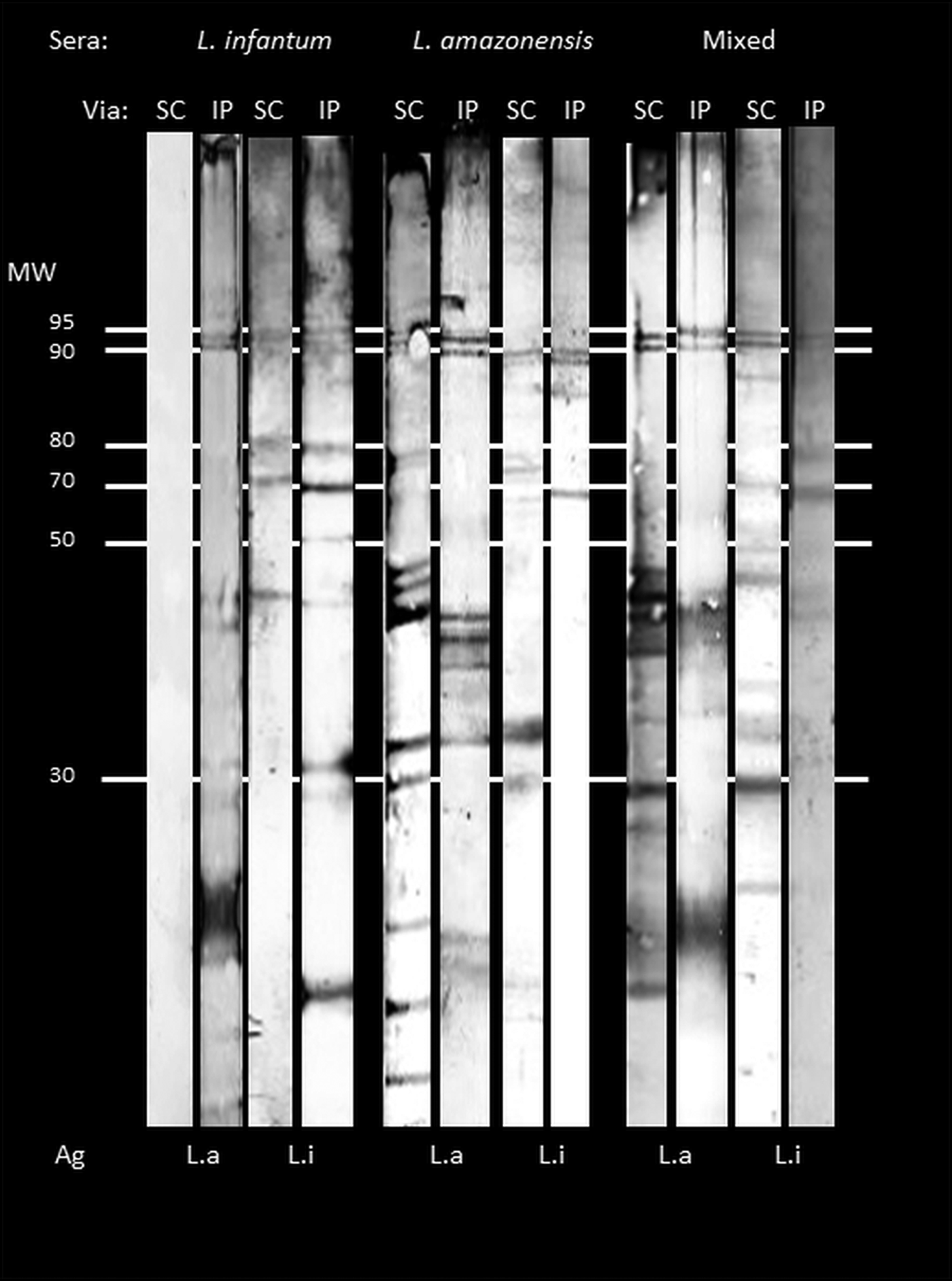
In order to contribute to humoral immune response data, a Western blot was conducted similar to the ELISA, except that pooled serum (not individual) was used. The bands were most clearly defined using serum from 90 d.p.i. in all groups (data not shown). By ELISA, the IgG reactivity was found to be greater against L. amazonensis antigens; in the Western blot, the bands were more defined for L. infantum antigens, except with serum from mixed infections in which both antigens resulted in well-defined bands. Similar to the ELISA results, the Western blot using serum from hamsters with L. infantum inoculated via the IP route showed a larger number of reactive bands than that from hamsters inoculated via the SC route, whereas hamsters infected with L. amazonensis SC showed greater reactivity following IP inoculation (Fig. 5).
Western blot – L. infantum (L.i.) and L. amazonensis (L.a.) antigens (Ag) were fractionated in a 12% SDS–PAGE, transferred to nitrocellulose strips, incubated with sera from hamsters experimentally infected with L. amazonensis, L. infantum, and mixed (both) via subcutaneous (SC) or intra-peritoneal (IP) routes. Western blots were developed with anti-total IgG antibody conjugated with horseradish peroxidase. Molecular weights (MW) in kDa. The molecular masses of bands were obtained using Gel Analyzer 2010a software (http://www.gelanalyzer.com).

The use of antigens from L. infantum and L. amazonensis was important to verify that the humoral immune response, represented by IgG, was induced by several antigenic proteins, of which some are shared and some are specific to either L. amazonensis or L. infantum extract. In other words, it is possible that some antigens can be used to differentiate between infections with L. infantum and L. amazonensis. Only serum from hamsters infected with L. amazonensis reacted with proteins with molecular masses lower than 50 kDa. On the other hand, serum from hamsters infected with L. infantum reacted with proteins with molecular masses of 80, and 70 kDa in the L. infantum protein extract. These antigens (80, 70 and >50 kDa) were also reactive with serum from hamsters with mixed infections. The proteins with molecular masses of 95 and 90 kDa showed higher immunogenicity once they were reactive in both extracts and in a majority of sera groups. No bands were visualized using sera from hamsters SC-inoculated with L. infantum and using L. amazonensis antigen (Fig. 5).
DISCUSSION
In endemic regions, multiple species of Leishmania may coexist, however, the extent to which mixed infections are maintained in a natural host population is not known. Therefore, mixed infections in leishmaniasis could be a frequent phenomenon. Mixed infection might also account for differences in the clinical course of leishmaniasis in many patients and may be a reason for treatment failures (Antoniou et al. Reference Antoniou, Doulgerakis, Pratlong, Dedet and Tselentis2004).
The results demonstrated that mixed infections were associated with more severe clinical disease than either L. amazonensis or L. infantum alone, regardless of the route of infection (SC or IP). Nonetheless, liver and spleen parasitic load in the animals from mixed infection via IP presented higher values and lower dispersion than in the other groups, however with a significant difference only when compared with single infection with L. amazonensis. Besides, infection with L. infantum was more successful via the IP route, whereas L. amazonensis was more successful via the SC route. In infections with Leishmania spp., the IP route has been shown to better with species that cause the visceral form of the disease, whereas the SC or intradermic routes were shown to be more appropriate for dermotropic species (Evans, Reference Evans1989). We also observed that mixed infections were associated with ulcers in the scrotum and earlier enlargement of the spleen, probably as a result of L. amazonensis dissemination, as demonstrated by PCR–RFLP assay. We hypothesized that mixed infections could better suppress host immunity, allowing the parasites to multiply and impairing the macrophage effector function causing more severe disease. The other clinical findings described in the present work are in agreement with data from earlier studies (Abreu-Silva et al. Reference Abreu-Silva, Calabrese, Cupolilo, Cardoso, Souza and Gonçalves Da Costa2004; Bruzual et al. Reference Bruzual, Arcay and Parte-Pérez2008; das Moreira et al. Reference das Moreira, Vitoriano-Souza, Roatt, de Vieira, Ker, de Oliveira Cardoso, Giunchetti, Carneiro, de Lana and Reis2012).
L. amazonensis is able to disseminate to the skin and cause lesions, and we found that in 100% of cases, the parasite was detected earlier in the spleen and liver than with L. infantum infection, mainly when inoculated via the IP route. Likewise, SC inoculation of L. amazonensis caused progressive, localized lesions more commonly in mixed infections. This was also observed in rodents with L. amazonensis in spleens and livers (Abreu-Silva et al. Reference Abreu-Silva, Calabrese, Cupolilo, Cardoso, Souza and Gonçalves Da Costa2004; Bruzual et al. Reference Bruzual, Arcay and Parte-Pérez2008), however, in present work, is the first evidence that the visceral tropism is associated with faster dissemination of L. amazonensis than L. infantum.
As demonstrated by PCR–RFLP, in spleens from hamsters with mixed infections, L. infantum outcompeted L. amazonensis in tissues after 90 d.p.i., but not in cultures from tissues. Likewise, results of the PCR–RFLP from mixed infections were not the same for culture or tissue samples. In culture, L. amazonensis was more frequently detected than L. infantum in the skin, spleen and liver. Previous in vitro work has shown that in cells with mixed infections of L. amazonensis and L. infantum, L. amazonensis is dominant (Abdullah et al. Reference Abdullah, Flath and Presber1998). In addition, it has been suggested that L. amazonensis is better adapted than L. infantum to grow in culture medium and will become the predominant species in mixed infections (Martinez et al. Reference Martinez, Mollinedo, Torrez, Muñoz and Bañuls2002).
Finally, we used two techniques to evaluate humoral immune responses to infection with these species. We demonstrated increased levels of IgG in animals with L. infantum; this effect was more apparent in animals that were inoculated via the IP rather than the SC route. On the other hand, increased levels of IgG detected in animals infected with L. amazonensis were more apparent using L. amazonensis antigen. Increased IgG levels in hamsters infected with L. infantum have already been described, but different inoculation routes were not used or compared (Requena et al. Reference Requena, Soto, Doria and Alonso2000). Nevertheless, in mixed infections, the number of reactive bands in the Western blot depended on the antigen. If antigen from L. infantum was used, sera from hamsters infected IP showed more bands, whereas if antigen from L. amazonensis was used, sera from hamsters infected SC produced more bands. Furthermore, IgG may be induced by several antigenic proteins corresponding to the species of Leishmania, and this may be influenced by the route of inoculation. Although, more than 30 bands were revealed in the Western blot, no bands were visualized using sera from hamsters SC infected with L. infantum and L. amazonensis antigen. In contrast, by ELISA, sera from hamsters SC infected with L. infantum reacted with L. amazonensis antigen. We speculate that the negative Western blot could be a result of low molecular weight proteins that did not transfer and/or migrate on the gel.
Another interesting result was that the proteins with molecular masses of 95 and 90 kDa showed higher immunogenicity; these proteins were reactive to both antigens and with the majority of sera groups. Moreover, among more than 30 proteins, the proteins with molecular masses lower than 50 kDa were reactive only with serum from hamsters infected with L. amazonensis. On the other hand, the proteins with molecular masses of 80, and 70 kDa were reactive only with serum from hamsters infected with L. infantum. Taken together, these data suggest that more studies must be carried out to determine whether these antigens are candidates to differentiate between infection with L. infantum and L. amazonensis.
CONCLUSION
To the best of our knowledge, this is the first report of experimental mixed infections of L. infantum and L. amazonensis in hamsters. The findings presented here have important implications regarding the biology of Leishmania and humoral immune response to infection with these organisms. Many aspects of mixed infections still need to be explored, such as infections with different species at different times and the species specificity of co-infection in vectors.
SUPPLEMENTARY MATERIAL
To view supplementary material for this article, please visit https://doi.org/10.1017/S0031182017000464.
ACKNOWLEDGEMENTS
We would like to acknowledge and dedicate this work to Ellen Marcia, in memory, for her attention and care to the animals used in experimental infections, for her invaluable collaboration, and for her unforgettable kindness.
FINANCIAL SUPPORT
This work was supported by Rede Mineira de Biomoléculas (CCB-RED00012-14) and Pro-reitoria de Pesquisa da Universidade Federal de Minas Gerais (PRPq/UFMG). H.M.A. is a CNPq fellow (PQ), J.L.L.C. is a CAPES fellow, and A.P.M.V. is a UNIMONTES fellow. The funding parties had no role in the study design, data collection or analysis, decision to publish or preparation of the manuscript.