INTRODUCTION
Microglia are the mesodermal origin immune effector cells of the brain, first described by Rio-Hortega as a distinct population of glial cells (Rio-Hortega, Reference Rio-Hortega1920, Reference Rio-Hortega1921a,Reference Rio-Hortegab). In the process of development, microglia infiltrate the brain during embryogenesis and early postnatal development where they play important roles in shaping neuronal maturation. Following infiltration, they differentiate from the typical ameboid appearance of their macrophage siblings to a unique ramified morphology (Cuadros and Navascues, Reference Cuadros and Navascues1998; Chan et al., Reference Chan, Kohsaka and Rezaie2007; Ransohoff and Perry, Reference Ransohoff and Perry2009). Microglia stay in this ramified ‘resting’ state until challenged by immune pathogens or other stimuli. However, resting microglia are not dormant. They have intermittent periods of high motility interspersed with static pauses. The motility involves extension and retraction of their numerous and extensive processes (Nimmerjahn et al., Reference Nimmerjahn, Kirchhoff and Helmchen2005), as if they are surveying and testing their micro-environment. In response to a range of stimuli associated with pathological changes, such as inflammation or tissue injury, microglia become rapidly activated, and their random resting movements become directed by selectively extending their processes to the injured site (Davalos et al., Reference Davalos, Grutzendler, Yang, Kim, Zuo and Jung2005). Activated microglia release various neurotrophic factors and cytokines that can modify neuronal circuits (Merrill, Reference Merrill1992; Biber et al., Reference Biber, Neumann, Inoue and Boddeke2007) and conversely express a range of receptors and signaling molecules to respond to neuronally released substances (Biber et al., Reference Biber, Neumann, Inoue and Boddeke2007). Microglia express α-amino-3-hydroxy-5-methyl-4-isoxazolepropionic acid (AMPA) receptors to respond to glutamate (Noda et al., Reference Noda, Nakanishi, Nabekura and Akaike2000), P2 purinoceptors to respond to ATP and other nucleotides (Tsuda et al., Reference Tsuda, Shigemoto-Mogami, Koizumi, Mizokoshi, Kohsaka and Salter2003; Koizumi et al., Reference Koizumi, Shigemoto-Mogami, Nasu-Tada, Shinozaki, Ohsawa and Tsuda2007), cytokine receptors to respond to tumour necrosis factor (TNF)-α and interleukin (IL)-1β (Sawada et al., Reference Sawada, Kondo, Suzumura and Marunouchi1989) and receptors for the major histocompatibility complex (MHC) class antigens and chemokines (Finsen et al., Reference Finsen, Jorgensen, Diemer and Zimmer1993; Bessis et al., Reference Bessis, Bechade, Bernard and Roumier2007; Cullheim and Thams, Reference Cullheim and Thams2007). Activation of these receptors induces signaling cascades in microglia that has been shown to be involved in microglia chemotaxis, activation and phagocytosis. Conversely microglia can release glutamate and ATP, a range of cytokines and growth factors that can affect neural circuits and brain homeostasis or disease. Glutamate receptor activation, for example, triggers TNF-α release from microglia that can be both neuro-protective in ischemic and Alzheimer brain, or neurotoxic in demyelination diseases (Lambertsen et al., Reference Lambertsen, Clausen, Babcock, Gregersen, Fenger and Nielsen2009; Saha et al., Reference Saha, Ghosh, Palencia, Fung, Dudek and Pahan2009). Neutrotrophic factors such as nerve growth factor (NGF), brain-derived neurotrophic factor (BDNF) and neurotrophin-3 are also released by microglia in response to phyiological and pathological stimulation (Coull et al., Reference Coull, Beggs, Boudreau, Boivin, Tsuda and Inoue2005; Biber et al., Reference Biber, Neumann, Inoue and Boddeke2007). ATP and glutamate release can have more acute effects on synaptic function as further discussed below. These are just a few examples to highlight the potential tight relationships between neurons and microglia that have been already demonstrated in several neuroimmune and neurodegenerative diseases (Fuhrmann et al., Reference Fuhrmann, Bittner, Jung, Burgold, Page and Mitteregger2010; Liu et al., Reference Liu, Condello, Schain, Harb and Grutzendler2010; Schwartz and Shechter, Reference Schwartz and Shechter2010). However, such close neuron–microglia interactions may extend beyond those in just pathological conditions, and in this review we will highlight interactions between microglia and neurons and synapses under more physiological conditions, although at times extending to implications for disease pathogenesis or treatment.
MICROGLIA–NEURON INTERACTIONS DURING DEVELOPMENT AND NEUROGENESIS
One of the phagocytic roles of microglia is to remove apoptotic cells from tissues and this is particularly important in the extensive programmed cell death that occurs during development (Ashwell, Reference Ashwell1990; Marin-Teva et al., Reference Marin-Teva, Cuadros, Calvente, Almendros and Navascues1999; Rakic and Zecevic, Reference Rakic and Zecevic2000). Microglia are more, however, than simply cleaning up the apoptoptic mess – activated microglia actively contribute to this neuronal death (Bessis et al., Reference Bessis, Bechade, Bernard and Roumier2007). In developing chick retina, microglia migrate into the neurogenesis zone and trigger apoptosis. Similarly, in rodents, programmed cell death via apoptosis is stimulated by microglia (Marin-Teva et al., Reference Marin-Teva, Dusart, Colin, Gervais, van Rooijen and Mallat2004; Bessis et al., Reference Bessis, Bechade, Bernard and Roumier2007). Microglia produce a wide variety of factors, including NGF, TNF-α, IL-1β, NO (nitric oxide) or glutamate, which can trigger apoptosis in cultured neurons (Bessis et al., Reference Bessis, Bechade, Bernard and Roumier2007). In the cerebellum, microglia stimulate Purkinje cell apoptosis via release of diffusible superoxide ions as a result of a respiratory burst (Marin-Teva et al., Reference Marin-Teva, Dusart, Colin, Gervais, van Rooijen and Mallat2004). In the hippocampus, neurons undergoing apoptosis are in contact with activated microglia expressing both the integrin CD11b and the immunoreceptor DAP12, which are involved in causing superoxide ion production and release that kills the developmental neurons (Wakselman et al., Reference Wakselman, Bechade, Roumier, Bernard, Triller and Bessis2008). Identifying the nature of the interactions between neurons and microglia that mediate these effects in different brain regions may open up new avenues for treating disorders of neuronal development. The hereditary disease, ataxia-telangiectasia, for example, is characterized by immune deficiency and an early onset of debilitating cerebellar motor co-ordination in young patients (Lavin et al., Reference Lavin, Gueven, Bottle and Gatti2007). In animal models the disease is associated with enhanced Purkinje neuron programmed cell death and later degeneration. As in mouse cerebellum, the majority of Purkinje cells undergoing developmental apoptosis are engulfed or contacted by activated microglia (Marin-Teva et al., Reference Marin-Teva, Dusart, Colin, Gervais, van Rooijen and Mallat2004) and the depletion of microglia rescues Purkinje cells from programmed cell death (Marin-Teva et al., Reference Marin-Teva, Dusart, Colin, Gervais, van Rooijen and Mallat2004). In ataxia-telangiectasia, the mutated gene is involved in DNA repair and neuronal death likely occurs via similar apoptotic mechanisms. Indeed, anti-oxidant free radical scavengers are being trialed as a therapy for delaying the debilitating and gradually worsening symptoms in these patients (Lavin et al., Reference Lavin, Gueven, Bottle and Gatti2007).
Apoptotic programmed cell death also occurs in adult neurogenesis, where the majority of the neural progenitor cells that arise from the subgranular zone of the hippocampal dentate gyrus die in the first few days of life (Mattocks and Tropepe, Reference Mattocks and Tropepe2010). Surviving neurons mature and integrate into neuronal circuits where they can become involved in memory formation and learning. An increased proliferation of neural stem cells is observed in certain epilepsies (Parent, Reference Parent2002; Parent et al., Reference Parent, Valentin and Lowenstein2002; Kuruba et al., Reference Kuruba, Hattiangady and Shetty2009), in Huntington's disease (Curtis et al., Reference Curtis, Faull and Eriksson2007) and in stroke (Jin et al., Reference Jin, Minami, Lan, Mao, Batteur and Simon2001; Koketsu et al., Reference Koketsu, Furuichi, Maeda, Matsuoka, Miyamoto and Hisatsune2006), while decreased number of neural stem cells in the subgranular zone is observed in AD (Rodriguez et al., Reference Rodriguez, Jones and Verkhratsky2009). New-born neurons undergoing apoptosis in the adult central nervous system (CNS) are, as in development, engulfed and phagocytosed by microglia (Sierra et al., Reference Sierra, Encinas, Deudero, Chancey, Enikolopov and Overstreet-Wadiche2010). Interestingly, new-born neurons are efficaciously engulfed by ramified or unchallenged ‘resting’ microglia, as opposed to the activated microglia typically involved in phagocytosis. This observation represents a novel neuronal interaction by ‘resting’ microglia and it will be of interest to compare the signals and mechanisms involved with activated microglial phagocytosis. The Cd11b integrin, for example, was not expressed above basal levels in unchallenged microglia, pointing to distinct pathways involved (Wakselman et al., Reference Wakselman, Bechade, Roumier, Bernard, Triller and Bessis2008; Sierra et al., Reference Sierra, Encinas, Deudero, Chancey, Enikolopov and Overstreet-Wadiche2010). Furthermore, phagocytosis by the resting ramified microglia involved the formation of a phagocytic pouch en passent in distal extensions, as opposed to engulfment by the ameboid cell soma.
Interactions between microglia and new-born hippocampal neurons could have implications for learning and memory under normal and pathological conditions, as suggested by various experiments. Environmental enrichment results in enhanced spatial memory tasks, increased neurogenesis and microglia proliferation and activation (Ziv et al., Reference Ziv, Ron, Butovsky, Landa, Sudai and Greenberg2006; Choi et al., Reference Choi, Veeraraghavalu, Lazarov, Marler, Ransohoff and Ramirez2008). It was proposed that immune T cells infiltrated the neurogenic niches (sub-ventricular and sub-granular zones) to activate microglia and enhance neurogenesis. In immune-deficient mice neurogenesis, microglia proliferation and spatial learning tasks were all impaired (Ziv et al., Reference Ziv, Ron, Butovsky, Landa, Sudai and Greenberg2006). The mechanisms involved in this T cell–microglia–neurogenesis–circuit formation signaling pathway were not investigated, although there was some correlation with increased neuronal BDNF. More recently, genetic knockout of the microglial chemokine receptor, Cx3CR1 resulted in decreased neurogenesis and spatial learning under basal conditions (Rogers et al., Reference Rogers, Morganti, Bachstetter, Hudson, Peters and Grimmig2011). Signaling between neuronally derived fractalkine (CxRCL1) and its specific microglial recptor CXC3R1 typically helps maintains microglia in a resting state, and an increase in activated microglia was reported in this study based on an increased volume of IBA+ fluorescence (a specific marker of microglia). Although an increase in microglia proliferation was clearly demonstrated in the Cx3CR1 −/ − , whether increased apoptosis via the phagocytic pouch in unchallenged microglia may contribute to the reduced neurogenesis phenotype of this mouse remains an interesting possibility.
A decrease in neurogenesis and impairments of spatial learning and memory is also a hallmark of AD. Mutations in the presenilin 1 gene are associated with some familial early onset AD cases, the same mutations overexpressed in mice are also associated with a decreased capacity for enrichment-induced increases in neurogenesis in the subgranular zone (Choi et al., Reference Choi, Veeraraghavalu, Lazarov, Marler, Ransohoff and Ramirez2008). In microglial cultures isolated from these same enriched presenilin 1 mice, there was a different repoitoire of upregulated and secreted substances that could drive proliferation of neural progenitors, as compared with the control mice. The conclusions were that alterations in these signaling cytokines and other factors secreted by microglia were responsible for the defective neurogenesis in these familial AD cases (Choi et al., Reference Choi, Veeraraghavalu, Lazarov, Marler, Ransohoff and Ramirez2008), and by extension possibly the associated cognitive decline.
Microglia have been implicated in other aspects of the AD pathogenesis, and there is debate as to whether their contribution is neurotoxic (by secreting toxic NO or peroxide ions as above), or neuroprotective, by phagocytosing Aβ or sectreting neurotrophic factors. Accumulation of microglia is seen surrounding β-amyloid deposits (Frautschy et al., Reference Frautschy, Walicke and Baird1991), which in culture at least can directly stimulate microglial-induced neuronal phagocytosis (Neniskyte et al., Reference Neniskyte, Neher and Brown2011). Fuhrmann et al. (Reference Fuhrmann, Bittner, Jung, Burgold, Page and Mitteregger2010) directly visualized both microglia and dying cortical layer II neurons using in vivo time-lapse two-photon imaging in a mouse model of AD. Microglia migrated to, and proliferated around, sites of dying neurons. When the Cx3CR1 signaling pathway was knocked out, there was a marked decrease in the extent of neuronal death while Aβ phagocytosis was unaffected. The results suggested an active role, via the CxCR31 pathway, of microglia in neuronal death. Importantly, the increased microglia surveillance, migration and proliferation all occurred prior to the neuronal elimination, rather than after or co-incidentally as may be expected by a passive phagocytosis.
In conclusion, microglia can affect neurogenesis through multiple mechanisms, phagocytotic activity to mediate apoptosis and by secreting molecules that can affect the proliferation and differentiation of neural progenitors.
MICROGLIA–SYNAPSE INTERACTIONS IN DEVELOPMENT AND DISEASE
Development of neural circuits involves an initial excess of synapses and a subsequent refinement over postnatal development. The refinement involves an activity-dependent loss of many synapses and a strengthening of the remaining ones. Involvement of immune cells such as microglia is implicated by the observation that the classical complement cascade system is needed for this developmental pruning (Stevens et al., Reference Stevens, Allen, Vazquez, Howell, Christopherson and Nouri2007). Activated microglia are known to accumulate in regions undergoing high rates of developmental synapse turnover (Tremblay et al., Reference Tremblay, Lowery and Majewska2010) and to accumulate around brainstem facial nuclei in response to motorneuron axotomy during the initial period that precedes loss of synapses onto the axotomized motorneurons. In both these instances, microglia are thought to contribute to synapse loss. Axotomy of the facial nerve indeed was one of the first demonstrations of interactions between microglia and neural circuits (Blinzinger and Kreutzberg, Reference Blinzinger and Kreutzberg1968). Morphological studies showed microglia processes becoming interspersed between the afferent terminals and the motorneuron suggesting that they literally stripped off the synapses (Blinzinger and Kreutzberg, Reference Blinzinger and Kreutzberg1968). Subsequent studies in an immune (MCSF deficient) compromised mouse failed to affect loss of synapses (Kalla et al., Reference Kalla, Liu, Xu, Koppius, Imai and Kloss2001). To examine whether microglia activation in response to CNS inflammation can trigger synapse loss, Trapp et al. (Reference Trapp, Wujek, Criste, Jalabi, Yin and Kidd2007) injected a priming dose of heat-inactivated bacteria (BCG) into the cerebral cortex of mice, followed by a peripheral injection to evoke a cerebral immune reaction. This led to an extensive and prolonged microglia proliferation and activation, and the activated microglia closely opposed neuronal soma and processes. Although there was no detectable subsequent neuronal loss, the proportion of neuronal surface opposed by synapses was markedly reduced by 45%. There was no difference in the extent of microglia activation or this ‘synaptic stripping’ in mice null for monocyte chemoattractant protein 1, a chemokine apparently implicated in the facila nerve axotomy model (Trapp et al., Reference Trapp, Wujek, Criste, Jalabi, Yin and Kidd2007). In another different model of neuronal pathology, Maezawa and Jin (Reference Maezawa and Jin2010) have shown that excessive release of glutamate by Mecp-2-deficient microglia in a mouse model of Rett syndrome results in excitotoxicity of postsynaptic specializations.
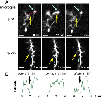
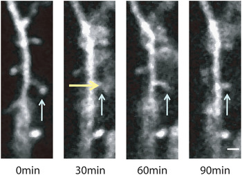
Interactions between synapses and microglia in the postnatal brain have been recently directly visualized (Wake et al., Reference Wake, Moorhouse, Jinno, Kohsaka and Nabekura2009; Paolicelli et al., Reference Paolicelli, Bolasco, Pagani, Maggi, Scianni and Panzanelli2011). Using in vivo two-photon microscopy, we observed that resting microglia made frequent contacts with synapses in the sensory cortices (Fig. 1), and the duration of these contacts increased markedly (from ~5 to ~60 min) in the penumbra of the ischemic cortex, where the brain constructs are intact but have higher frequency of synaptic turnover (Wake et al., Reference Wake, Moorhouse, Jinno, Kohsaka and Nabekura2009). Prolonged time-lapse imaging indicated that some of these contacted synapses were subsequently lost. We proposed that microglial surveillance detected synapse health and could identify synapses that needed to be lost as part of post-ischemia circuit remodeling. Paolicelli et al. furthermore directly demonstrated that microglia actually engulf synapses in the postnatal mouse hippocampus (Paolicelli et al., Reference Paolicelli, Bolasco, Pagani, Maggi, Scianni and Panzanelli2011). Both presynaptic (SNAP-25) and postsynaptic (PSD95) immunogold reactivity could be detected inside microglia using electron microscopy (Paolicelli et al., Reference Paolicelli, Bolasco, Pagani, Maggi, Scianni and Panzanelli2011). The interaction between synapses and microglia was decreased in a Cx3CR1 knockout mouse, indicating this chemokine receptor was involved (Paolicelli et al., Reference Paolicelli, Bolasco, Pagani, Maggi, Scianni and Panzanelli2011). The Cx3CR1 knockout mouse also displayed a transient increase in PSD and spine density, along with a phenotype of functional synaptic properties resembling an immature state, all suggesting that the microglia–synapse interactions formed part of the synaptic pruning process important for circuit maturation. We have also recently observed prolonged microglia–synapse contact during normal cortical development that is associated with a change in the morphology of the postsynaptic spine (Fig. 2), supporting this role in circuit maturation. Mice devoid of the classic complement proteins genes Cq1 and C3 fail to undergo developmental synapse loss, and at retinogeniculate synapses at least, remain multi-innervated (Stevens et al., Reference Stevens, Allen, Vazquez, Howell, Christopherson and Nouri2007). By analogy with the innate immune system, the C1q protein may initiate a cascade in which C3 tags synapses for phagocytosis by microglia. Interestingly, without such synapse elimination the excessive excitatory connections can render the brain hyperexcitable and more vulnerable to seizures (Chu et al., Reference Chu, Jin, Parada, Pesic, Stevens and Barres2010).
Microglial process contacts with synapses. (A) Microglial process (yellow arrow in each image) contacts with presynaptic bouton (blue arrows in top row) and postsynaptic spines (blue arrow in middle row). Images in each rows (-pre, -post) were obtained from single plane free-scanning images at a higher resolution (scale bar 2 µm). (B) The tip of glial process is enlarged during the contact. Left, middle and right panels in each row were obtained before, during and after the contact of the microglial process with synaptic structure. (Modified from Wake et al., Reference Wake, Moorhouse, Jinno, Kohsaka and Nabekura2009.)

Prolonged connection between microglia and synapse change synapse morphology in normal development. Microglia (yellow allow) have prolonged connection with dendritic spine (blue allow) in normal development. After the prolonged connection, some dendritic spine changes their morphology (scale bar 2 µm).

In summary, close interactions between synapses and microglia occur in the brain with implications for synapse homeostasis, development and disease. Microglia contacts with synapses can modify synapse number, and a number of different mechanisms seem to be involved under different conditions. Elucidating these mechanisms and determining the consequences of the interactions for synapse and circuit homeostasis under physiological conditions will be key questions to resolve.
NEURONAL CIRCUIT ACTIVITY AND MICROGLIA–NEURON INTERACTIONS
In vivo imaging technology with fluorescently tagged neuronal elements (Denk et al., Reference Denk, Strickler and Webb1990) has enabled detection and quantification of microglia behavior in the living mouse in real time (Denk et al., Reference Denk, Strickler and Webb1990). Resting microglia were highly motile under normal physiological conditions, extending and retracting their processes within the parenchyma (Nimmerjahn et al., Reference Nimmerjahn, Kirchhoff and Helmchen2005). The speed of process motility observed in different studies in vivo regions is rather variable, from 0.2 to 6.5 µm/min, with an average 2.5 µm/min (Tremblay et al., Reference Tremblay, Lowery and Majewska2010). Processes paused briefly during otherwise continuous motility (Nimmerjahn et al., Reference Nimmerjahn, Kirchhoff and Helmchen2005), which may correspond to the briefer transient contacts with presynaptic boutons and/or postsynaptic spines (Wake et al., Reference Wake, Moorhouse, Jinno, Kohsaka and Nabekura2009; Tremblay et al., Reference Tremblay, Lowery and Majewska2010). Synaptic contacts were prolonged in the ischemic brain (Wake et al., Reference Wake, Moorhouse, Jinno, Kohsaka and Nabekura2009), when synapses may be sub-optimal. In addition, microglial processes preferentially contact smaller dendritic spines, and these smaller contacted spines could either grow marginally or show an increased chance to disappear following microglial contact (Tremblay et al., Reference Tremblay, Lowery and Majewska2010). Again these results point to microglia, somehow detecting subtle differences in the functional state of neuronal elements.
The question arises as to whether some signal is being released by neurons to flag their activity or functional integrity. ATP, for example, locally applied or released from injured tissues is known to act as a chemotractant for microglia (Davalos et al., Reference Davalos, Grutzendler, Yang, Kim, Zuo and Jung2005).
Acute changes in neuronal activity by surface application of ion channel blockers have given inconsistent results: tetrodotoxin had no effect on motility while the γ-aminobutyric acid A receptor blocker bicuculline increased motility (Nimmerjahn et al., Reference Nimmerjahn, Kirchhoff and Helmchen2005). Microglia motility did not change even after long-term potentiation was induced by high-frequency stimulation in the hippocampus in vitro (Wu and Zhuo, Reference Wu and Zhuo2008). However, alterations of sensory evoked neuronal activity have more consistently shown effects on microglia dynamics. Injection of TTX into the eye or a decrease in body temperature (to 32°C) both decreased synaptic activity in the visual cortex in vivo and decreased the frequency of microglia–synapse contacts (from ~1/h to ~0.4/h), without marked changes in process motility speed (Wake et al., Reference Wake, Moorhouse, Jinno, Kohsaka and Nabekura2009). Following a chronic period of sensory deprivation, dark adaptation for 8–10 days, microglia motility was decreased, although with no marked differences in contact frequency (Tremblay et al., Reference Tremblay, Lowery and Majewska2010). Even sensory experience modifies microglia dynamics, it remains open just how this may be signaled to the surrounding microglia – a simple correlation with neuronal activity has not been readily identified.
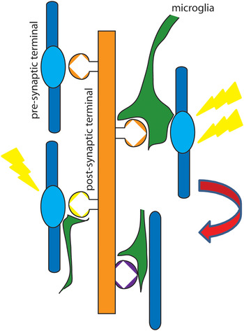
Conversely, what impact may microglia interaction have on synapses and circuits under typical physiological behaviors in a non-injured mature brain? As described above, there is some indication that contact with microglia, particularly if prolonged, may signal an increased chance for subsequent elimination of the synaptic element. Just how resting microglia contacts achieve this, or how this interaction acutely affects neuronal activity, has not been addressed. In contrast, activated microglia have been shown to affect neuronal activity and circuit function and a reasonably clear example of this regards the plasticity of spinal dorsal horn circuits during chronic pain. ATP is released in the spinal cord in response to peripheral nerve crush or injury and the subsequent activation of microglial P2X4 receptors is necessary for tactile allodynia to develop (Tsuda et al., Reference Tsuda, Shigemoto-Mogami, Koizumi, Mizokoshi, Kohsaka and Salter2003). The release of BDNF from spinal microglia activated by ATP signals a decrease in neuronal K+ Cl− cotransporter expression in dorsal horn neurons which results in a neuronal anion gradient shift and a reduced inhibitory efficacy of GABA and glycine signaling that is causative for neuropathic pain (Coull et al., Reference Coull, Beggs, Boudreau, Boivin, Tsuda and Inoue2005). Pascual et al. (Reference Pascual, Ben Achour, Rostaing, Triller and Bessis2012) have recently examined the acute effects of microglia activation by the pro-inflammatory molecule lipo-polysacharide (LPS) on synaptic transmission in isolated hippocampal neurons. Bath application of LPS resulted in an increase in spontaneous excitatory postsynaptic currents and a decrease in the threshold for seizure like bursting. Using a variety of pharmacological and genetic manipulations to elucidate mechanisms, they report that microglia activation results in ATP release that subsequently activates astrocyte purinoceptors (P2Y12) to release glutamate and activate presynaptic mGluRs to enhance glutamate activation of AMPA receptors (Pascual et al., Reference Pascual, Ben Achour, Rostaing, Triller and Bessis2012). The study not only provides one of the few examples of acute effects of microglia activation on neurons, but also demonstrates the important role of glial cells in neuronal circuits. Indeed, the notion of the tripartite synapse comprising astrocytes, spines and presynaptic terminals may need to be elevated to a quadripartite synapse to recognize also the close interactions that microglia play with these synaptic elements (Fig. 3).
Schematic diagram of microglial processes monitoring the state of synapses. In intact brain, microglial processes activity dependently monitor and touch the synapses with a constant duration (~5 min). In the remodeling brain, the contact duration between synapses and microglial processes are markedly prolonged. Prolonged contact by microglia determines the fate of synapses, to disappear or to remain.

DISCUSSION
In this review, we briefly summarized some recent data on the physiological and pathological roles of microglia with a focus on direct interactions between neurons and microglia under physiological conditions. In the developing nervous system, microglia closely interact with neurons to contribute to the developmental apoptosis of excessive neurons and the stripping of synapses. There is evidence to indicate that microglia can direct apoptosis rather than only passively phagocytosing the debris. Microglia can similarly contribute to apoptosis of excess progenitor neurons during neurogenesis in the adult brain, although the underlying mechanisms seem to differ from those observed in development. Understanding these neuron–microglia interactions may have implications for understanding developmental diseases and for diseases where neurogenesis is altered. Abnormal microglial function, especially at early developmental stages, may cause hyper-excitability of neural networks that may contribute to diseases such as Alzheimer's disease and Parkinson's disease. In the healthy mature brain, microglia may contribute to homeostasis of synapses, with recent evidence indicating that their surveillance of synapses can be influenced by sensory activity and seems to be directed toward identifying synaptic elements with less than optimal functional capacity. Indeed, microglia may come to be seen as a fourth member of a quadripartite synapse comprising also boutons, spines and astrocytes. Further elucidating role of microglia in synapse homeostasis and the distinct molecular mechanism that mediate microglia–neuron communication in health and disease are important and exciting challenges.
ACKNOWLEDGEMENTS
We gratefully acknowledge R. Douglas Fields and Olena Bukalo for critical reading. This work was supported by the intramural research program at the National Institute of Child Health and Human Development and by a Japan Society for the Promotion of Science fellowship for H. Wake.
Statement of interest
None.