Introduction
Electroconvulsive therapy (ECT) is an effective and rapid antidepressant treatment for major depressive disorder (MDD) (Group, Reference Group2003; Spaans et al., Reference Spaans, Sienaert, Bouckaert, van den Berg, Verwijk, Kho and Kok2015), but it is only typically used in patients who attempted suicide or failed with numerous medications. Moreover, it has been frequently reported to cause deficits in cognitive functions, especially memory (Vasavada et al., Reference Vasavada, Leaver, Njau, Joshi, Ercoli, Hellemann and Espinoza2017). Although several studies suggested that the side effect of ECT is short term and self-limited (Fujita et al., Reference Fujita, Nakaaki, Segawa, Azuma, Sato, Arahata and Furukawa2006; Semkovska & McLoughlin, Reference Semkovska and McLoughlin2010), there is also a study that reported cognitive deficits persisting in about 6-month after the last ECT (Sackeim et al., Reference Sackeim, Prudic, Fuller, Keilp, Lavori and Olfson2007). The much-feared side effects made ECT to be under-utilized for patients who may otherwise benefit from it to alleviate the clinical symptoms. Moreover, a recent systematic review of neuroimaging studies on ECT in depression reported that the accuracy rate to predict ECT response was 78–90% (Enneking, Leehr, Dannlowski, & Redlich, Reference Enneking, Leehr, Dannlowski and Redlich2020), and most of them lack of validation with independent datasets. Therefore, to uncover accurate prognostic prediction of ECT response is essential to fine-tune ECT to enhance treatment effectiveness and to develop personalized treatment.
Accumulating evidence of structural plasticity in the hippocampus and amygdala induced by ECT in patients with MDD has been widely reported (Bouckaert et al., Reference Bouckaert, De Winter, Emsell, Dols, Rhebergen, Wampers and Vandenbulcke2016; Joshi et al., Reference Joshi, Espinoza, Pirnia, Shi, Wang, Ayers and Narr2016; Ota et al., Reference Ota, Noda, Sato, Okazaki, Ishikawa, Hattori and Kunugi2015; Sartorius et al., Reference Sartorius, Demirakca, Bohringer, Clemm von Hohenberg, Aksay, Bumb and Ende2016; Takamiya et al., Reference Takamiya, Chung, Liang, Graff-Guerrero, Mimura and Kishimoto2018), suggesting potential roles to predict ECT outcomes. Specifically, a longitudinal pilot study on therapy refractory depression showed ECT-related increases in hippocampal and amygdala volumes (Tendolkar et al., Reference Tendolkar, van Beek, van Oostrom, Mulder, Janzing, Voshaar and van Eijndhoven2013). Another similar study revealed increased volume in both hippocampal and amygdala related to symptom improvement, as well as pronounced morphometric changes in the anterior hippocampus and basolateral and centromedial amygdala after ECT in patients with MDD (Joshi et al., Reference Joshi, Espinoza, Pirnia, Shi, Wang, Ayers and Narr2016). In addition, some literature revealed that the structural plasticity of the hippocampus and amygdala determines the therapeutic outcome (Gbyl et al., Reference Gbyl, Rostrup, Raghava, Andersen, Rosenberg, Larsson and Videbech2021; Leaver et al., Reference Leaver, Vasavada, Kubicki, Wade, Loureiro, Hellemann and Narr2020), whereas other studies did not find any associations between structural plasticity and clinical improvements in depressed patients (Gbyl & Videbech, Reference Gbyl and Videbech2018; Oltedal et al., Reference Oltedal, Narr, Abbott, Anand, Argyelan, Bartsch and Dale2018; Sartorius et al., Reference Sartorius, Demirakca, Bohringer, Clemm von Hohenberg, Aksay, Bumb and Ende2019). For example, a previous study revealed that the amygdala volume could well predict depression scores after ECT, but hippocampal volume alone cannot predict depression scores (Ten Doesschate, van Eijndhoven, Tendolkar, van Wingen, & van Waarde, Reference Ten Doesschate, van Eijndhoven, Tendolkar, van Wingen and van Waarde2014). However, using machine learning and data mining, Jiang and colleagues identified six gray matter regions including the hippocampus as predictors of ECT response, and validated in three independent datasets (Jiang et al., Reference Jiang, Abbott, Jiang, Du, Espinoza, Narr and Calhoun2018). These inconsistencies may be resulted from several factors, such as differences in sample size, number of treatments, electrode position, and anatomical locations of the two areas. Since the hippocampus and amygdala were considered as single, homogeneous structures in most studies, the potentially useful information about their sub-regions has been discarded. In fact, the hippocampus and amygdala containing distinct sub-regions responding for different functions, especially for emotion and memory have been well investigated (Kedo et al., Reference Kedo, Zilles, Palomero-Gallagher, Schleicher, Mohlberg, Bludau and Amunts2018; Palomero-Gallagher, Kedo, Mohlberg, Zilles, & Amunts, Reference Palomero-Gallagher, Kedo, Mohlberg, Zilles and Amunts2020; Robinson et al., Reference Robinson, Barron, Kirby, Bottenhorn, Hill, Murphy and Fox2015). However, most studies were performed only focusing on hippocampal sub-regions (Bai et al., Reference Bai, Wei, Xie, Wang, Wang, Ji and Tian2019; Cao et al., Reference Cao, Luo, Fu, Du, Qiu, Yang and Qiu2020; Gbyl et al., Reference Gbyl, Rostrup, Raghava, Andersen, Rosenberg, Larsson and Videbech2021; Laroy et al., Reference Laroy, Emsell, Germann, Dols, Stek, Chakravarty and Bouckaert2019; Leaver et al., Reference Leaver, Vasavada, Kubicki, Wade, Loureiro, Hellemann and Narr2020) rather than amygdala sub-regions or both together. The amygdala is a key structure in the regulation of emotion and memory, and reduced volume of the amygdala has been widely reported to be linked with MDD (Rubinow et al., Reference Rubinow, Mahajan, May, Overholser, Jurjus, Dieter and Stockmeier2016; Wang et al., Reference Wang, Wei, Bai, Zhou, Sun, Becker and Kendrick2017; Yang et al., Reference Yang, Yin, Svob, Long, He, Zhang and Yuan2017). A recent study using high-resolution 7.0T MRI revealed that MDD severity was negatively correlated with decreased volume of amygdala nuclei, i.e. smaller amygdala volume corresponding to worse depressive symptoms (Brown et al., Reference Brown, Rutland, Verma, Feldman, Alper, Schneider and Balchandani2019). Given the diverse functions of the amygdala, delineating the changing pattern of the hippocampus and amygdala at the sub-regional level and their relationship with clinical effects is important to reveal its neural basis for ECT.
Recently, a statistical atlas of the hippocampal (Iglesias et al., Reference Iglesias, Augustinack, Nguyen, Player, Player, Wright and Alzheimer's Disease Neuroimaging2015) and amygdala (Saygin et al., Reference Saygin, Kliemann, Iglesias, van der Kouwe, Boyd, Reuter and Alzheimer's Disease Neuroimaging2017) at the sub-regional level using ultra-high resolution ex vivo MRI combined with in vivo data, as well as a longitudinal segmentation (Iglesias et al., Reference Iglesias, Van Leemput, Augustinack, Insausti, Fischl, Reuter and Alzheimer's Disease Neuroimaging2016), were constructed and available in the version of Freesurfer 6 and above version (https://surfer.nmr.mgh.harvard.edu/fswiki/rel7downloads). The newly developed longitudinal atlas, which removes the confounding inter-subject variability and provides higher sensitivity than the cross-sectional counterparts, is extremely ideal for investigating ECT induced structural plasticity of hippocampal and amygdala at the sub-regional level. Using this pipeline, a recent longitudinal analysis was performed to investigate changes of hippocampal and amygdala sub-fields in patients with treatment-resistant depression undergoing ECT (Gryglewski et al., Reference Gryglewski, Baldinger-Melich, Seiger, Godbersen, Michenthaler, Klobl and Lanzenberger2019). However, the findings of this study were extremely needed to be validated before any conclusions can be made since the sample size is relatively small (n = 14). Moreover, no further analysis was performed to investigate whether these sub-regions could be useful to predict treatment response of ECT.
Therefore, in the current study, we used structural T1 images obtained from two independent samples (Dataset 1: 23 MDD patients before and after ECT from Anhui Medical University, and Dataset 2: 41 MDD patients before and after ECT from the University of Science and Technology of China) and aimed to explore: (1) whether and how ECT induces structural plasticity of hippocampus and amygdala at sub-region level, (2) the specific mechanism underlying antidepressant effects of ECT, and (3) whether these sub-regions are useful to predict treatment response at individual level.
Material and methods
Participants
We presented a prospective study with two independent samples of MDD patients. All these patients were recruited from Anhui Mental Health Center, and the diagnosis, inclusion and exclusion criteria for them assigned to ECT were the same. Specifically, patients showing resistance to drug therapy or a severe suicidal tendency were assigned to ECT. Diagnosis of MDD was evaluated according to the Diagnostic and Statistical Manual of Mental Disorders-IV (DSM-IV) criteria (American Psychiatric Press, 1994). Moreover, we also excluded patients who have: (1) the dependency of any psychoactive substances, (2) a physical or neurological condition affecting brain structure, (3) life-threatening somatic disease, (4) a previous head trauma followed by unconsciousness for more than five minutes, (5) MRI-related contraindications, (6) ECT in the past six months, and (7) any compulsory treatment. At last, a total of 23 MDD remained in dataset 1 and 41 MDD remained in dataset 2 for this study. Since ethical requirement, a majority of patients (61/63) continued to take anti-depression drugs during ECT administrations. All patients provided written informed consent and the study was approved by the local ethics committees of the Anhui Medical University (Approval number: 20140072).
Clinical measurements
The severity of MDD was assessed using the 17-item Hamilton Rating Scale for Depression (HAMD) (Hamilton, Reference Hamilton1960) at two time points: 12–24 h before the first ECT and 24–72h after the last ECT. ECT remitters were defined as >50% reduction in HAMD and final HAMD ⩽7. Otherwise, they were classified into non-remitters. Based on these criteria, 23 remitters and 0 non-remitter were included in dataset 1, whereas 26 remitters and 15 non-remitters were included in dataset 2.
ECT procedures
All patients underwent modified bi-frontal ECT using a Thymatron System IV Integrated ECT Instrument (Somatics, Lake Bluff, IL, USA). Particularly, the first three sessions occurred on consecutive days in the first week, and the remaining was conducted every other day with a break of weekends until patients' HAMD score was no more than 7. During ECT, the initial percent energy was set based on the age of each patient (e.g. 50% for a 50-year-old patient), the stimulation strength was adjusted with an increment of 5% of the maximum charge (approximately 1000 millicoulombs), and the percent energy was increased until seizure was visually observed. All patients were anesthetized with propofol and paralyzed with succinylcholine and atropine to relax the musculature.
MRI data acquisition
All patients underwent two MRI scans at 12–24 h before the first ECT and 24–72 h after the last ECT. The sites, MRI scanners, scanning time, and scanning parameters were not the same. For detail, Dataset 1 was scanned at Anhui Medical University from 2012 to 2014 using a Signa HDxt, GE 3.0T MRI, and Dataset 2 was scanned at the University of Science and Technology of China from 2017 to 2020 using a Discovery GE 750 3.0T MRI. The parameters of the T1 images for dataset 1 were: repetition time (TR) = 8.676 ms, echo time (TE) = 3.184 ms, flip angle = 8°, field of view = 256 × 256 mm2, matrix size = 256 × 256, voxel size = 1 × 1 × 1 mm3, and slices = 188. For dataset 2, T1 images with 188 slices were also acquired with TR = 8.16 ms; TE = 3.18 ms; flip angle = 12°; field of view = 256 × 256 mm2; slice thickness = 1 mm; and voxel size = 1 × 1 × 1 mm3.
Hippocampal and amygdala sub-regions
All T1 images were processed using the standard longitudinal segmentation pipeline available in the Freesurfer 7.1.1 (https://surfer.nmr.mgh.harvard.edu/fswiki/rel7downloads). First, data from the two time-points were longitudinally processed to generate an unbiased within-subject template. This step can significantly reduce the confounding effect of inter-individual variability to avoid biases with respect to any time point. The remaining steps including skull stripping, Talairach registration and initialization of cortical surface reconstruction, cortical atlas registration and subcortical parcellation were initialized using this template (Reuter, Rosas, & Fischl, Reference Reuter, Rosas and Fischl2010). Moreover, gray matter volume for sub-cortical regions, including thalamus, putamen, caudate, pallidum, amygdala, and hippocampus were calculated. Second, we used the longitudinal hippocampal and amygdala subfields tool to delineate sub-regions within the same subject and across all time points. The segmentations of the scans at two time points are jointly computed using Bayesian inference, where each voxel is labeled combing the probabilistic atlas and image intensities (Iglesias et al., Reference Iglesias, Augustinack, Nguyen, Player, Player, Wright and Alzheimer's Disease Neuroimaging2015; Saygin et al., Reference Saygin, Kliemann, Iglesias, van der Kouwe, Boyd, Reuter and Alzheimer's Disease Neuroimaging2017). The probabilistic atlas for amygdala and hippocampus was built with ultra-high resolution ex vivo MRI data (~0.1 mm isotropic) and in vivo manually segmented data. Then, these atlases are mapped to the subject image using an affine, robust registration (Reuter et al., Reference Reuter, Rosas and Fischl2010). Any voxels outside the atlas were not considered for further segmentation. All these procedures are automatically performed in one step with default parameters. Then, we visually inspected the results of subfield segmentation and found no errors in segmentation. More detailed information on the segmentation method could be found in the literature (Iglesias et al., Reference Iglesias, Augustinack, Nguyen, Player, Player, Wright and Alzheimer's Disease Neuroimaging2015, Reference Iglesias, Van Leemput, Augustinack, Insausti, Fischl, Reuter and Alzheimer's Disease Neuroimaging2016; Saygin et al., Reference Saygin, Kliemann, Iglesias, van der Kouwe, Boyd, Reuter and Alzheimer's Disease Neuroimaging2017). Finally, Freeview (https://surfer.nmr.mgh.harvard.edu/fswiki/FreeviewGuide/FreeviewIntroduction) was used to show the amygdala and hippocampal sub-regions.
Longitudinal volume changes
Paired two-samples t tests were used to determine the longitudinal volume changes of subcortical regions, as well as all hippocampal and amygdala sub-regions using SPSS Statistics (Armonk, NY: IBM Corp., v.19.0). The results were corrected by Bonferroni correction (p < 0.05/12 = 4.18 × 10⁻³ for subcortical regions, and p < 0.05/56 = 8.92 × 10⁻⁴ for hippocampal and amygdala sub-regions), respectively.
Correlation analyses
Moreover, for all sub-cortical regions, hippocampal, and amygdala sub-regions related to ECT, correlation analyses between their baseline volume, volume changes and changes of HAMD scores were performed using partial correlation controlling age, gender, and education level. All these analyses were separately performed in two datasets using SPSS. The significance level was set at p < 0.05.
Predictions based on support vector machine
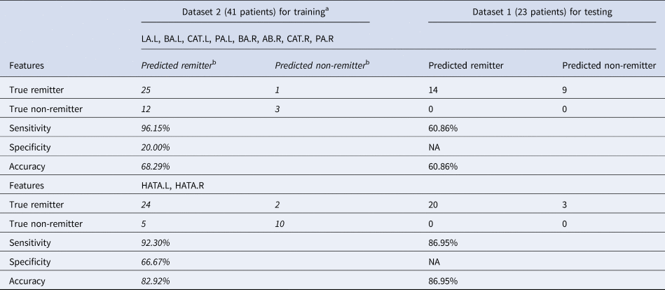
Furthermore, we investigated whether the gray matter volume of ECT-related sub-regions before ECT could predict the treatment response at an individual level. Thus, we used Least Squares Support Vector Machines (LSSVM) with radial basis function (RBF) kernel in MATLAB as a prediction model. Since no non-remitter was included in dataset 1, we used dataset 2 as training and dataset 1 as testing. The normalized gray matter volume ranged to (−1 1) before ECT of those regions showed consistent longitudinal changes were used as input features, including 2 features (HATA.L and HATA.R) for hippocampal subfields and 8 features for amygdala subfields (LA.L, BA.L, CAT.L, PA.L, BA.R, AB.R, CAT.R and PA.R), respectively. The main parameters (gam, sig2) were optimized by using grid research with leave-one-out-cross-validation to train the model. Finally, the model was evaluated by calculating the sensitivity, specificity, and accuracy for both datasets.
Results
The sample and clinical effect of ECT
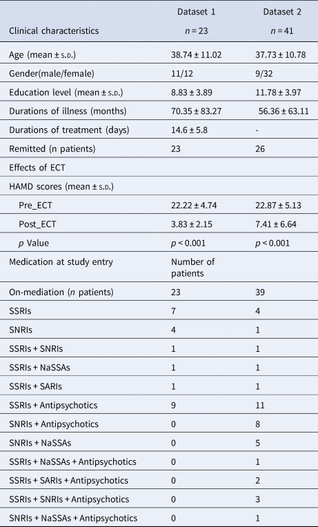
The two samples comprise 64 MDD patients (23 for dataset 1 and 41 for dataset 2) enrolled for ECT (Table 1). Paired t tests were performed on the HAMD, which was significantly decreased in the MDD after ECT treatments in both datasets.
Clinical characteristics of the two samples and the clinical effects of ECT

s.d., standard deviation; ECT, electroconvulsive therapy; HAMD, Hamilton Rating Scale for Depression; SSRIs, selective serotonin reuptake inhibitors; SNRIs, serotonin-norepinephrine reuptake inhibitors; NASSAs, norepinephrine and specificity serotonergic antidepressants; SARIs, serotonin antagonist/reuptake inhibitors.
Hippocampal and amygdala segmentation
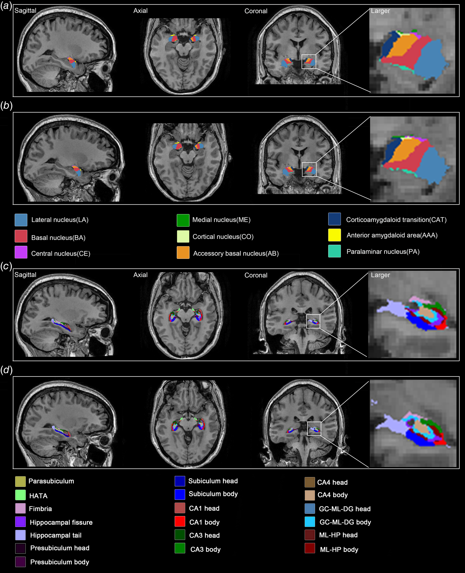
Amygdala segmentation yielded 9 subfields for each hemisphere (Fig. 1a and b). These sub-regions including the bilateral lateral nucleus (LA), basal nucleus (BA), central nucleus (CE), medial nucleus (ME), cortical nucleus (CO), accessory basal nucleus (AB), corticoamydaloid transition (CAT), anterior amygdaloid area (AAA), and paralaminar nucleus (PA).
Segmentation results of amygdala and hippocampus on T1 MRI scans of a major depressive disorders (MDD) patient before and after electroconvulsive therapy (ECT). (a) Nine amygdala sub-regions before ECT, (b) Nine amygdala sub-regions after ECT, (c) Nineteen hippocampal sub-regions before ECT, and (d) Nineteen hippocampal sub-regions after ECT. Abbreviations: HATA, hippocampus-amygdala-transition-area; CA, cornu ammonis; GC-ML-DG, granule cell molecular layer of dentate gyrus; and ML-HP molecular layer hippocampus.

Hippocampal segmentation yielded 19 subfields for each hemisphere including the hippocampal tail, subiculum body, subiculum head, cornu ammonis (CA) 1 body, CA1 head, CA3 body, CA3 head, CA4 head, CA4 body, hippocampal fissure, presubiculum head, presubiculum body, parasubiculum, molecular layer hippocampal (ML-HP) head, ML-HP body, granule cell molecular layer of dentate gyrus (GC-ML-DG) head, GC-ML-DG body, fimbria, and hippocampus-amygdala-transition-area (HATA) (Fig. 1c and d).
Longitudinal volume changes
Gray matter volume of bilateral hippocampus and amygdala were higher in MDD patients after ECT in both datasets (online Supplementary Table S1).
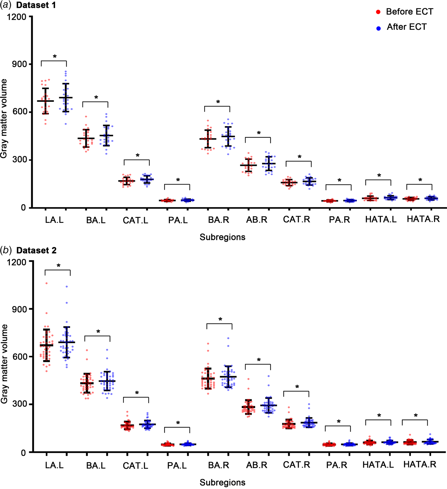
At the sub-regional level, MDD patients after ECT showed significantly increased gray matter volume in the LA.L, AB.L, CAT.L, PA.L, AB.R, BA.R, CAT.R, and PA.R of the amygdala, as well as increased gray matter volume in the HATA.L and HATA.R of the hippocampus (Fig. 2 and online Supplementary Table S1) in both datasets. Moreover, gray matter volume of bilateral head of GC-ML-DG, bilateral head of CA4, and left head of CA3 were significantly increased in dataset 2, but only marginally increased in dataset 1.
The longitudinal gray matter volume changes of the amygdala and hippocampal sub-regions in MDD patients before and after ECT for dataset 1 (a) and dataset 2 (b). Abbreviations of amygdala sub-regions were listed in the Fig. 1. The stars represent a significant difference after Bonferroni corrections (p < 0.05/56 = 8.9 × 10⁻⁴). L for left, and R for right. *Represents significant results.

Correlation between longitudinal changes and clinical effects
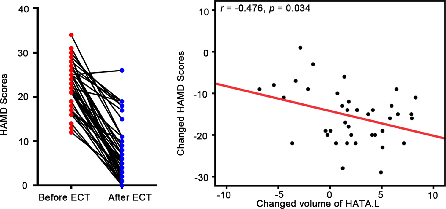
No significant correlation was identified between the volume of the whole hippocampus, amygdala and changed HAMD score. Increased volume of HATA.L was correlated with decreased HAMD score in MDD patients before and after ECT (r = −0.476, p = 0.034, not corrected, Fig. 3) in dataset 2.
Relationship between changes of gray matter volume and clinical measure in MDD patients were mapped. HAMD, the Hamilton Rating Scale for Depression.

Correlation between baseline volumes and clinical effects
No significant correlation was found between baseline volumes of the whole hippocampus, amygdala, or sub-regions and change of HAMD scores.
ECT response and remitters prediction
In addition, the classification of MDD patients as ECT remitters or non-remitters achieved a high degree of precision using gray matter volume of HATA.L and HATA.R before ECT (sensitivity 86.96%, and accuracy 86.96% for dataset 1, and sensitivity 92.30%, specificity 66.67%, and accuracy 82.92% for dataset 2) (Table 2).
Prediction accuracy of ECT remitters for two sites

a The predictive models were trained on Dataset 2 and the sensitivity, specificity and accuracy of Dataset 2 were obtained from leave-one-out-cross-validation. Thus, they were not independent and very likely to be inflated.
b Italic represents Results for training data (Dataset 2).
Discussion
In the current study, we used structural T1 imaging of MDD patients from two independent datasets to explore longitudinal volume changes of hippocampal and amygdala sub-regions induced by ECT, and to further test whether these sub-regions were useful to predict ECT response and remitters at the individual level using machine learning. We found that gray matter volumes of bilateral hippocampus and amygdala were higher in MDD patients after ECT in both datasets. At the sub-regional level, MDD patients after ECT showed selectively and consistently increased volume in the LA.L, bilateral AB, bilateral CAT, bilateral PA, and BA.R of the amygdala, as well as bilateral HATA for both datasets. Further correlation analyses revealed that increased volumes of HATA.L were significantly associated with antidepressant effects after ECT. Moreover, gray matter volumes of these regions in the MDD patients before ECT could serve as potential biomarkers to predict ECT response and remitters with high accuracy in both datasets (i.e. the high accuracy of 86.96% and 82.92% with features of HATA.L and HATA.R).
Effects of ECT on hippocampus and amygdala
As expected, both amygdala and hippocampus showed increased gray matter volume induced by ECT in the present study, which is in line with pathophysiological models of depression (Schmaal et al., Reference Schmaal, Veltman, van Erp, Samann, Frodl, Jahanshad and Hibar2016). In addition, the findings were highly supported by many previous studies with consistent evidence of volume increases for the hippocampus–amygdala complex after ECT compared to baseline (Dukart et al., Reference Dukart, Regen, Kherif, Colla, Bajbouj, Heuser and Draganski2014; Gryglewski et al., Reference Gryglewski, Baldinger-Melich, Seiger, Godbersen, Michenthaler, Klobl and Lanzenberger2019; Jorgensen et al., Reference Jorgensen, Magnusson, Hanson, Kirkegaard, Benveniste, Lee and Jorgensen2016; Ota et al., Reference Ota, Noda, Sato, Okazaki, Ishikawa, Hattori and Kunugi2015; Sartorius et al., Reference Sartorius, Demirakca, Bohringer, Clemm von Hohenberg, Aksay, Bumb and Ende2016). Further meta- and mega-analyses also reported robust structural plasticity of the hippocampus and amygdala following ECT (Oltedal et al., Reference Oltedal, Narr, Abbott, Anand, Argyelan, Bartsch and Dale2018; Ota et al., Reference Ota, Noda, Sato, Okazaki, Ishikawa, Hattori and Kunugi2015; Takamiya et al., Reference Takamiya, Chung, Liang, Graff-Guerrero, Mimura and Kishimoto2018). Moreover, these findings were much in line with previously reported changes following bi-temporal ECT (Cao et al., Reference Cao, Luo, Fu, Du, Qiu, Yang and Qiu2018), but partly similar to studies which showed increased volume only in the right hemisphere following right unilateral ECT (Gryglewski et al., Reference Gryglewski, Baldinger-Melich, Seiger, Godbersen, Michenthaler, Klobl and Lanzenberger2019), further supporting the conclusion that the effect of ECT may be sensitive to electrode placement.
Longitudinal segmentation of hippocampus and amygdala
In the current study, 19 hippocampal subfields and 9 amygdala nuclei were segmented, and gray matter volume of these regions was calculated. The segmentation of 9 amygdala nuclei is extremely consistent with previous studies (Morey et al., Reference Morey, Clarke, Haswell, Phillips, Clausen, Mufford and LaBar2020; Tesli et al., Reference Tesli, van der Meer, Rokicki, Storvestre, Rosaeg, Jensen and Haukvik2020), whereas 19 hippocampal subfields are not always. To our knowledge, hippocampal subfields were mapped according to the main three additional sets of volumes. Specially, 19 hippocampus subfields were summarized into head, body and tail in the first one, no head/body subdivision for second one, and GC-ML-DG and molecular layer are absorbed by the CA subfields in the third one. The majority of previous studies used 12 hippocampus subfields with no head/body subdivision (Myrvang et al., Reference Myrvang, Vangberg, Stedal, Ro, Endestad, Rosenvinge and Aslaksen2018; Postel et al., Reference Postel, Viard, Andre, Guenole, de Flores, Baleyte and Guillery-Girard2019; Wannan et al., Reference Wannan, Cropley, Chakravarty, Van Rheenen, Mancuso, Bousman and Bartholomeusz2019; Xu et al., Reference Xu, Hu, Jiang, Zhang, Wang and Zeng2020), whereas increasing studies adopted 19 hippocampus subfields (Gryglewski et al., Reference Gryglewski, Baldinger-Melich, Seiger, Godbersen, Michenthaler, Klobl and Lanzenberger2019; Phillips et al., Reference Phillips, De Bellis, Brumback, Clausen, Clarke-Rubright, Haswell and Morey2021). In particular, 19 hippocampal subfields and 9 amygdala nuclei were adopted in a recent longitudinal analysis of patients with treatment-resistant depression undergoing ECT (Gryglewski et al., Reference Gryglewski, Baldinger-Melich, Seiger, Godbersen, Michenthaler, Klobl and Lanzenberger2019). To further verify and extent their results, we used the same atlas.
Effects of ECT on amygdala sub-regions
Although increased volume in the amygdala has been widely reported in the MDD patients after ECT (Joshi et al., Reference Joshi, Espinoza, Pirnia, Shi, Wang, Ayers and Narr2016; Takamiya et al., Reference Takamiya, Chung, Liang, Graff-Guerrero, Mimura and Kishimoto2018; Wang et al., Reference Wang, Wei, Bai, Zhou, Sun, Becker and Kendrick2017), fewer investigations were performed at the sub-regional level. In the current study, we found that ECT selectively induced increased volume in the LA.L, AB.L, CAT.L, PA.L, AB.R, BA.R, CAT.R, and PA.R of the amygdala. These findings were consistent with a previous study which showed increased volume in the BA, LA, and CAT of the right amygdala after a series of right unilateral ECT in the treatment-resistant depression (Gryglewski et al., Reference Gryglewski, Baldinger-Melich, Seiger, Godbersen, Michenthaler, Klobl and Lanzenberger2019). It is well known that these regions mainly belong to the basolateral complex (Aghamohammadi-Sereshki et al., Reference Aghamohammadi-Sereshki, Hrybouski, Travis, Huang, Olsen, Carter and Malykhin2019), which is thought to represent an integration center for coordinating inputs from certain cortical and subcortical regions, and is involved in learning and memory (Roozendaal, McEwen, & Chattarji, Reference Roozendaal, McEwen and Chattarji2009). Through the intimate interconnection with the prefrontal cortex, it is a likely source of disturbances in amygdala-prefrontal connectivity in depression (Rubinow et al., Reference Rubinow, Mahajan, May, Overholser, Jurjus, Dieter and Stockmeier2016). Recently, structural MRI at 7 T revealed a significantly negative correlation between right basolateral complexes and depressive symptoms (Brown et al., Reference Brown, Rutland, Verma, Feldman, Alper, Schneider and Balchandani2019). Moreover, a post-mortem study on patients with MDD also reported decreased density of total glia due to oligodendrocytes in the basolateral complex (Hamidi, Drevets, & Price, Reference Hamidi, Drevets and Price2004). The evidence suggested that the basolateral complex may be preferentially altered structurally and functionally in patients with MDD. A recent animal depression model-based study showed that electroconvulsive shocks (ECS) can not only attenuate dendritic arborization (Khaleel, Roopa, Smitha, & Andrade, Reference Khaleel, Roopa, Smitha and Andrade2013), but also selectively down-regulate the expression of voltagegated calcium channels in the basolateral complex (Maigaard, Hageman, Jorgensen, Jorgensen, & Wortwein, Reference Maigaard, Hageman, Jorgensen, Jorgensen and Wortwein2012) to improve neuronal survival (Wildburger, Lin-Ye, Baird, Lei, & Bao, Reference Wildburger, Lin-Ye, Baird, Lei and Bao2009). Taken together, it is likely to speculate that ECT was able to reverse pathological changes in glial cells in patients with depression, which might account for the volume increase in the amygdala sub-regions with ECT.
Effects of ECT on hippocampal sub-regions
In this study, several hippocampal sub-regions showed significantly increased gray matter volume induced by ECT (i.e. bilateral GC-ML-DG head, bilateral CA4 head, and left CA3 head in dataset 2), only the increased volume of HATA.L and HATA.R were consistent in both datasets. The HATA, locating in the medial region of the hippocampus and closely connecting to the amygdala (Amunts et al., Reference Amunts, Kedo, Kindler, Pieperhoff, Mohlberg, Shah and Zilles2005), was considered to have no direct functional implications for a long time (Prasad et al., Reference Prasad, Shah, Bhalsing, Kumar, Saini, Ingalhalikar and Pal2019). Until recently, it has been segmented and defined as a distinct sub-region of the hippocampus available in Freesurfer 6.0 and above versions (Iglesias et al., Reference Iglesias, Augustinack, Nguyen, Player, Player, Wright and Alzheimer's Disease Neuroimaging2015) serving as one of the main targets of the hippocampal-amygdala projection originating in the CA1 (Fudge, deCampo, & Becoats, Reference Fudge, deCampo and Becoats2012). Thus, recently, it has been widely adopted to investigate structural changes in different disorders, such as Parkinson's disease (Wang, Zhang, Yang, Luo, & Fan, Reference Wang, Zhang, Yang, Luo and Fan2019), classic trigeminal neuralgia (Vaculik, Noorani, Hung, & Hodaie, Reference Vaculik, Noorani, Hung and Hodaie2019), essential tremor (Prasad et al., Reference Prasad, Shah, Bhalsing, Kumar, Saini, Ingalhalikar and Pal2019), and amyotrophic lateral sclerosis (Christidi et al., Reference Christidi, Karavasilis, Rentzos, Velonakis, Zouvelou, Xirou and Bede2019), and showed changed volume associated with disease progression. The first study of the new hippocampal sub-regions on patients with MDD might be a gene study, which showed loss volume associated with the FKBP5 gene within the HATA (Mikolas et al., Reference Mikolas, Tozzi, Doolin, Farrell, O'Keane and Frodl2019). Recently, a longitudinal study reported a significant volume increase in bilateral HATA immediately after the ECT series as well as at a six-month follow-up (Gbyl et al., Reference Gbyl, Rostrup, Raghava, Andersen, Rosenberg, Larsson and Videbech2021) providing direct evidence to support our findings. Another study using right unilateral ECT on treatment-resistant depression also showed increased volume in the HATA.L (Gryglewski et al., Reference Gryglewski, Baldinger-Melich, Seiger, Godbersen, Michenthaler, Klobl and Lanzenberger2019). Moreover, correlation analysis in our study also revealed that changed volume of HATA.L was positively correlated with changed HAMD scores after ECT (i.e the more the volume of HATA.L increased, the lower the HAMD scores achieved, and representing better outcomes). This result was supported by a recent publication showing similar correlations in most of the hippocampal sub-regions including HATA (Gbyl et al., Reference Gbyl, Rostrup, Raghava, Andersen, Rosenberg, Larsson and Videbech2021). These findings collectively demonstrated an important role of HATA in depression and response during ECT.
Predicting response and remitters
Individualized prediction of ECT response is the most useful strategy to establish steady biomarkers for clinical applications (Gabrieli, Ghosh, & Whitfield-Gabrieli, Reference Gabrieli, Ghosh and Whitfield-Gabrieli2015). Previous studies focusing on prior presumed regions (i.e hippocampus and amygdala) (Ten Doesschate et al., Reference Ten Doesschate, van Eijndhoven, Tendolkar, van Wingen and van Waarde2014), whole-brain voxel-wise data mining (Jiang et al., Reference Jiang, Abbott, Jiang, Du, Espinoza, Narr and Calhoun2018; Redlich et al., Reference Redlich, Opel, Grotegerd, Dohm, Zaremba, Burger and Dannlowski2016), and brain network (Leaver et al., Reference Leaver, Vasavada, Kubicki, Wade, Loureiro, Hellemann and Narr2020; Qi et al., Reference Qi, Abbott, Narr, Jiang, Upston, McClintock and Calhoun2020) using machine learning have made great progress and reached an accuracy rate nearly 90%. Similar to the current investigation, Cao et.al showed that hippocampal subfield volumes at baseline were able to predict the change in depressive symptoms, and predict robust remission with AUC = 0.90 (Cao et al., Reference Cao, Luo, Fu, Du, Qiu, Yang and Qiu2018). However, the sample size is relatively small with 24 patients with MDD at one site. The model with small samples may not work well for other data sets since over-fitting usually occurs especially when the training samples are limited while features are high dimensional. Since no non-remitter was included in dataset 1, we used dataset 2 as training and further validated in dataset 1. It was really surprising that the prediction accuracy with only two features (i.e. gray matter volume of HATA.L and HATA.R) is comparable with the previous studies using whole-brain gray matter and bran network connectivity as features (Jiang et al., Reference Jiang, Abbott, Jiang, Du, Espinoza, Narr and Calhoun2018; Qi et al., Reference Qi, Abbott, Narr, Jiang, Upston, McClintock and Calhoun2020). Although many potential factors could possibly attribute to the higher accuracy, it is hard to deny the contribution of the newly refined segmentation of hippocampal subfields. This finding provides potential biomarkers (especially HATA) for more effective and timely interventions for ECT in clinical applications.
Limitations
Several major limitations should be stressed in the current study. First, the majority of patients was ongoing psychopharmacological treatment, and took several medications before and during ECT administrations. The potential effects of medications on our results are hard to tell, even though these patients showed resistance to drug therapy. Another issue is that the structural T1 images were scanned using GE 3.0T MRI scanner but with different versions and different scanning parameters. However, the final voxel-level resolution for both datasets is the same (1 × 1 × 1 mm3). Given the Freesurfer processing steps including intensity correction, the results obtained by the two datasets should be the same even though it may exclude the confound effects. Moreover, future studies using ultra-high field MRI (i.e. 7.0T) with higher resolution are warranted for more accurate segmentation, especially for sub-regions with relatively fewer voxels. Finally, we can not train models in dataset 1 and generalized to dataset 2 since no non-remitter was included in dataset 1. The generalization of models to each other needs to be further verified even though we achieved a relatively high accuracy using dataset 2 as training and generalized to dataset 1.
Conclusions
Our attempts were to explore ECT-induced structural changes and predict ECT response and remitters at the individual level using hippocampal and amygdala sub-regions resulted positive outcomes, and further validated in a relatively larger dependent dataset. These results not only suggested that ECT could selectively induce structural plasticity of the amygdala and hippocampal sub-regions associated with antidepressant effects of ECT in MDD patients, but also provided potential biomarkers (especially HATA) for effectively and timely interventions for ECT in clinical applications.
Supplementary material
The supplementary material for this article can be found at https://doi.org/10.1017/S0033291722001337
Financial support
This work was supported by the National key research program (No. 2018YFB1105600), National Natural Science Foundation of China (NO. 62176044, 32071054, 62006220), Anhui Provincial Science Fund for Distinguished Young Scholars (1808085J23), Shenzhen Science and Technology Research Program (No. JCYJ20200109114816594), and the Sichuan Science and Technology Program (No. 2021YJ0186).
Conflict of interest
All authors have no competing interests to declare.