Introduction
Chagas disease and leishmaniasis are considered neglected tropical diseases (NDTs) caused by trypanosomatid parasites that affect millions of people mostly living in the poorest condition areas afflicted by the vector-borne parasites (Nagle et al., Reference Nagle, Khare, Kumar, Supek, Buchynskyy, Mathison, Chennamaneni, Pendem, Buckner and Gelb2014; Zulfiqar et al., Reference Zulfiqar, Jones, Sykes, Shelper, Davis and Avery2017). Moreover, these trypanosomatid diseases, caused respectively by Trypanosoma and Leishmania species, represent one of the global health threats for millions of people especially those originated from Latin American, African and South Asian communities (WHO, 2018a, 2018c). More than 6 million people suffered from Chagas disease, resulting in approximately 10 000 deaths each year in Latin America (WHO, 2018a). On the other hand, among the 97 countries where leishmaniasis is endemic, 90% of new cases were reported in the above-mentioned areas (WHO, 2018b). These diseases became a healthcare problem in the affected communities because of drug-resistance, severe toxicity and long-term treatment of available drugs (Nagle et al., Reference Nagle, Khare, Kumar, Supek, Buchynskyy, Mathison, Chennamaneni, Pendem, Buckner and Gelb2014). The use of plant-based traditional remedy for therapeutic purposes, dates back to millenary and natural products occupy the front line in pharmaceutical drugs discovery (Cheuka et al., Reference Cheuka, Mayoka, Mutai and Chibale2016). Nature biodiversity is an important source of innumerable structural diversity.
Thus, various secondary metabolites previously prospected for their antiparasitic effects led to interesting response to the parasites responsible for these diseases (Izumi et al., Reference Izumi, Ueda-Nakamura, Dias Filho, Júnior and Nakamura2011; Cheuka et al., Reference Cheuka, Mayoka, Mutai and Chibale2016). For instance, the intracellular form of Trypanosoma cruzi was sensitive to naphthyl isoquinolines, biflavanones, acyclic and cyclic diterpenes, sesquiterpenes, monoterpenes, xanthones, anthraquinones and polyacetylenic hydrocarbons (Izumi et al., Reference Izumi, Ueda-Nakamura, Dias Filho, Júnior and Nakamura2011; Cheuka et al., Reference Cheuka, Mayoka, Mutai and Chibale2016). Moreover, remarkable antiparasitic activity was obtained with fisetin (3,7,3′,4′-tetrahydroxyflavone) which inhibited the growth of L. donovani with IC50 value 0.6 µg mL−1 and with high selectivity index (SI of 38.5) towards L6 cells (Tasdemir et al., Reference Tasdemir, Kaiser, Brun, Yardley, Schmidt, Tosun and Rüedi2006). In turn, betulinic acid (IC50 of 73.43 µ m) was more active than benznidazole (IC50 of 218 µ m) on T. cruzi epimastigote after 24 h treatment. An opposite feature was obtained with T. cruzi trypomastigote, which was more sensible to benznidazole than the natural acid. Interestingly, this triterpene showed selectivity towards the host cell LLC-MK2 higher than that of the control (Sousa et al., Reference Sousa, da Silva Souza, Tessarolo, Sampaio, Canuto and Martins2017). On the basis of the above-mentioned factors, it can be observed that plant metabolites from diverse skeletal structures were found with interesting antiprotozoal effects (Izumi et al., Reference Izumi, Ueda-Nakamura, Dias Filho, Júnior and Nakamura2011; Cheuka et al., Reference Cheuka, Mayoka, Mutai and Chibale2016). Therefore, we randomly selected 20 plant secondary metabolites (belonging to flavonoids, triterpenes, furoquinolines, lignans, polyacetylenic thiophenes, cyclohexenes and acetogenins) and evaluated their antiparasitic activities. To the best of our knowledge, the metabolites herein reported have never been evaluated for their trypanocidal and leishmanicidal activities. Therefore, the study describes the antiprotozoal effect of 20 secondary metabolites bearing different chemical skeletons on intracellular T. cruzi and Leishmania amazonensis amastigotes. We also report the in silico interactions observed for abyssinone IV (1) and atalantoflavone (2) as well as their in vitro effect on trypanothione reductase (TR). Moreover, the depolarization effects of the mitochondrial membrane of 1 and 2 are also described.
Materials and methods
General
Vacuum liquid chromatography, column chromatography (CC) and thin layer chromatography (TLC) were performed on silica gel 60 (particle size 90%, <45 mm), 200–300 mesh silica gel and silica gel GF254 (Merck, Darmstadt, Germany), respectively. Medium pressure liquid chromatography (MPLC) was performed on an FMI Pump QSY (Fluid Metering Inc., New York, USA) with silica RP C18 (Merck, Darmstadt, Germany). A solution of vanillin and sulphuric acid was used to unveil the TLC spots. NMR spectra were recorded on a 300 MHz Ultrashield (Bruker, Massachusetts, USA). The mass value was measured on a Waters (Massachusetts, USA) Xevo G2-S QToF ESI mass spectrometer.
Secondary metabolites’ isolation and identification
Abyssinone IV (1) and atalantoflavone (2) were obtained from Erythrina sigmoidea (Kuete et al., Reference Kuete, Sandjo, Djeussi, Zeino, Kwamou, Ngadjui and Efferth2014), eriodictyol (3) was isolated from Vernonanthura tweedieana (da Silva et al., Reference Da Silva, Faqueti, Reginatto, Dos Santos, Barison and Biavatti2015), while compound 4 was identified from Thecacoris annobonae (Guedem et al., Reference Guedem, Sandjo, Opatz, Schollmeyer and Ngadjui2012) and donellanic acids A (5) and B (6) were isolated from Donella ubanguiensis (Djoumessi et al., Reference Djoumessi, Sandjo, Liermann, Schollmeyer, Kuete, Rincheval, Berhanu, Yeboah, Wafo and Ngadjui2012). Taraxerol (7), omphalocarpoidone (8) and spinasterol (10) were identified from Tridesmostemon omphalocarpoides (Fru et al., Reference Fru, Sandjo, Kuete, Liermann, Schollmeyer, Yeboah, Mapitse, Abegaz, Ngadjui and Opatz2013) while β-sitosterol (9) was obtained from the aerial parts of Mimosa invisa (Nana et al., Reference Nana, Sandjo, Keumedjio, Kuete and Ngadjui2012). While, the phytochemical study of Ficus conraui (Kengap et al., Reference Kengap, Kapche, Dzoyem, Simo, Ambassa, Sandjo, Abegaz and Ngadjui2011) and Omphalocarpum elatum (Sandjo et al., Reference Sandjo, Fru, Kuete, Nana, Yeboah, Mapitse, Abegaz, Efferth, Opatz and Ngadjui2014a) furnished 6β-hydroxystigmasta-4,22-dien-3-one (11) and elatumic acid (12), respectively, euphol-3-O-cinnamate (13) was identified from Ficus polita (Kuete et al., Reference Kuete, Kamga, Sandjo, Ngameni, Poumale, Ambassa and Ngadjui2011). A chemical investigation previously performed on Echinops giganteus conducted to the identification of compounds 14 and 15 (Sandjo et al., Reference Sandjo, Kuete, Siwe, Poumale and Efferth2016b), while teclearverdoornine (16) was identified from Zanthoxylum buesgenii (Sandjo et al., Reference Sandjo, Kuete, Tchangna, Efferth and Ngadjui2014b).
Acetogenins (17–20) were obtained from Persea americana seeds (collected in the Douala-Littoral region of Cameroon, voucher specimen no. 16628/SFR/CAM). The organic extract (MeOH/DCM 1/1, v/v, 50 mg) was separated by flash chromatography using hexane (F1), DCM (F2) and MeOH (F3). F1 and F2 were pooled together based on the TLC profile to afford F1′ (46 mg). F1′ was purified by successive CC on silica gel using gradient condition of hexane and ethyl acetate. The hexane fraction (45 mg) was purified by using MPLC on silica RP C18. The separation was performed with a gradient of MeOH and H2O affording heptadec-16-yne-1,2,4-triol (15.3 mg, 17), 2,4-dihydroxyheptadec-16-yn-1-yl acetate (8.9 mg, 18), heptadec-16-ene-1,2,4-triol (12.2 mg, 19) and 2,4-dihydroxyheptadec-16-en-1-yl acetate (6.5 mg, 20), confirmed by comparison of spectroscopic data with the literature (Adikaram et al., Reference Adikaram, Ewing, Karunaratne and Wijeratne1992; Oberlies et al., Reference Oberlies, Rogers, Martin and McLaughlin1998).
Heptadec-16-yne-1,2,4-triol (17): 1H NMR (CDCl3, 300 MHz, TMS); δ H (ppm) J (Hz); 3.57–3.70 (1H, m, H-1a), 3.42–3.54 (1H, m, H-1b), 3.82–3.98 (1H, m, H-2), 1.43–1.63 (6H, m, H-3, H-5 and H-13), 3.93–4.04 (1H, m, H-4), 1.22–1.31 (14H, m, H-6 to H-12), 1.33–1.43 (2H, m, H-14), 2.17 (2H, dt, J = 7.0; 2.6, H-15), 1.94 (1H, t, J = 2.6, H-17) and 2.95 (3OH, brs, OH-1, OH-2 and OH-4). HRESI(+)-QTof MS: m/z 307.2241 [C17H32O3 + Na]+ (calcd. for m/z 307.2249).
2,4-Dihydroxyheptadec-16-yn-1-yl acetate (18): 1H NMR (CDCl3, 300 MHz, TMS); δ H (ppm) J (Hz); 4.11 (1H, dd, J = 12.0; 3.4, H-1a), 3.99 (1H, dd, J = 12.0; 7.6, H-1b), 4.06–4.11 (1H, m, H-2), 1.43–1.61 (6H, m, H-3, H-5 and H-13), 3.82–3.93 (1H, m, H-4), 1.26–1.45 (16H, m, H-6 to H-12), 1.32–1.43 (2H, m, H-14), 2.17 (2H, dt, J = 7.0; 2.6, H-15), 1.94 (1H, t, J = 2.6, H-17), 2.10 (3H, s, COCH3), 3.45 (OH, brs, OH-2) and 2.76 (OH, brs, OH-4). HRESI(+)-QTof MS: m/z 349.2342 [C19H34O4 + Na]+ (calcd. for m/z 349.2355).
Heptadec-16-ene-1,2,4-triol (19): 1H NMR (CDCl3, 300 MHz, TMS); δ H (ppm) J (Hz); 3.59–3.69 (1H, m, H-1a), 3.45–3.53 (1H, m, H-1b), 3.93–4.02 (1H, m, H-2), 1.44–1.54 (2H, m, H-3), 3.82–3.92 (1H, m, H-4), 1.54–1.64 (2H, m, H-5), 1.22–1.35 (16H, m, H-6 to H-13), 1.34–1.43 (2H, m, H-14), 2.03 (2H, dd, J = 13.9; 6.8, H-15), 5.73–5.89 (1H, m, H-16), 4.95–5.03 (1H, m, H-17a), 4.89–4.95 (1H, m, H-17b), 1.87 (OH, brs, OH-1), 3.04 (OH, brs, OH-2) and 2.68 (OH, brs, OH-4). HRESI(+)-QTof MS: m/z 309.2418 [C17H34O3 + Na]+ (calcd. for m/z 309.2406).
2,4-Dihydroxyheptadec-16-en-1-yl acetate (20): 1H NMR (CDCl3, 300 MHz, TMS); δ H (ppm) J (Hz); 3.06–4.15 (2H, m, H-1), 3.95–4.04 (1H, m, H-2), 1.44–1.54 (2H, m, H-3), 3.84–3.94 (1H, m, H-4), 1.54–1.64 (2H, m, H-5), 1.22–1.35 (16H, m, H-6 to H-13), 1.34–1.43 (2H, m, H-14), 2.03 (2H, dd, J = 13.9; 6.8, H-15), 5.73–5.89 (1H, m, H-16), 4.95–5.03 (1H, m, H-17a), 4.89–4.95 (1H, m, H-17b), 2.11 (3H, s, COCH3), 3.41 (OH, brs, OH-2) and 2.69 (OH, brs, OH-4). HRESI(+)-QTof MS: m/z 351.2473 [C19H36O4 + Na]+ (calcd. for m/z 351.2511).
Antiparasitic assays
The biological assays were performed on intracellular amastigote form of the parasites L. amazonensis clinical cutaneous form (MHOM/BR/77/LTB0016) and the standard T. cruzi (Tulahuen strain found in Chilean triatomine bugs) as we previously reported (Pollo et al., Reference Pollo, de Moraes, Cisilotto, Creczynski-Pasa, Biavatti, Steindel and Sandjo2017). Therefore, T. cruzi trypomastigotes expressing β-galactosidase were grown in infected L929 cell line maintained at 37 °C, 5% CO2 in RPMI-1640 medium supplemented with 5% FBS while L. amazonensis promastigotes expressing β-galactosidase were grown at 26 °C in Schneider's insect medium (Sigma Chemical Co., St. Louis, MO, USA) supplemented with heat inactivated 5% FBS and 2% human urine .
The human leukaemic THP-1 (ATCC TIB202) cell line used as a host cell was cultivated according to the protocol reported in the same previous work (Pollo et al., Reference Pollo, de Moraes, Cisilotto, Creczynski-Pasa, Biavatti, Steindel and Sandjo2017). This cell line was grown at 37 °C, 5% CO2, in RPMI-1640 without phenol red (Sigma-Aldrich, Co., St. Louis, MO, USA) supplemented with 10% FBS, HEPES 12.5 mm, penicillin (100 U mL−1), streptomycin (100 µg mL−1) and Glutamax (2 mm).
For antiparasitic assays, cells were cultivated in 96 well microplates (4.0 × 10⁴ cells/well) in supplemented RPMI-1640 and treated with phorbol 12-myristate 13-acetate 100 ng mL−1 for 72 h (37 °C, 5% CO2) to reach cell differentiation into non-dividing macrophages. Four days old culture promastigotes of L. amazonensis (4.0 × 10⁶ parasites mL−1) was washed with phosphate buffered saline (PBS, pH 7.4) and incubated in RPMI-1640 supplemented with 10% of heat inactivated human B+ serum for 1 h (34 °C) to parasite opsonization. THP-1 cells were then incubated with a parasite/cell ratio of 10:1 for 3 h (34 °C, 5% CO2) for L. amazonensis and with a parasite/cell ratio of 2:1 overnight (37 °C, 5% CO2) for T. cruzi. Thereafter, non-adherent parasites were removed by two washes with PBS and infected cells were incubated with 180 µL of full supplemented RPMI-1640 medium for another 24 h (34 °C or 37 °C, 5% CO2) to allow the transformation of promastigotes/trypomastigotes into intracellular amastigotes. The infected cells were treated with 20 µL of each compound serially diluted in dimethylsulphoxide (DMSO) 50 µ m to 1.56 µm in triplicate, followed by incubation for 48 h (34 °C or 37 °C, 5% CO2). Subsequently, cells were carefully washed with PBS and incubated for 16 h (34 °C, 5% CO2) with 250 µL of chlorophenol red-β-d-galactopyranoside (Sigma-Aldrich Co., St. Louis, MO, USA) at 100 µm and 0.1% Nonidet P-40 (Amresco Inc., Solon, Ohio, USA) (NP-40). Optical density was read at 570/630 nm using an Infinite M200 TECAN, Austria. Amphotericin B (Bristol-Myers, Squibb) and benznidazole (Sigma Aldrich) were used as positive controls and 1% DMSO as a negative control. The concentration of each sample that reduced parasite viability by 50% when compared to untreated control (IC50) was estimated by non-linear regression of concentration–response curves using the GraphPad Prism program.
Cytotoxicity assay
THP-1 cell line was cultivated in 96 well microplates (4.0 × 10⁶ cells/well), and treated with the compounds serially diluted in concentrations ranging from 500 to 15.6 µ m and incubated for 72 h (37 °C, 5% CO2). Cell viability was assessed by the MTT assay. 1% DMSO (Merck), benznidazole (Sigma Aldrich) and amphotericin B (Squibb) were used as negative and positive controls, respectively. The optical density was read at 540 nm using an Infinite M200 TECAN microplate reader immediately after the dissolution of formazan crystals with DMSO. 1% DMSO (v/v) and 50% DMSO (v/v) were used as negative and positive controls, respectively. The concentration of each sample that reduced cells viability by 50% (CC50) was calculated by a non-linear regression using the GraphPad Prism program. The SI of each active compound to THP-1 (human monocytic cell line) was found by using the following ratio (SI = CC50/IC50).
Molecular docking
The structure of TR of T. cruzi (PDB ID: 4NEW) (Persch et al., Reference Persch, Bryson, Todoroff, Eberle, Thelemann, Dirdjaja, Kaiser, Weber, Derbani and Brun2014) in complex with the inhibitor analogue (5-{5-[1-(pyrrolidin-1-yl)cyclohexyl]-1,3-thiazol-2-yl}−1H-indole, PCTI) of 1-[1-(benzo[b]thien-2-yl)cyclohexyl]piperidine was downloaded from the Protein Data Bank (PDB). The enzyme and compound structures were prepared using the same default parameter settings in the Molegro Virtual Docker, v. 6.0.1 software package (Thomsen and Christensen, Reference Thomsen and Christensen2006) (Score function: MolDock Score; Ligand evaluation: Internal ES, Internal hydrogen bond, Sp2–Sp2 Torsions, all checked; number of runs: 10 runs; algorithm: MolDock s.e.; maximum interactions: 1500; max. population size: 50; max. steps: 300; neighbour distance factor: 1.00; max. number of poses returned: 5). The docking procedure was performed using a GRID of 15 Å in radius and 0.30 Å in resolution to cover the ligand-binding site.
Trypanothione reductase enzymatic assay
Trypanosoma cruzi recombinant TR (TcTR) obtained by using the experimental protocol previously described by Borges et al. (Reference Borges, Cunningham, Tovar and Fairlamb1995). The enzymatic assay was performed in 96-well microplates with TcTR in a final volume of 240 µL containing the reaction buffer (40 mm HEPES, 1 mm EDTA, pH 7.5), TcTR (0.005 mU), 1 µ m T[S]2 (Sigma-Aldrich, Missouri, USA), 150 µm NADPH (Sigma-Aldrich, Missouri, USA) and compounds at 50 µm. 1% DMSO and 100 µ m clomipramine were used as negative and positive controls, respectively. The reaction was incubated at 27 °C for 30 min before addition of 25 µm 5,5-dithiobis-(2-nitrobenzoic acid) (Sigma-Aldrich, Missouri, USA). The enzyme activity was monitored by increase in absorbance at 412 nm using a spectrophotometer (Infinite M200 TECAN, Männedorf, Switzerland) (Hamilton et al., Reference Hamilton, Saravanamuthu, Eggleston and Fairlamb2003).
Mitochondrial membrane potential assay (ΔΨm)
The analysis of changes in mitochondrial membrane potential (ΔΨm) of T. cruzi (Y strain) was carried out by flow cytometry using Rhodamine R123 (Sigma-Aldrich, Missouri, USA). Three concentrations used were chosen with respect of doses that can inhibit 100% of the T. cruzi amastigote and show no or less toxicity towards THP-1 cells. In other words, the chosen concentrations (three times IC50, six times IC50 and ten times IC50) were comprised between IC50 and CC50 values of the active compounds (1 and 2). Trypanosoma cruzi epimastigotes (2 × 10⁶ mL−1) were seeded in 96-well plates and treated with increasing concentrations which were three, six and ten times the IC50 values of compounds 1 (20, 40, 70 µm) and 2 (10, 20, 40 µm) for 2 h. After treatment, parasites were centrifuged at 1,500 g for 10 min and incubated with 100 µL of R123 (5 µg mL−1) in PBS for 15 min at 27 °C. After three washes with PBS, the marker R123 was measured in a FACSCanto (BD Biosciences, New Jersey, USA). A total of 50 000 events were acquired and relative mitochondrial membrane potential was determined by considering the fold change in the geometric mean of fluorescence intensity. The data were subsequently analysed using flowing software. All experiments were performed in triplicate, using 1% DMSO as a negative control and 20 µm carbonyl cyanide-4-(trifluoromethoxy)phenylhydrazone (FCCP) (Sigma-Aldrich, Missouri, USA) as a positive control. One-way ANOVA followed by the Tukey post-test were used to evaluate the differences between the treatment and the DMSO group.
Results
Trypanocidal activity
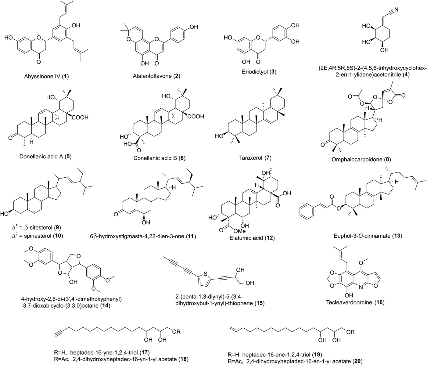
Nine terpenoids, three flavonoids, four acetogenins, one furoquinoline, one lignan, one cyclohexylacetonitrile and one thiophene (Fig. 1) were evaluated against intracellular T. cruzi and L. amazonensis amastigote. Diseases caused by both parasites namely Chagas’ disease and leishmaniasis are clinically treated in Brazil with benznidazole and amphotericin B, respectively (Brasil, 2010). Because in vitro antiparasitic assays with macrophage-amastigotes models are closely related to the pathophysiological conditions of leishmaniasis and trypanosomiasis, the THP-1 cell line was used as the host cell. These cell differentiations mimic the primary macrophages and alternatively represent a good choice to evaluate activities of small molecules on the intracellular amastigote forms of these two parasites (Jain et al., Reference Jain, Sahu, Walker and Tekwani2012).
Compounds evaluated for their antiparasitic effect.

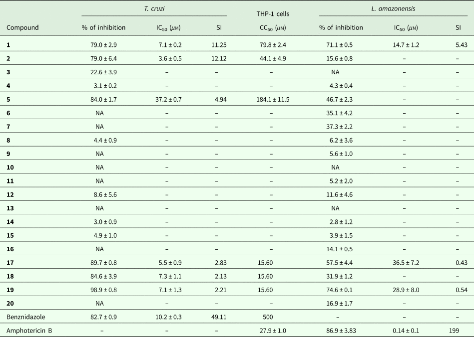
Among them, six compounds showed antitrypanosomal activity with IC50 values of 7.1 ± 0.2 µm for abyssinone IV (1), 3.6 ± 0.5 µm for atalantoflavone (2), 37.2 ± 0.7 µm for donellanic acid A (5), 5.5 ± 0.9 µm for heptadec-16-yne-1,2,4-triol (17), 7.3 ± 1.1 µm for 2,4-dihydroxyheptadec-16-yn-1-yl acetate (18) and 7.1 ± 1.3 µm for heptadec-16-ene-1,2,4-triol (19) (Table 1).
IC50, CC50 and the SI values of the metabolites tested on T. cruzi and L. amazonensis amastigotes.

NA, not active.
Values of IC50 and CC50 are expressed in μ m of mean ± standard deviation (n = 3).
Five of these metabolites presented a lower IC50 value than benznidazole, the reference control (IC50 of 10.2 ± 0.3 µm). Abyssinone IV (1) and atalantoflavone (2) revealed a moderate toxicity to the host cell THP-1 and their SI (11.25 and 12.12, respectively), was almost 4-fold lower than the value (SI of 49.11) obtained for the control. The remaining four active metabolites revealed strong toxicity to the host THP-1 cell.
Eight of the terpenoids including donellanic acid B (6), taraxerol (7), omphalocarpoidone (8), β-sitosterol (9), spinasterol (10), 6β-hydroxystigmasta-422-dien-3-one (11), elatumic acid (12) and euphol-3-O-cinnamate (13) did not show activity on T. cruzi. No antitrypanosomal activity was also observed for eriodictyol (3), cyclohexenylacetonitrile (4), lignan (14), the polyacetylenic thiophene (15) and the furoquinoline (16).
Leishmanicidal activity
Among the tested compounds, three showed leishmanicidal activity against the intracellular L. amazonensis amastigote. Abyssinone IV (1) presented an IC50 of 14.7 ± 1.2 µm and SI of 5.43 (Table 1). Heptadec-16-yne-1,2,4-triol (17) and heptadec-16-ene-1,2,4-triol (19) were weakly active on L. amazonensis with an IC50 of 36.5 ± 7.2 and 28.9 ± 8.0 µm, respectively. Moreover, both acetogenins revealed low selectivity to THP-1 (SI of 0.43 and 0.54, respectively for 17 and 19). No leishmanicidal activities were observed for the terpenoids 5–13 and for the acetogenins 18 and 20, as well as for compounds 2–4 and 14–16.
Molecular docking
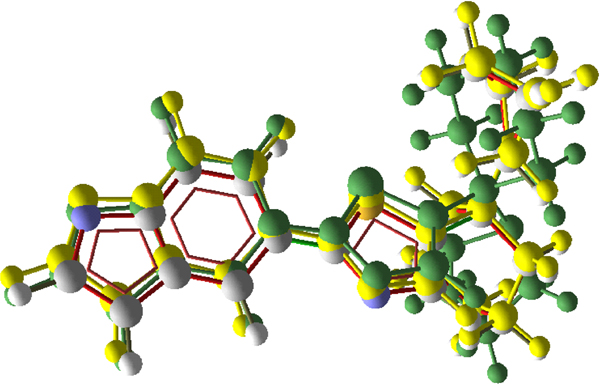
A molecular docking study was considered to understand the trypanocidal effect of compounds 1 and 2. The molecular docking was validated by performing redocking of the original ligand (PCTI) for eight of T. cruzi protein used in this study. Superposition of some poses is represented in Fig. 2, and showed almost perfect match, being the root mean square deviation (RMSD) between the lowest MolDock energy pose and the original ligand of only 1.28.
Superposition of crystal pose (grey) and docking poses (green and yellow) for the inhibitor 5-{5-[1-(pyrrolidin-1-yl)cyclohexyl]-1,3-thiazol-2-yl}−1H-indole (PCTI).
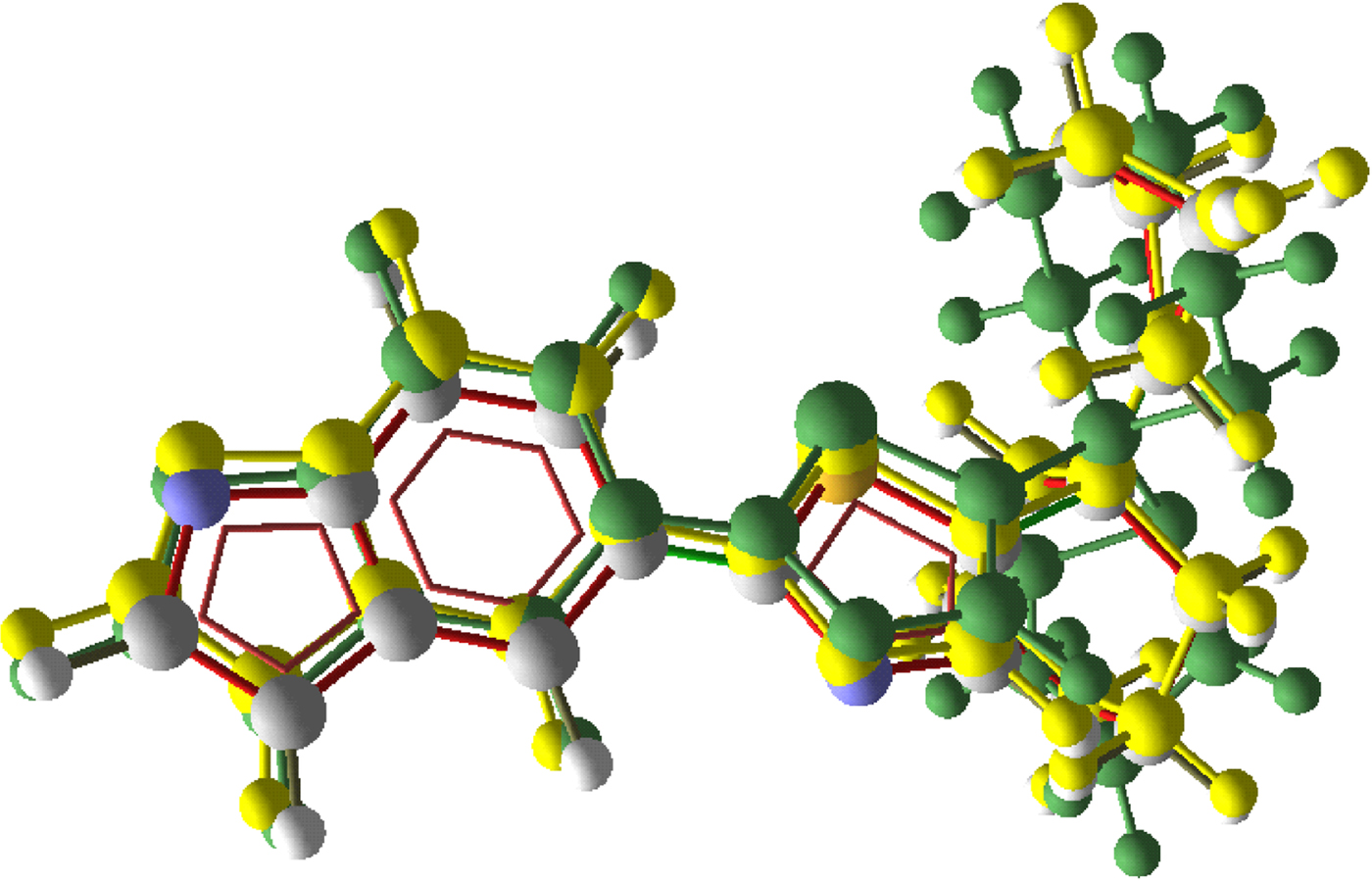
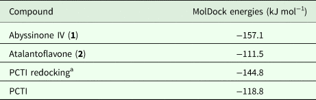
The results presented in Table 2 indicated that both compounds abyssinone IV (1) and atalantoflavone (2) exhibited potential inhibitory of TR since the energies of the crystalized ligand and of its redocking pose were similar. Moreover, the abyssinone IV (1) poses turned to have the lowest MolDock energy (−157.1).
Summary of the MolDock energies of abyssinone IV (1), atalantoflavone (2), PCTI and PCTI redocking from the PDB structure 4NEW

a Redocking RMSD = 1.28. PCTI: 5-{5-[1-(pyrrolidin-1-yl)cyclohexyl]-1,3-thiazol-2-yl}−1H-indole.
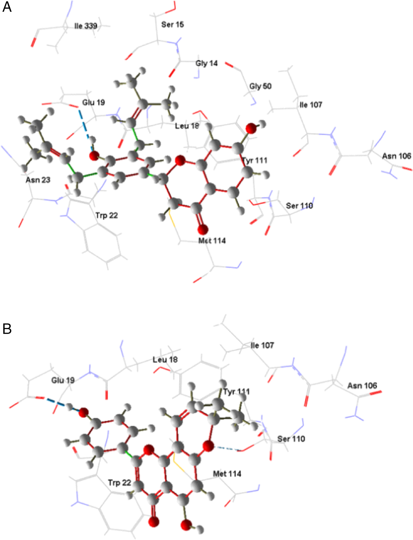
Considering the docking poses for the compounds 1 and 2 with T. cruzi TR, the hydroxyl group of B ring of both compounds present H-bond (hydrogen bond) with Glu18 (Fig. 3a and b), while atalantoflavone (2) interacts with Ser110 through a H-bond of oxygen of pyran ring (Fig. 3b).
Docking poses of abyssinone IV (1, a) and atalantoflavone (2, b) with T. cruzi TR (PDB ID: 4NEW).
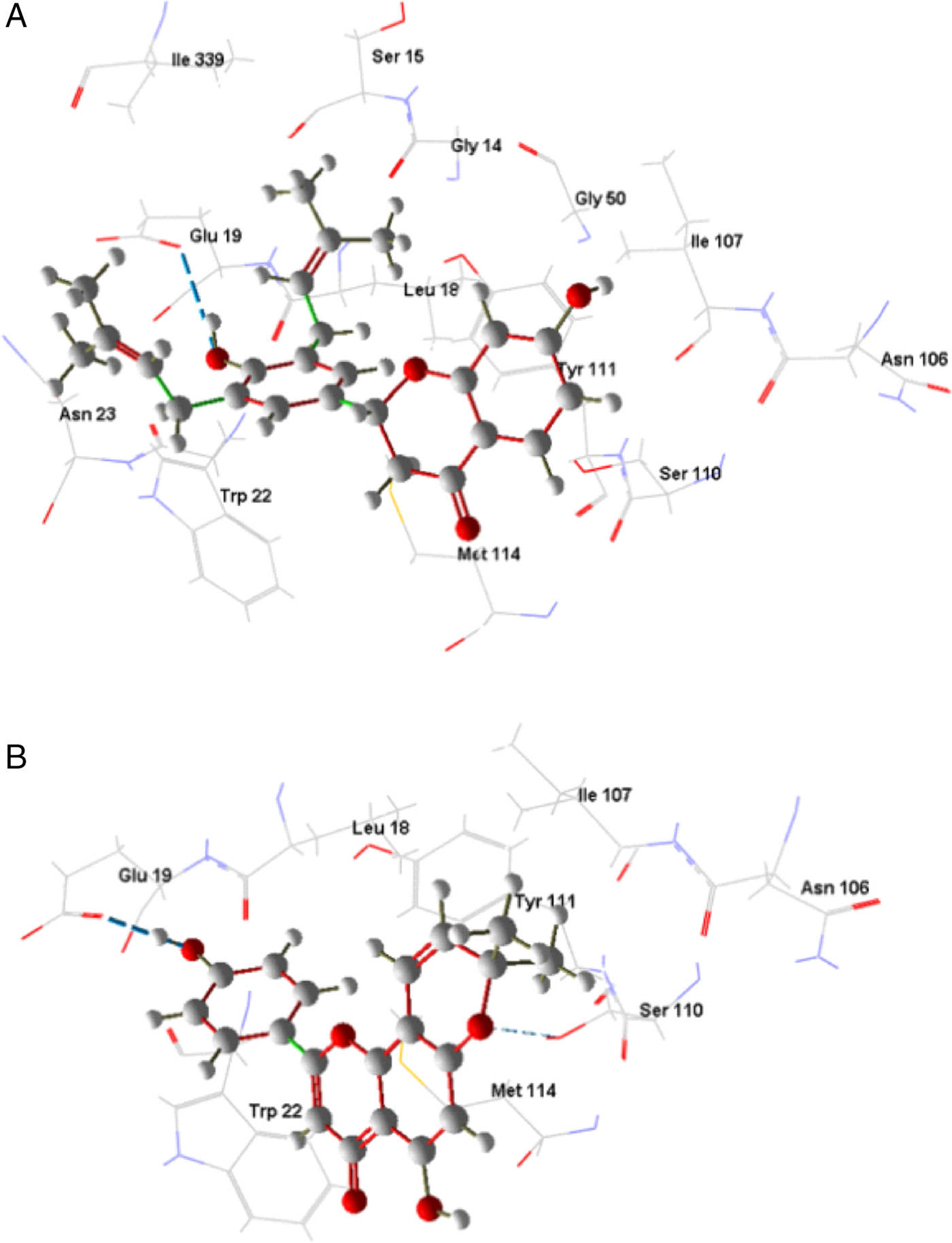
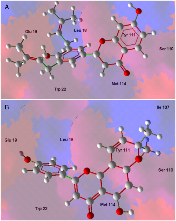
These residues were reported by Persch and co-workers as closers contact with PCTI (Persch et al., Reference Persch, Bryson, Todoroff, Eberle, Thelemann, Dirdjaja, Kaiser, Weber, Derbani and Brun2014), even more, the hydrophobic residues in the binding site located close to both studied compounds comprising Leu18, Trp22, Tyr111 and Met114 were already highlighted in the same previous study. Abyssinone IV (1) and atalantoflavone (2) ring B are directed to the hydrophobic surface (blue colour) of the binding site (Fig. 4a and b).
Docking poses of abyssinone IV (1, a) and atalantoflavone (2, b) with T. cruzi TR (PDB ID: 4NEW). The coloured surface represents the hydrophobic regions (blue) and hydrophilic regions (red).
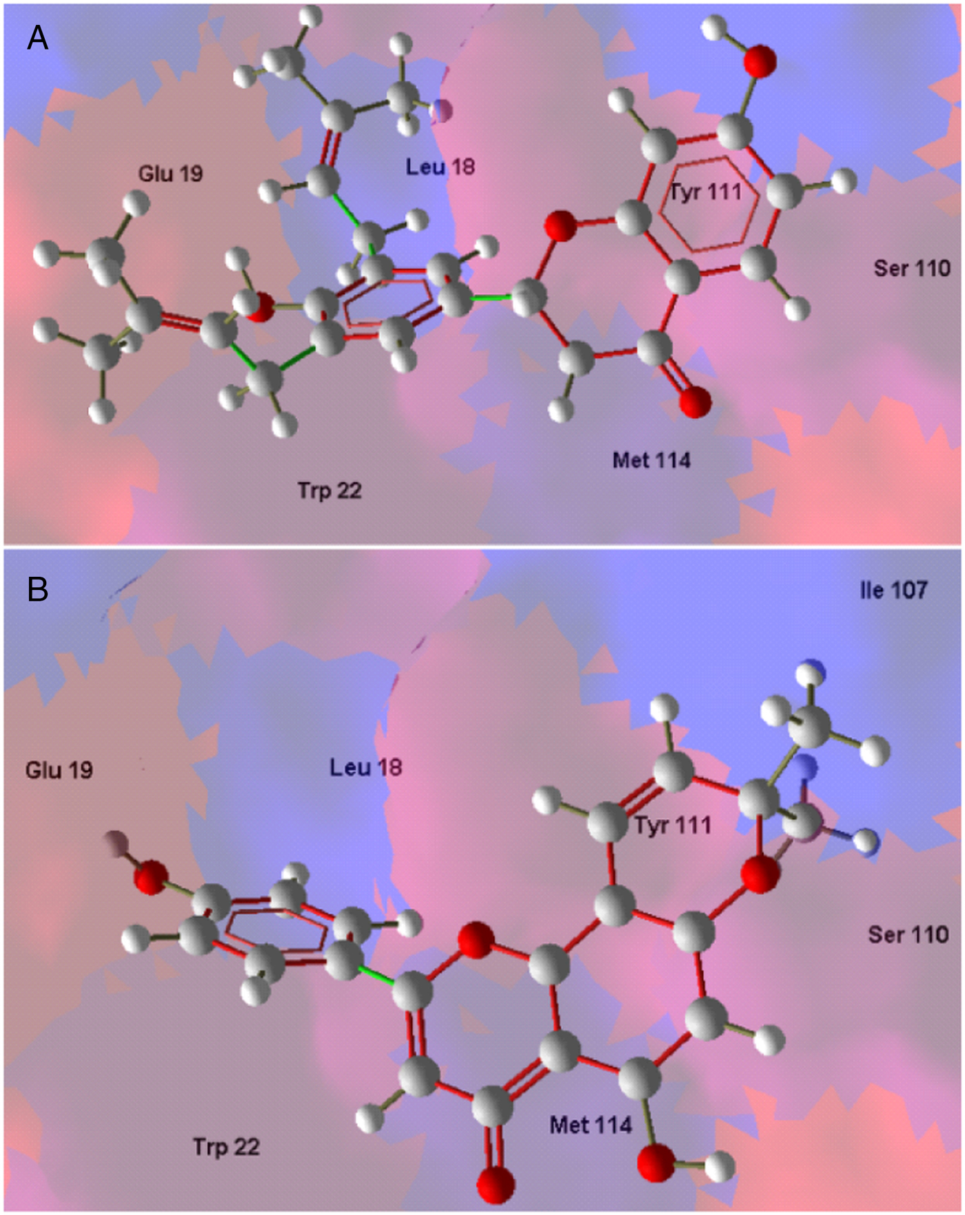
The presence of two hydrophobic groups (3-methylbut-2-en-1-yl) at positions 3 and 5 of ring B in 1 is responsible for higher hydrophobic interactions. However, abyssinone IV (1) showed the lowest MolDock energy when compared to that of atalantoflavone (2) (Table 2).
Enzymatic activity
On the basis of the interaction observed in the molecular docking, abyssinone IV (1) and atalantoflavone (2) were assayed on recombinant TR in vitro. Both metabolites revealed no inhibition on TR.
Mitochondrial membrane depolarization assay
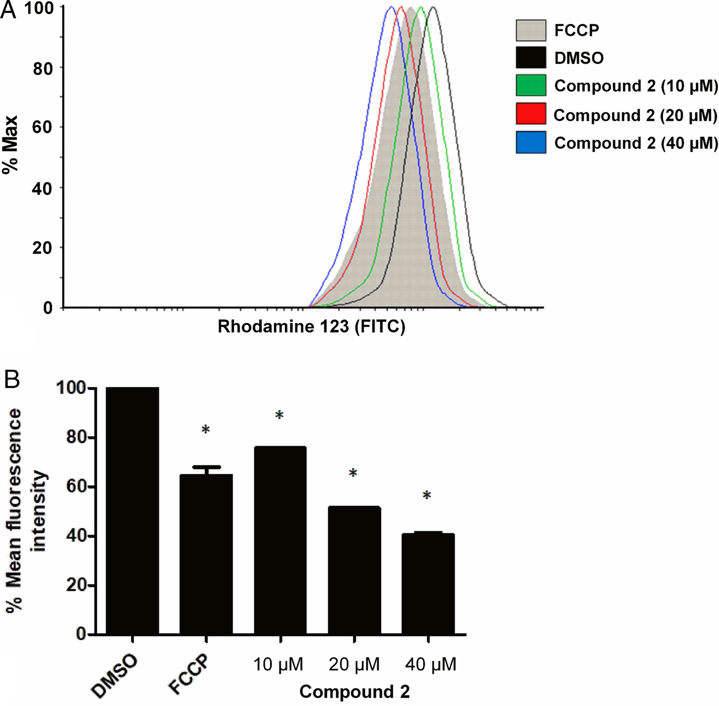
Since compounds 1 and 2 failed to inhibit in vitro the TR action, another investigation focused on the mitochondrial function study was considered. Thus, compound 1 showed no alteration of the mitochondrial membrane potential at the tested concentrations. In contrast, compound 2 displayed ability to alter the mitochondrial membrane potential of T. cruzi epimastigote after 2 h of incubation at different concentrations (10, 20 and 40 µm) (Fig. 5a and b).
Mitochondrial membrane depolarization assay for T. cruzi epimastigotes incubated with different concentrations of atalantoflavone (2, a). Fluorescence intensity measurement for atalantoflavone (2) at different concentrations in mitochondrial membrane depolarization assay (b). Values shown are expressed in mean percentage of fluorescence intensity ± standard deviation of three independent experiments. FCCP: carbonyl cyanide-4-(trifluoromethoxy)phenylhydrazone at 20 µm (positive control); DMSO: 1% dimethylsulphoxide (negative control); *indicates significant difference relative to the DMSO group (P < 0.0001).
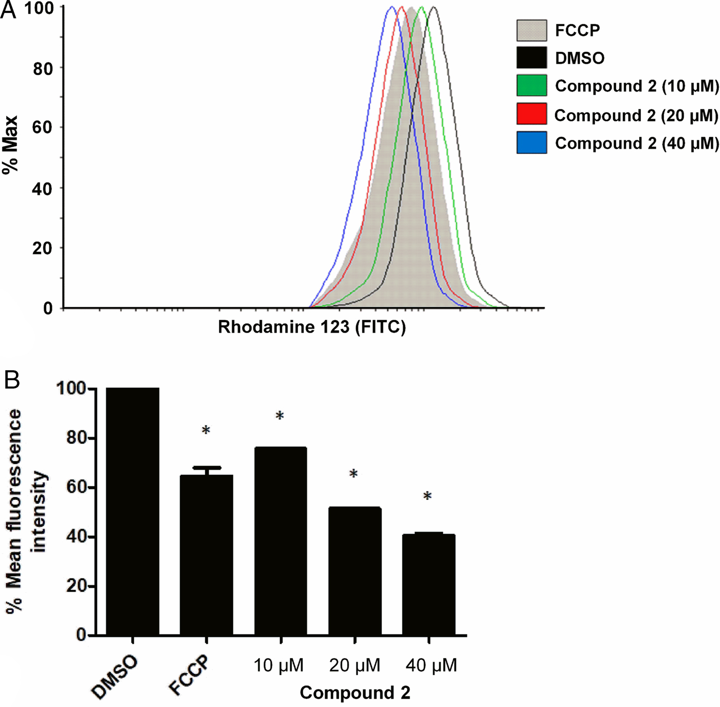
The tested concentrations of compound 2 showed a decrease in total Rhodamine R123 fluorescence intensity, indicating a mitochondrial depolarization. At 10 µm, compound 2 was slightly less potent than the positive control FCCP. However, the loss of ΔΨm caused by atalantoflavone (2) was dose-dependent, as depicted in Fig. 5b, which depicted a pronounced change of ΔΨm at 20 and 40 µ m when compared to the control (DMSO).
Discussion
The lack of potent and affordable drugs to treat parasitic tropical diseases has led to a high demand in new drugs models. Unfortunately, pipeline working on developing drugs against NDTs are still scanty which makes any contribution in this particular problematic as a rewarding task. Mitochondrion has a fundamental role to T. cruzi survival and regulation of the energy balance and apoptosis modulation. Moreover, mitochondrial metabolism is characterized by oxygen consumption and the mitochondrial membrane potential (ΔΨm). The protozoan programmed cell death progression depends on the variations of this mitochondrial membrane potential. Thus, among the investigated metabolites, abyssinone IV (1) showed trypanocidal activity with an IC50 of 7.1 µ m. This compound presented excellent activity compared to 6,8-(di-3-methyl-but-2-enyl)eriodictyol which was inactive on the same parasite despite the fact that both metabolites share the same skeleton and differ by the position of the two prenyl groups (Sandjo et al., Reference Sandjo, de Moraes, Kuete, Kamdoum, Ngadjui and Steindel2016a). 8-Prenylnaringenin, a flavanone related to abyssinone IV showed trypanocidal activity on T. cruzi epimastigote with an IC50 value at 18 µm (Uchiyama, Reference Uchiyama2009). Abyssinone IV (1) has previously shown antiprotozoal effect on two different Plasmodium strains as well as its ability to disrupt the mitochondrial membrane potential and trigger the ROS production in the leukaemia CCRF-CEM cancer cell line (Yenesew et al., Reference Yenesew, Induli, Derese, Midiwo, Heydenreich, Peter, Akala, Wangui, Liyala and Waters2004; Kuete et al., Reference Kuete, Sandjo, Djeussi, Zeino, Kwamou, Ngadjui and Efferth2014). Based on former studies, the increase of ROS plays an important role in T. cruzi proliferation (Goes et al., Reference Goes, Rocha, Diniz, Aguiar, Machado and Vieira2016) suggesting that the antiparasitic effect of abyssinone IV (1) might be a series of events as shown for CCRF-CEM.
Atalantoflavone (2) was more potent on T. cruzi (IC50 at 3.6 µm) than other tested metabolites (Table 1). Its high conjugated ketone seems to increase the activity against T. cruzi amastigote when compared to related compounds bearing flavanone core (Tasdemir et al., Reference Tasdemir, Kaiser, Brun, Yardley, Schmidt, Tosun and Rüedi2006). Moreover, the presence of a chromenone core turned to be beneficial for the trypanocidal activity. Apigenin, a metabolite related to compound 2 presented in a former study antitrypanosomal effect on T. cruzi trypomastigote (IC50 at 21.8 µm) (Tasdemir et al., Reference Tasdemir, Kaiser, Brun, Yardley, Schmidt, Tosun and Rüedi2006). As outlined above, oxidative burst, which is a rapid release of ROS might play a crucial role in T. cruzi survival and the disruption of this cellular activity might reduce or inhibit the parasite growth (Goes et al., Reference Goes, Rocha, Diniz, Aguiar, Machado and Vieira2016). Interestingly, atalantoflavone (2) displayed significant effects on the oxidative burst of the whole blood with an IC50 of 10 ± 0.2 µm (Fomani et al., Reference Fomani, Happi, Bisoue, Ndom, Waffo, Sewald and Wansi2016). Compounds 1 and 2 showed moderate cytotoxicity on the THP-1 cell line. These cells were 1.8-fold more susceptible to 2 than to 1. There is less evidence on the cytotoxicity of compounds 1 and 2 against human and other mammalian normal cells. However, both showed no cytotoxicity on AML12 (hepatocytes Alpha Mouse Liver 12) at IC50 up to 110 µm in a previous study (Kuete et al., Reference Kuete, Sandjo, Djeussi, Zeino, Kwamou, Ngadjui and Efferth2014). In addition, 2 showed weak cytotoxicity (CC50 246.8 µm) on a clone human T leukaemia C8166 cell line (Feng et al., Reference Feng, Wang, Cai, Zheng and Luo2010). Although the presence of chromene moieties in 2 favoured the trypanocidal activity, their planarity as well as the presence of an α,β-unsaturation ketone also seemed to affect the viability of host cells.
Heptadec-16-yne-1,2,4-triol (17), 2,4-dihydroxyheptadec-16-yn-1-yl acetate (18) and heptadec-16-ene-1,2,4-triol (19) were as active as the flavonoids, however, these acetogenins also inhibited the growth of THP-1 cells. A previous study reported the three compounds with trypanocidal activity on T. cruzi epimastigote indicating that the parasites and the host cells might be both sensitive to these acetogenins (Abe et al., Reference Abe, Nagafuji, Okawa, Kinjo, Akahane, Ogura, Martinez-Alfaro and Reyes-Chilpa2005). The observed cytotoxic effects were supported by previous reports that described the activity of these compounds on a panel of cancer cell lines (Oberlies et al., Reference Oberlies, Rogers, Martin and McLaughlin1998; Abe et al., Reference Abe, Nagafuji, Okawa, Kinjo, Akahane, Ogura, Martinez-Alfaro and Reyes-Chilpa2005).
Triterpenes are one of the metabolites widespread in plants kingdom with various biological properties among them antiparasitic activities (Al Musayeib et al., Reference Al Musayeib, Mothana, Gamal, Al-Massarani and Maes2013). In this study, donellanic acid A (5), a cyclopropane triterpene revealed a moderate antitrypanosomal activity (IC50 of 37.2 µ m) while its congener donellanic acid B (6) was not active. Both structures differ by the presence of a ketone in 5, while 6 contains a secondary alcohol group. Moreover, donellanic acid A (5) is a 23-nortriterpene while C-23 in donellanic acid B (6) is a carboxylic acid function. Comparing the functional groups in terpenoids 5–13, it turned out that those highly hydroxylated (6, 8 and 12) and the highly lipophilic ones (7–11 and 13) showed no trypanocidal activity. As presented in the literature, ursolic, oleanolic and betulinic acids were from moderately to weakly trypanocidal metabolites on T. cruzi trypomastigotes (Izumi et al., Reference Izumi, Ueda-Nakamura, Dias Filho, Júnior and Nakamura2011).
Compound 1 showed interesting antileishmanial activity against L. amazonensis (IC50 of 14.7 µ m) while atalantoflavone was weakly active. Flavanones such as sakuranetin devoid of prenyl groups showed weak antileishmanial against L. amazonensis promastigotes (IC50 of 181 µm) (Grecco et al., Reference Grecco, Reimão, Tempone, Sartorelli, Cunha, Romoff, Ferreira, Fávero and Lago2012). Other flavanones such as (S)-naringenin and 5,3′-hydroxy-7,4′-dimethoxyflavanone, also devoid of prenyl group showed from moderate to weak antileishmanial activity against L. donovani amastigotes (IC50 of 18.4 µ m) and L.(V.) panamensis amastigotes (EC50 at 53.7 µm), respectively (Tasdemir et al., Reference Tasdemir, Kaiser, Brun, Yardley, Schmidt, Tosun and Rüedi2006; Robledo et al., Reference Robledo, Cardona, Ligardo, Henao, Arbeláez, Montoya, Alzate, Pérez, Arango and Vélez2015).
As shown in Table 1, acetogenins 17 and 19 showed moderate leishmanicidal effect on amastigote form of L. amazonensis. These polyols have never been evaluated on Leishmania species, however, similar metabolites bearing more than one olefinic bond revealed good antileishmanial activity against L. donovani promastigote (Dharmaratne et al., Reference Dharmaratne, Tekwani, Jacob and Nanayakkara2012).
The most trypanocidal metabolites with better selectivity to THP-1 namely abyssinone IV (1) and atalantoflavone (2) were subjected to molecular docking using TR as the target. This in silico study revealed that both compounds maintained different interactions with TR. In contrast, both flavones showed no inhibition effect for the in vitro assay on TR. The opposite outcome between both assays indicated that the interactions observed in silico were not enough to disrupt the enzyme action as shown in vitro. Moreover, it also means that the antiparasitic effects occur through other mechanisms. So, a further study on the mitochondrial membrane potential interestingly revealed that atalantoflavone (2) caused mitochondrial membrane depolarization which is a mechanism related to its ability of displaying effect on the oxidative burst (Goes et al., Reference Goes, Rocha, Diniz, Aguiar, Machado and Vieira2016). Quercetin, a flavonol, exerted its antileishmanial activity via the same mechanism as demonstrated in a previous study (Fonseca-Silva et al., Reference Fonseca-Silva, Inacio, Canto-Cavalheiro and Almeida-Amaral2011), suggesting that flavonoids with flavone skeleton might have the ability of disrupting the mitochondrial membrane. On the basis of the obtained results and the fact that none of the active metabolites gave Pan-Assay Interference Compounds (PAINS) alert (Baell and Nissink, Reference Baell and Nissink2017; Daina et al., Reference Daina, Michielin and Zoete2017), 1 and 2 could be considered for further chemical modifications in order to develop more potent trypanocidal derivatives.
Author ORCIDs
Louis Pergaud Sandjo, 0000-0001-5840-7733
Financial support
The authors are grateful to CNPq/CAPES (Conselho Nacional de Desenvolvimento Científico e Tecnológico/Coordenação de Aperfeiçoamento de Pessoal de Nível Superior) for their financial support. LALS would like to thank CAPES for the scholarship and LPS and MWB would like to thank PNPD/CAPES (Programa National de Pós-doutorado/Coordenação de Aperfeiçoamento de Pessoal de Nível Superior). The work is part of the activities of the Research Network Natural Products against Neglected Diseases (ResNetNPND; http://www.ResNetNPND.org).
Conflict of interest
The authors declare no conflicts of interest.
Ethical standards
Not applicable.