

Introduction
Benign paroxysmal positional vertigo (BPPV) is a disorder that can be quickly and easily assessed and treated in the clinical setting. It is characterised by brief episodes of vertigo when the head is moved into certain positions (Herdman & Tusa, Reference Herdman, Tusa and Herdman2007). Vertigo being the illusion of movement of the self or the environment (Tusa, Reference Tusa and Herdman2007). The episodes of vertigo can be triggered by everyday activities such as rolling over in bed or turning the head (Bhattacharyya et al., Reference Bhattacharyya, Baugh, Orvidas, Barrs, Bronston, Cass and Haidari2008; Herdman & Tusa, Reference Herdman, Tusa and Herdman2007). BPPV is the most common cause of vertigo in adults, with 20–30% of all cases of vestibular vertigo likely to be caused by BPPV (Bhattacharyya et al., Reference Bhattacharyya, Baugh, Orvidas, Barrs, Bronston, Cass and Haidari2008; Furman & Cass, Reference Furman and Cass1999; Herdman, Reference Herdman1997; Herdman & Tusa, Reference Herdman, Tusa and Herdman2007; Von Brevern et al., Reference Von Brevern, Radtke, Lezius, Feldmann, Ziese, Lempert and Neuhauser2007). A lifetime prevalence of BPPV of 2.4% has been reported for the general population, with a 1-year incidence of 0.6% (Von Brevern et al., Reference Von Brevern, Radtke, Lezius, Feldmann, Ziese, Lempert and Neuhauser2007).
BPPV has been associated with changes in both static and dynamic balance, unsteady gait, sensitivity to head movement and falls, and can lead to significant changes in the way a person moves (Celebisoy, Bayam, Gulec, Kose, & Akyurekli, Reference Celebisoy, Bayam, Gulec, Kose and Akyurekli2009; Herdman, Reference Herdman1997; Oghalai, Manolidis, Barth, Stewart, & Jenkins, Reference Oghalai, Manolidis, Barth, Stewart and Jenkins2000). It can impact on the way a person is able to perform everyday activities, with increased impairments in daily living (Herdman & Tusa, Reference Herdman, Tusa and Herdman2007; Oghalai et al., Reference Oghalai, Manolidis, Barth, Stewart and Jenkins2000; Von Brevern et al., Reference Baloh, Honrubia and Jacobson2007). Von Brevern et al. (Reference Von Brevern, Radtke, Lezius, Feldmann, Ziese, Lempert and Neuhauser2007) reported that one-third of their group of 80 participants with BPPV had a fear of falling, 24% of the participants gave up driving during episodes of BPPV and 18% avoided leaving the house. BPPV has been associated with reduction in health-related quality of life and increases in depression and clinical anxiety (Lopez-Escamez, Gamiz, Fernandez-Perez, & Gomez-Finana, Reference Lopez-Escamez, Gamiz, Fernandez-Perez and Gomez-Finana2005; Magliulo, Bertin, Ruggieri, & Gagliardi, Reference Magliulo, Bertin, Ruggieri and Gagliardi2005; Oghalai et al., Reference Oghalai, Manolidis, Barth, Stewart and Jenkins2000). In the general population, for most cases of BPPV the cause is idiopathic; however, where a preceding event can be identified, head trauma is the most prevalent (17–20% of cases) (Baloh, Honrubia, & Jacobson, Reference Baloh, Honrubia and Jacobson1987; Furman & Cass, Reference Furman and Cass1999).
The pathophysiology of BPPV is complex, but there are potentially two separate pathophysiological processes that may also co-exist, canalithiasis and cupulolithiasis. Calcium carbonate crystals, known as otoconia, are generally embedded in the gelatinous matrix of the utricular macula. They may become dislodged from this otoconial layer due to degeneration or head trauma, and gravitate as debris in the endolymph (Buckingham, Reference Buckingham1999; Fife, Reference Fife2009; Furman & Cass, Reference Furman and Cass1999; Korres & Balatsouras, Reference Korres and Balatsouras2004). They may remain as free-floating debris, causing changes in the endolymphatic currents and consequent changes in the stimulation of the crista ampularis of the affected canal (canalithiasis) (Buckingham, Reference Buckingham1999; Fife, Reference Fife2009). Alternatively, they may lodge on the cupula itself, changing the deflection of the cupula on to the crista ampularis in response to gravity or accelerating forces (cupulolithiasis) (Buckingham, Reference Buckingham1999). These changes in stimulation lead to vertigo, nausea and nystagmus (Baloh, Reference Baloh2005; Bhattacharyya et al., Reference Bhattacharyya, Baugh, Orvidas, Barrs, Bronston, Cass and Haidari2008; Herdman & Tusa, Reference Herdman, Tusa and Herdman2007).
A number of techniques are used to diagnose BPPV and the best clinical practice guidelines for BPPV have been presented by Bhattacharyya et al. (Reference Bhattacharyya, Baugh, Orvidas, Barrs, Bronston, Cass and Haidari2008). Diagnostic positional tests for BPPV, such as the Hallpike manoeuvre and the supine roll test, assess the onset and duration of vertigo and associated eye movements (nystagmus) after particular positioning of the head. Bhattacharyya et al. (Reference Bhattacharyya, Baugh, Orvidas, Barrs, Bronston, Cass and Haidari2008) make strong recommendations that BPPV should be diagnosed with these clinically based positional tests. An accurate diagnosis of BPPV is important as it will determine the choice of the most appropriate treatment modality and decrease the number of treatment interventions required. Clinicians should be aware of some limitations to bedside clinical examination, such as the time of day it is performed, speed of movement during the test, accuracy of the angle of the plane of the occiput and the ability of the assessor to determine visually the direction, rotation and duration of the nystagmus. In addition, there are a number of contraindications and difficulties to assessment using positional tests, such as limited cervical range of movement, significant vascular disease, lower back dysfunction and pain. Bhattacharyya et al. (Reference Bhattacharyya, Baugh, Orvidas, Barrs, Bronston, Cass and Haidari2008) state that further radiological or vestibular testing may be required if the diagnosis remains uncertain, or there is an atypical presentation. However, in many cases, particularly those involving the posterior canal, BPPV can be quickly, accurately and easily diagnosed by clinicians without the need for expensive or time-consuming apparatus.
BPPV can be treated using a number of particle-repositioning manoeuvres (Bhattacharyya et al., Reference Bhattacharyya, Baugh, Orvidas, Barrs, Bronston, Cass and Haidari2008; Herdman & Tusa, Reference Herdman, Tusa and Herdman2007). The canalith repositioning manoeuvre, otherwise known as the Epley manoeuvre, has been demonstrated to be the most effective and reliable method of treating posterior canal BPPV (Fife et al., Reference Fife, Iverson, Lempert, Furman, Baloh, Tusa and Gronseth2008; Herdman & Tusa, Reference Herdman, Tusa and Herdman2007). Most trials indicate a high rate of success with this treatment technique, with all symptoms of BPPV resolved in 70–90% of cases after just one treatment and close to 100% resolution after consequent treatment (Epley, Reference Epley1992; Furman & Cass, Reference Furman and Cass1999; Herdman & Tusa, Reference Herdman, Tusa and Herdman2007). Results for an alternative repositioning procedure for posterior canal BPPV, the liberatory manoeuvre, also known as the Semont manoeuvre, are less well documented with poorer-quality studies (Bhattacharyya et al., Reference Bhattacharyya, Baugh, Orvidas, Barrs, Bronston, Cass and Haidari2008; Fife et al., Reference Fife, Iverson, Lempert, Furman, Baloh, Tusa and Gronseth2008). However, it has been suggested to be potentially effective in the treatment of posterior canal BPPV where the otoconia particles may not be free floating but may be adhered to the cupula (cupulolithiasis) (Herdman & Tusa, Reference Herdman, Tusa and Herdman2007). The roll manoeuvre (Lempert manoeuvre or barbeque roll) is the most effective way of treating horizontal-canal BPPV and though there is limited evidence, anterior canal BPPV appears to have a good response to specific particle repositioning manoeuvres (Bhattacharyya et al., Reference Bhattacharyya, Baugh, Orvidas, Barrs, Bronston, Cass and Haidari2008; Fife et al., Reference Fife, Iverson, Lempert, Furman, Baloh, Tusa and Gronseth2008). All the canalith repositioning procedures can potentially be performed by clinicians in a clinical setting.
While brain injury is cited as a potential cause of BPPV, there has been little information presented in the literature regarding the actual incidence of BPPV in individuals with a brain injury. Although Gordon, Joffe, Levite and Gadoth (Reference Gordon, Joffe, Levite and Gadoth2002) suggest that 11% of their group of individuals with potentially mild brain injury had BPPV, there is no evidence to date of incidence in groups with more severe injuries. The purpose of this study, therefore, was to determine the incidence of BPPV in an inpatient rehabilitation setting, in people with more severe brain injury. In addition, the study aimed to determine the types of BPPV presenting and to record the response to treatment.
Method
A retrospective audit was conducted on all individuals admitted to a metropolitan-based acquired brain injury (ABI) unit over a 1-year period from March 2009 to March 2010. The unit provides an inpatient service for individuals with an acquired brain injury (traumatic or non-traumatic), particularly those requiring a contained environment for severe behavioural disturbance and post-traumatic amnesia (PTA) management.
Participants
Consecutive individuals admitted to the unit were included in the audit. They were excluded if they could not indicate verbally or non-verbally whether they were experiencing dizziness or vertigo.
Procedures
Data on pre-injury demographics, the brain injury diagnosis and, in the case of traumatic injuries, the severity of brain injury were collected. Severity of traumatic brain injury (TBI) was based on the length of post-traumatic amnesia (PTA), as described by Jennett and Teasdale (Reference Jennett, Teasdale and Plum1981). Therefore severity was classified as very mild (<5 min of PTA), mild (5–60 min), moderate (1–24 h), severe (1–7 days), very severe (1–4 weeks) and extremely severe (>4 weeks of PTA).
Physiotherapy staff used a standardised process for recording all assessment and treatment details with regard to dizziness and/or vertigo. As part of this process, all patients were routinely asked whether they were experiencing any dizziness. The term ‘dizziness’ was initially used with each individual as a general layman's term to determine whether they were experiencing any unusual sensations such as giddiness, whirling, light headedness, disorientation, disequilibrium, bewilderment or confusion. If they reported yes, or, if at any stage during their inpatient stay they reported any dizziness, a full, clinically based vestibular assessment was initiated. As part of this assessment process they were subjectively asked whether they were experiencing a sensation of themselves or the world moving, to determine whether they were specifically experiencing vertigo. If the person reported transient vertigo in relation to changes in head or body position, the tests for BPPV were undertaken. The Hallpike manoeuvre was used to assess for posterior and anterior canal BPPV and the roll test was used to assess horizontal canal BPPV (Appendix 1). The physiotherapist would make a clinically based decision of the appropriate treatment technique based on the assessment findings (Appendix 2). All physiotherapists working on the ABI unit had routinely received additional training on assessing and treating BPPV, as part of orientation to the unit. Any queries regarding diagnosis or treatment were referred to the senior physiotherapist on the unit for further clinical clarification. Data were collected based purely on a bedside clinical examination; it was not within the scope of this study for individuals to be referred for a more detailed vestibular assessment to establish canal involvement.
Data Analysis
Specific details regarding the presence of vertigo and results of BPPV testing were collated from the physiotherapy assessments. In addition, if BPPV was diagnosed, information regarding the canals involved, treatment type, frequency and response to treatment was collected.
The data were extracted from the recording forms by the senior physiotherapist and stored on SPSS 19 as non-identifiable information. Descriptive statistics were used to analyse the data. This study was approved by Austin Health Human Research Ethics Committee.
Results
Participant Characteristics
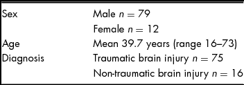
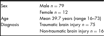
Ninety-one individuals were admitted to the ABI unit over the 1-year period (Table 1). Of the 89 individuals for whom data were collated, 74 had suffered a traumatic brain injury. Only three participants were classified in the very mild to moderate range for severity of brain injury. All others were in the severe to extremely severe range.
Participant Characteristics
Prevalence of Dizziness and BPPV
Forty-six individuals did not report any dizziness at any point during their inpatient stay and therefore no further data were collected. The remaining 43 did report a sensation of dizziness and a comprehensive subjective and objective vestibular assessment took place, including questioning to differentiate potential BPPV from other otological conditions. All the individuals reporting a sensation of dizziness described it as a sense of the world moving, transitory and related to changes in body/head position, therefore, unless contraindicated, a full BPPV assessment was performed. Thirty-two participants undertook a detailed vestibular assessment, of which 13 were clinically diagnosed with BPPV (Figure 1). A large group of 11 could not be assessed using the diagnostic positional tests for BPPV due to contraindications to testing.
The incidence of dizziness and BPPV in consecutive patients admitted to the ABI unit over 1 year. Two participants were excluded from the overall data collection as it was not possible to determine whether they were experiencing dizziness due to the severity of their brain injuries.

Analysis of the diagnostic groups indicated that all cases of BPPV occurred in those individuals who had suffered a TBI, no BPPV was diagnosed in participants with a non-traumatic injury (Figure 1). Of all 74 individuals admitted to the unit with a TBI over the 1-year period, 17.6% of this particular diagnostic group were confirmed to have BPPV.
Bilateral canals were affected in more than half of the group with BPPV, with seven of the 13 individuals diagnosed with bilateral posterior canal BPPV. Two individuals had horizontal or anterior canal involvement. They were both initially diagnosed with bilateral posterior canal BPPV, then diagnosed at a later stage with horizontal canal BPPV and additionally, in one of the cases, with anterior canal BPPV.
Response to Treatment
Ten of the individuals received treatment for the BPPV. Seven of the participants required multiple interventions while three required just one treatment. There was full resolution of symptoms of vertigo in all individuals who received treatment interventions. Three individuals could not be treated due to an inability to be rolled on to their side because of rib fractures, or due to unplanned discharge from the unit.
Discussion
This study demonstrates a high incidence of BPPV in a group of people with largely severe to extremely severe TBI. The finding of 17.6% of all individuals admitted with a TBI to our unit suffering BPPV is considerably higher than the 1-year incidence for a general adult population of 0.6% (Von Brevern et al., Reference Von Brevern, Radtke, Lezius, Feldmann, Ziese, Lempert and Neuhauser2007). This finding has not been described in the previous literature for a similar cohort of individuals. Gordon et al. (Reference Gordon, Joffe, Levite and Gadoth2002) found an incidence of BPPV of 10.7% in 75 consecutive traumatic head injury admissions to an Emergency Department. This population had received relatively mild brain injuries, with only two individuals suffering a brief loss of consciousness. Our population, with more severe TBI, appears to show an even greater incidence of BPPV. With head trauma being described as an identifiable cause of BPPV, our results support the concept by Baloh (Reference Baloh2005) that a more severe blow further increases the likelihood of BPPV occurring.
Tomaz et al. (Reference Tomaz, Gananca, Ganaca, Ganaca, Caovilla and Harker2009) state that BPPV most commonly occurs unilaterally, with only 10.9% of cases as bilateral. Katsarkas (Reference Katsarkas1999) reports that the incidence of bilateral BPPV is increased following head trauma, as a blow to the head can equally affect both ears. Katsarkas found that 14.5% of individuals with BPPV diagnosed following a traumatic brain injury had bilateral symptoms, in comparison with 6.3% of a group with idiopathic BPPV. Our findings were considerably higher, with more than half (53.8%) of the individuals diagnosed with BPPV post traumatic brain injury having bilateral symptoms. The severity of head injury in the Katsarkas study is unknown; however, the population was drawn from a dizziness clinic. Our inpatient population potentially had more severe brain injuries and was also potentially closer in time to the onset of the head injury. As yet there is no evidence in the literature supporting the concept that a more severe blow to the head may lead to a further increase in bilateral symptoms. This proposal would require further systematic investigation. The relatively recent occurrence of the injury may have contributed to the high proportion of participants with bilateral BPPV in this study, and over time there may be a natural resolution of symptoms in some cases. In addition, the bedside clinical assessment process in this study may have led to some inaccuracy in the initial diagnosis. The results do, however, emphasise the need for clinicians to be aware of the high potential for bilateral BPPV in individuals following a traumatic brain injury.
The posterior canal is claimed to be the most commonly affected canal (76–95% of cases), with the horizontal canal reported in 5–17% of cases and the anterior canal the least common presentation (Bhattacharyya et al., 2008; Fife et al., Reference Fife, Iverson, Lempert, Furman, Baloh, Tusa and Gronseth2008). Multiple canal involvement is very rare (1.5%) (Tomaz et al., Reference Tomaz, Gananca, Ganaca, Ganaca, Caovilla and Harker2009). Two of our 13 participants with BPPV developed horizontal canal BPPV and one of these also anterior canal BPPV in addition to posterior canal BPPV. Further investigation would be required to determine whether presentations of lateral, anterior or multiple canal involvement are more prevalent in people with more severe brain injuries.
It was interesting to note that individuals did not necessarily report dizziness or vertigo as a problem unless specifically asked by the clinician whether they were experiencing symptoms. The individuals tended to believe that feeling vertiginous was just part of recovering from a head injury, particularly as the symptoms were transient, and therefore did not volunteer the information themselves. This highlights the need for clinicians to be aware of the high potential for the presence of vertigo and to enquire about symptoms specifically, as part of their general assessment processes.
A large group of individuals (25.6%) who had reported symptoms of transient vertigo could not be fully assessed for BPPV with any positional testing manoeuvres. This was due to the wearing of neck collars for cervical fractures, severe pain from rib fractures or severe behavioural disturbance. There was therefore a large group whose vertigo symptoms remained undiagnosed and untreated as an inpatient. If this group could have been assessed, there may have been a significant change to the overall incidence of BPPV found. Considering the high incidence of BPPV in the remainder of the cohort, it is likely that a proportion of this group may also have been suffering from BPPV. It would be important to follow this group and perform appropriate assessments as the contraindications to the assessment process resolved.
In this study, no individual with a non-traumatic brain injury (n = 16) was diagnosed with BPPV. With such small numbers we are unable to determine whether the incidence of BPPV in this group is any different to that of the general population. A larger investigation would be of benefit as longer-term clinical observations within our unit suggest that individuals with non-traumatic brain injury also present with signs of BPPV.
Two individuals in this study were assessed as having involvement in other canals, both horizontal and anterior, after the initial diagnosis and treatment of posterior canal BPPV. It may be that the treatment process itself moved particles into other canals (Fife et al., Reference Fife, Iverson, Lempert, Furman, Baloh, Tusa and Gronseth2008; Herdman, Reference Herdman1997). Bhattacharyya et al. (Reference Bhattacharyya, Baugh, Orvidas, Barrs, Bronston, Cass and Haidari2008) state that approximately 6% of cases who initially present with posterior canal BPPV transition after treatment to lateral canal BPPV. Alternatively, multiple canals may have been affected initially but the clinicians were not able to discern the differences in nystagmus, particularly if both posterior and anterior canals were involved. It may be that the second canal's involvement became apparent as the symptoms of the first canal were resolved (Bhattacharyya et al., Reference Bhattacharyya, Baugh, Orvidas, Barrs, Bronston, Cass and Haidari2008). In our study the therapists used visual analysis to determine the direction of the nystagmus, which has the potential for error in recognition of subtle signs of nystagmus. The clinical assessment process could be improved in the future with the use of some easily attainable tools, such as Frenzel glasses. It is interesting to note that Jackson, Morgan, Fletcher and Krueger (Reference Jackson, Morgan, Fletcher and Krueger2007) felt that anterior canal BPPV was under-recognised when using clinical observation of nystagmus for diagnosis. They found anterior canal BPPV to be present in 21.2% of their population and that it was particularly prevalent after a head trauma. While it remains unclear why a proportion of our individuals presented at a later date with BPPV in other canals, our findings show the need for therapists to set up an ongoing assessment process if symptoms of vertigo persist. In addition, they highlight the need for therapists to be confident in treating the less common presentations of BPPV.
Seven of the 10 individuals who were treated required multiple treatments before a resolution of their symptoms of vertigo. This could be due to the high numbers of individuals with bilateral symptoms, the potential for multiple canal involvement, the severity of the head injuries or initial misdiagnosis of the canals involved from the bedside clinical assessment. None of these hypotheses can be substantiated by the literature; however, it should be noted that all individuals treated did ultimately achieve full resolution of their symptoms.
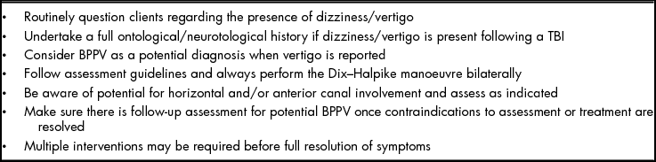
The information gained from our study may be important for the effective assessment and treatment of vertigo in individuals with more severe TBI. Almost half of the individuals admitted to our unit reported symptoms of dizziness at some stage during their admission. Forty-one per cent of those initially reporting dizziness were suffering from vertigo and went on to be diagnosed with BPPV, a potentially debilitating condition that can be readily assessed and successfully treated in the clinical setting (Bhattacharyya et al., Reference Bhattacharyya, Baugh, Orvidas, Barrs, Bronston, Cass and Haidari2008). Clinicians should therefore always consider BPPV as a potential diagnosis when investigating a subjective report of dizziness in individuals with a TBI (see Table 2 for clinical recommendations).
Benign Paroxysmal Positional Vertigo: Clinical Recommendations for Individuals with TBI

The major limitation of this study is the relatively small number of participants involved. Compounding this, of the 43 participants who reported vertigo, a group of 11 participants could not be assessed or treated for BPPV due to contraindications. If data from this relatively large group could be included, it may influence the outcomes we obtained. Our group was a very specific cohort of people receiving inpatient rehabilitation, with largely severe to extremely severe brain injuries. Therefore, there may be limitations in the application of the results to other groups of people with severe brain injuries, such as those in a later stage of recovery.
Conclusion
This study provides some important, previously unrecorded, information regarding the incidence of BPPV in a consecutive group of individuals with largely severe to extremely severe traumatic brain injury in the subacute period of their recovery. The high incidence of BPPV in those with a TBI highlights the need for clinicians to be aware of BPPV as a potential cause of dizziness in this group and to be adept at assessing and treating it. While, overall, the presentation of BPPV in our group was more complex than that seen in a general population with BPPV, it is important to note that full resolution of all symptoms of vertigo was still achieved in all cases treated.
Acknowledgements
This research received no specific grant from any funding agency, commercial or not-for-profit sectors. I wish to thank Zoe Langford, Catherine O'Brien, Dr Jannette Blennerhassett and Dr Ian Smith for their help with this project.
Declaration of interest
None.
APPENDIX 1
Diagnostic Tests for BPPV
Dix–Hallpike Test
Test procedure for a right Dix-Hallpike test:
• *Warn the patient that the test may reproduce his/her symptoms.
• Start in long sitting, extend the patient's neck 10–15 degrees and rotate approximately 30 degrees to the right.
• Lie the patient back as quickly as can be tolerated so that the head is over the end of the couch. If the patient is unable to extend his/her neck, lie the patient back over some pillows or tip the bed downwards so that the head and trunk are tilted downwards 10–15 degrees.
• The patient must keep his/her eyes open and fixed on your nose.
• You must be in a position to see his/her eyes.
• Hold this position for up to 1 min.
• Watch for nystagmus
For a left Dix–Hallpike, as above but rotate the head to the left approximately 30 degrees.
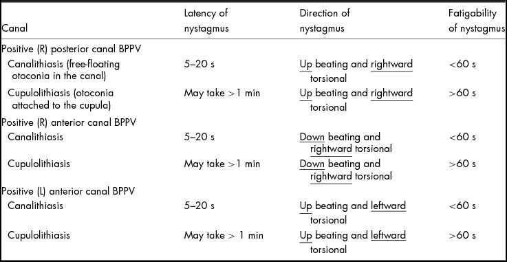
Test Results. Determining the canal involved: positive results are shown in Table A1 for a right Dix–Hallpike test. Reverse the left- or rightward direction of nystagmus for a positive left Hallpike test.
Horizontal Canal
• The patient lies in supine with his/her head flexed 20 degrees (this can be done by putting the head on a pillow).
• Roll his/her head quickly to one side, look for any nystagmus and ask the patient to report any symptoms of vertigo.
• Slowly roll the head back to midline.
• Quickly roll the head to the other side and again check for nystagmus and vertigo.
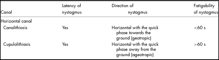
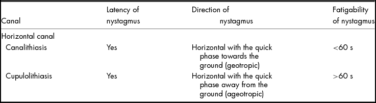
Test Results. For positive horizontal canal BPPV, the nystagmus (Table A2) will occur when turning the head in both directions. The affected ear is the side that shows more severe symptoms.
APPENDIX 2
Treatment Options for BPPV
Canalith Repositioning Treatment (CRT) orModified Epley's Manoeuvre for PosteriorBPPV: Canalithiasis
Positioning described for a right posterior canal BPPV.
• Begin as for a right Dix–Hallpike manoeuvre (head rotated 45 degrees to the right, extended 30 degrees and patient laid back over the edge of the bed).
• Hold this position for 1–2 min.
• Slowly rotate the head, maintaining moderate neck extension 45 degrees to the left and hold for 1–2 min.
• Roll the patient on to the left side, maintaining the head position so that the head is now rotated 45 degrees towards the floor and hold for 1–2 min.
• Keeping the head deviated, sit the patient up slowly over the side of the bed.
Semont Manoeuvre (Liberatory Manoeuvre) forPosterior Canal BPPV: Cupulolithiasis
• Positioning described for right posterior canal BPPV.
• The patient sits on the side of the bed and the therapist turns his/her head 45 degrees to the left.
• The patient is laid down quickly on to his/her right side while keeping the head turned to the left. He/she is are kept in this position for 2–3 min.
• The patient is then rapidly moved up through sitting and down on to the other side (the head is still turned to the left). The patient stays in this position for a few minutes before slowly sitting up.
Bar-B-Que Treatment for Horizontal CanalBPPV: Canalithiasis
• The patient lies supine with the head turned towards the affected ear.
• The patient's head is rolled slowly from the affected ear until the face is pointed upwards. The patient stays in this position for 15 s or until the dizziness stops.
• Roll the head slowly to the opposite side (affected ear up) and hold for 15 s or until dizziness settles.
• Keep rolling the head and the body in that same direction until the patient is lying face down.
• After 15 s the patient slowly sits up (by moving back from lying on his/her hands and knees) with the head level or tilted down 30 degrees.
Liberatory Manoeuvre for Horizontal CanalBPPV: Canalithiasis
• The patient starts in a sitting position looking straight ahead.
• He/she lies down quickly on to the unaffected side and holds this position for 2 min.
• In this position he/she quickly turns the head 45 degrees downwards and holds this podition for another 2 min before sitting up.
Liberatory manoeuvre for Horizontal CanalBPPV: Cupulolithiasis
Results for a Right Hallpike Manoeuvre
Results for Horizontal Canal Testing
• The patient starts in a sitting position looking straight ahead.
• He/she lies down quickly on to the affected side and holds this position for 2 min.
• He/she quickly turns the head 45 degrees downwards and holds this position for another 2–3 min before sitting back up.