Introduction
The use of assisted reproductive technologies (ARTs) has become an accepted medical practice. Conservative estimates indicate that over 1.5 million in vitro fertilization (IVF) cycles are carried out every year worldwide, resulting in approximately 400,000 babies born. In the United States and the United Kingdom, approximately 2 and 3% of all infants born every year are conceived using ART. Reference Luke1 Globally, ART babies represent about 0.3% of the total yearly live birth rate. While the vast majority of ART infants and children are healthy, there is evidence of increased complications using ART, including imprinting disorders, low birth weight (a marker of in utero stress and linked to long-term health problems), and malformations. Reference Feuer, Camarano and Rinaudo2,Reference Chen and Heilbronn3 Long-term effects described include a susceptibility to developing impaired glucose metabolism and insulin resistance and an increase in the risk of cardiovascular diseases. Reference Jiang, Wang, Lin, Xu, Ding and Huang4–Reference Duranthon and Chavatte-Palmer6
Initially, higher fasting glucose levels were observed in pubertal IVF children compared with age and gender matches spontaneously conceived control children. Reference Ceelen, van Weissenbruch, Vermeiden, van Leeuwen and Delemarre-van de Waal7 Later, the higher fasting glucose levels were also identified at the age of 5–6 years, among children conceived through ovulation induction and IVF-ICSI compared with children of fertile couples. Reference Pontesilli, Painter and Grooten8 Furthermore, reduced peripheral insulin sensitivity was observed using hyperinsulinemic–euglycemic clamp in a small cohort of IVF young adults. Reference Chen, Wu and Zhao9 Animal studies confirm these findings. Reference Scott, Yamazaki and Yamamoto10 Specifically, male mice conceived by IVF showed altered growth and glucose tolerance, highlighting a sexual dimorphic phenotype. Reference Donjacour, Liu, Lin, Simbulan and Rinaudo11,Reference Feuer, Liu and Donjacour12 Furthermore, a liver metabolomics study in mice showed clear male–female differences in hepatic tissue, Reference Feuer, Donjacour and Simbulan13 while IVF mice showed reduced insulin-stimulated Akt phosphorylation in the liver after 8 weeks of consuming either chow or high-fat diet (60%). Reference Chen, Wu and Zhao9
In addition, several studies suggested that ART may increase the risk of cardiovascular diseases. Systolic and diastolic blood pressure levels were higher in IVF children (8 to 18 years old) when compared to children who were conceived naturally by subfertile parents, independent of early life factors and parental characteristics. Reference Ceelen, van Weissenbruch, Vermeiden, van Leeuwen and Delemarre-van de Waal7 This finding has also been reported in children from 4 to 14 years old. Reference Sakka, Loutradis and Kanaka-Gantenbein14 Interestingly, ART conceived children showed cardiac and vascular remodeling already in fetal life, which persisted in postnatal life Reference Valenzuela-Alcaraz, Crispi and Bijnens15 with evidence of right ventricular dysfunction. Reference von Arx, Allemann and Sartori16 Animal studies showed that increased systolic blood pressure and activity of enzymatic regulators of cardiovascular and metabolic physiology, namely, serum angiotensin-converting enzyme and the gluconeogenesis controller, hepatic phosphoenolpyruvate carboxykinase, were significantly elevated in mice that had been cultured in vitro from the two-cell stage to the early blastocyst stage. Reference Watkins, Platt and Papenbrock17 Our prior study showed that male mice conceived by IVF had an enlarged left heart but normal blood pressure. Reference Donjacour, Liu, Lin, Simbulan and Rinaudo11
It is often believed that epigenetic changes underlie these phenotypic manifestations. In fact, the preimplantation embryos undergo significant changes in DNA methylation and histone marks, Reference Reik18 and embryos generated by IVF show changes in imprinted gene expression. Reference Doherty, Mann, Tremblay, Bartolomei and Schultz19 We recently showed that the inner cell mass (ICM) of embryos generated by IVF have changes in DNA methylation and histone marks in a sexually dimorphic pattern. Reference Ruggeri, Lira-Albarran and Grow20 Several metabolic pathways showed epigenetic changes in the early embryos. However, if global epigenetic changes exist in the liver of IVF conceived offspring is not known. This paper describes the epigenetic changes in the liver of offspring generated by IVF.
We found that the liver of IVF conceived mice shows global hypomethylation with alterations of several metabolic pathways, including hepatic fibrosis.
Materials and methods
Animals, IVF, embryo culture, and transfer
According to institutional regulations and after approval by a Review Committee, all animals were maintained under a constant 12-h light/dark cycle with ad libitum access to water and chow (PicoLab; 5058). Beginning at 24 weeks, all animals were placed on a high-fat diet (number D12492; Research Diets, Inc) until the time of death at 30 weeks. The high-fat diet was given during the last 6 weeks of life to understand the consequences of nutritional stress Reference Feuer, Liu and Donjacour12 , with the belief that a “second hit” would have induced more glucose alteration to mice conceived by IVF rather than in vivo.
IVF, embryo culture, and embryo transfer experiments were performed as previously described. Reference Feuer, Liu and Donjacour12,Reference Feuer, Donjacour and Simbulan13 Briefly, C57BL/6J females aged 6–8 weeks were injected with 5-IU pregnant mare’s serum gonadotropin (PMSG; Biovendor, RP1782725000) followed 46–48 h later by 5 IU human chorionic gonadotropin (hCG; Sigma-Aldrich, CG5) to induce superovulation. Cumulous–oocyte complexes were isolated from ampullae 13–15 h after hCG administration and incubated 4–6 h in HTF medium (Millipore Corp; MR-070-D) with sperm obtained from capacitated (1 h) cauda epididymal sperm of C57BL/6J males. Fertilized zygotes were washed and cultured to the blastocyst stage in potassium simplex optimization medium (KSOM MR-106-D; Millipore), at 37°C under Ovoil (10,029; Vitrolife) with 5% CO2 and 5% O2 in a modular humidified chamber. To generate postimplantation cohorts, pseudopregnancy was induced by mating naturally cycling CF-1 females to vasectomized CD-1 males, confirmed by the presence of a copulation plug the next morning (considered d 0.5). Late-cavitating blastocysts were transferred to the uterine horns of recipients on day 2.5 of pseudopregnancy, as routinely done in our laboratory. Reference Donjacour, Liu, Lin, Simbulan and Rinaudo11,Reference Nagy, Vintersten and Behringer21 For control experiments, C57BL/6J female mice were superovulated as described above and mated to C57BL/6J males overnight. Embryonic day 3.5 blastocysts (96 h after hCG administration) were flushed from the oviducts and transferred immediately to the uterine horns of CF-1 recipients, since this approach controls for litter size and the embryo transfer procedure. Reference Donjacour, Liu, Lin, Simbulan and Rinaudo11 Only animals derived from litters of 5–7 pups were used in this study. Recipient animals had similar weight at the time of transfer and gained the same weight during pregnancy.
Whole-Genome Bisulfite Sequencing (WGBS)
Nonfasted animals were killed by CO2 exposure followed by cervical dislocation in the morning, and tissues were harvested from animals generated by three separate IVF and three control experiments. At least three independent cohorts contributed to each analysis of liver tissue. After collection, whole liver (6 samples; n = 3 for each conception condition) samples were snap-frozen for whole-genomic bisulfite sequencing (WGBS). This technique was completed using the Methyl-MaxiSeq platform from Zymo Research. Reference Denomme, McCallie, Parks, Booher, Schoolcraft and Katz-Jaffe22 Liver samples of animal conceived by IVF or in vivo (FB group) were provided to Zymo for library preparation using the Pico Methyl Seq library preparation kit (Zymo Research, Catalog #D5455). Libraries were prepared directly from the liver tissue without any additional extraction. The DNA was subjected to bisulfite conversion using the EZ DNA Methylation-Lightning kit (Zymo, Research, Catalog #D5030). Briefly, the lightning reagent was mixed with the genomic DNA and incubated at 98°C for 8 min and 1 h at 54°C. Samples were subjected to a column, incubated with L-disulphonation buffer, and washed with M-wash buffer. Bisulfite-converted DNA was then eluted from the columns. After bisulfite conversion, samples were incubated with a PreAmp primer and polymerase. After the PreAmp polymerase reaction, samples were transferred to a Zymo-Spin IC column to be purified and concentrated using the DNA Clean & Concentrator-5 (Zymo Research, Catalog #D4003). After purification, samples were subjected to PCR to amplify the library for five cycles using the following PCR conditions: 94°C for 30 s; thermocycling at 94°C for 30 s, 45°C for 30 s, and 55°C for 30 s; followed by 68°C for 5 min. Following amplification, libraries were purified again using the Zymo-Spin IC column and eluted. Individual libraries were prepared with unique Illumina Index primers. The library was mixed with the LibraryAmp master mix and the designated index primer. Samples were subjected to PCR amplification for a total of 10 cycles using the following conditions: 94°C for 30 s; thermocycling at 94°C for 30 s, 58°C for 30 s, and 68°C for 1 min; followed by 68°C for 5 min. The final product was purified using the Zymo-Spin IC column and eluted. Library fragment size and concentration were evaluated using the Agilent 2200 TapeStation instrument and sequenced on the Illumina HiSeq 2500 platform.
Bioinformatics and statistical analyses
The ends of the raw fastq reads were trimmed at positions with a quality score of less than 30 using Fastq-mcf. Reference Aronesty23 The trimmed reads were aligned to the mouse genome build, mm10, using the bismark aligner Reference Krueger and Andrews24 that uses bowtie2 Reference Langmead and Salzberg25 following options --non_directional --score_min L,0,-0.4. The aligned reads were deduplicated using deduplicate_bismark. Reference Krueger and Andrews24 The genome-wide methylation levels were extracted using the bismark_methylation_extractor. Reference Krueger and Andrews24 For WGBS, the mapping efficiency of our samples was 69%.
The differentially methylated regions (DMRs) for IVF vs. FB were identified using the bsseq package Reference Hansen, Langmead and Irizarry26 in R. The ns (the minimum number of methylation loci in a smoothing window) and h (the minimum number of bases in the smoothing window) parameters that are part of the BSmooth function were set at 70 and 1000, respectively. Reference Hansen, Langmead and Irizarry26 Regions were identified as DMR, either in the bottom or the top 2.5% quantiles of the resulting t-statistics using the dmrfinder function. These regions were further filtered if the absolute value MaxStat parameter from this function’s output is ≥ 3.0. Significance was denoted as P ≤ 0.003. The annotations for the DMRs were assessed using the annotatr Reference Cavalcante and Sartor27 R package.
Once DMRs were determined, Genomic Regions for Enrichment Analysis Tool (GREAT version 4.0.4, http://great.stanford.edu) was utilized to associate genes with the regions, Reference McLean, Bristor and Hiller28 previous conversion to the Mus musculus genome assembly mm9 (https://genome.ucsc.edu/cgi-bin/ hgLiftOver). We decided to use the mm9 version since we wanted to compare liver methylation data to embryo methylation data (that we had generated using the mm9 version). Reference Ruggeri, Lira-Albarran and Grow20 Further, we needed to utilize the mm9 version for identifying methylation of imprinted genes, since the mm9 version was used to study imprinted control regions. Reference Bina29
The functional enrichment analysis was performed using g:Profilerβ (version e99_eg46_p14_55317af) with g:SCS multiple testing correction method applying significance threshold of P-adjusted value < 0.05 Reference Raudvere, Kolberg and Kuzmin30 uploading the list of genes within DMRs. The overrepresented biological processes (BPs) or molecular functions (MFs) were summarized with REVIGO (reduce + visualize Gene Ontology, http://revigo.irb.hr) to avoid redundant gene ontology (GO) terms. Reference Supek, Bosnjak, Skunca and Smuc31 The nomenclature of BPs and MFs uses the Gene Ontology Consortium. Reference Ashburner, Ball and Blake32 The predicted transcription factor binding motifs in regulatory regions were identified using the TRANSFAC database Reference Matys, Kel-Margoulis and Fricke33 as part of the functional enrichment analysis provided by g:Profilerβ. The statistically significant canonical pathways (CPs, P-value < 0.05, using the right-tailed Fisher’s exact test) were identified using Ingenuity® Pathway Analysis (IPA®, http://ingenuity.com).
For long repetitive element and CpG island analyses, the annotated regions associated with repeat elements and the CpG islands for the mouse genome build mm10 were downloaded from the UCSC genome browser Reference Karolchik, Hinrichs and Furey34,Reference Karolchik, Hinrichs and Kent35 (http://genome.ucsc.edu). The average methylation level for each of the samples in each annotated region was computed using the bedmap program that used the coverage file produced from the bismark_methylation_extractor program Reference Krueger and Andrews24 and the annotated regions as a bed file. Paired t-tests were used to determine differentially methylated repeat elements and CpG islands for the IVF vs. FB comparison, and significance was denoted as P-value < 0.05. The imprinted genes within the DMRs were identified using an imprinting atlas for mouse developmental stages and tissues. Reference Babak, DeVeale and Tsang36
Results
The liver of mice generated by IVF shows an increase in hypomethylated DMR distally to transcription start sites
Unsupervised principal component analysis shows that while the three IVF samples clustered together, the FB samples were more diverse in their methylation profile (Supplementary Fig. S1). Overall, only 4.91% of DMRs (n = 2692) were statistically different in the liver of IVF conceived mice when compared to in vivo generated mice (Fig. 1). Since 74.44% of DMRs contained the regulatory elements of two different genes (Supplementary Fig. S2A), we identified 2657 genes in the hypomethylated DMRs, 396 in proximity to hypermethylated DMRs, and 206 genes were located in DMRs with both evidence of hyper- and hypomethylation (Supplementary File S1A). Out of the total DMRs identified, the majority (n = 2286, i.e., almost 85%) was hypomethylated in IVF samples. DMRs were more frequently located 50–500 kb distal to the transcription start sites (Supplementary Fig. S2B), particularly in intronic and intergenic regions (Fig. 1). Surprisingly, only 0.16% of CpG islands were differentially methylated, and only a few DMRs were located on known gene promoters (n = 283) or enhancers (n = 190; Supplementary File S1B). Although 26 imprinted genes showed differentially methylation areas, none showed methylation changes in the imprinting control region (ICR, Supplementary File S1C). Reference Bina29
Distribution of differentially methylated regions and its genomic localization in liver samples from IVF vs. FB contrast.

Notably, the liver of IVF mice showed hypomethylation of transposable elements (TEs) (long-interspersed element (LINE), short-interspersed element (SINE), and long terminal repeat (LTR)) (Table 1).
Percentage of methylation (mean ± SD) of cytosine in CpG, CHG, or CHH context (where H can be either A, T, or C) of transposable elements in liver samples of mice conceived by IVF or in vivo (FB group).

DMRs in the liver of mice generated by IVF are enriched for genes involved in developmental and cellular metabolic processes
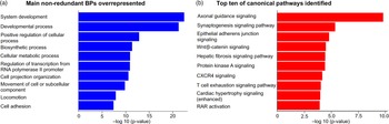
Functional enrichment analysis showed that BPs overrepresented included developmental, biosynthetic, and cellular metabolic processes and the regulation of transcription from RNA polymerase type II promoter (Fig. 2a and Supplementary File S1D). In this regard, the main MFs overrepresented included the RNA polymerase II transcription factor activity (sequence-specific DNA binding), MF regulator, and chromatin binding (Supplementary File S1E). Interestingly, a sub-analysis using the list of 473 genes differentially methylated in promoter and or enhancer regions identified the cellular metabolic process and the regulation of binding as the main BPs overrepresented. In addition, of the 110 unique transcription factors that had preferential binding on the promoters of differential methylated genes, nine (Ahr, Foxn4, Hdac1, Hes1, Max, Sp4, Sp5, Tbx5, and Tfap2a) were also differentially methylated in liver samples (Supplementary File S1F). The top 10 CPs identified by IPA in adult liver samples of IVF vs. FB (Fig. 2b) included epithelial adherens junction, Wnt/β-catenin, and protein kinase A signaling, and remarkably the hepatic fibrosis signaling pathway (Supplementary Fig. S3). Other metabolic relevant CPs overrepresented (Supplementary File S1G) included insulin receptor and insulin secretion signaling (Fig. 3 and Supplementary Fig. S4, respectively), and type II diabetes mellitus signaling (Supplementary Fig. S5).
a) Main non-redundant biological processes overrepresented and b) canonical pathways statistically significant, using the all list of 3259 differentially methylated genes in adult liver samples from IVF vs. FB contrast.

Canonical pathway insulin receptor signaling defined by the Ingenuity Knowledge Base. The genes highlighted in gray were differentially methylated in liver samples from male mice generated by IVF vs. FB.

One-fifth of differentially methylated genes in the ICM remain differentially methylated in the liver of IVF-generated offspring and are involved in the cellular metabolic process and the regulation of gene expression
To evaluate if epigenetic changes found in the ICM of embryos generated by IVF could persist into adulthood, we compared the list of differentially methylated genes (DMG) in the liver of adult mice and our recently published results from the ICM. Reference Ruggeri, Lira-Albarran and Grow20 We found that 22% (720/3259) of the DMG in the ICM of IVF embryos were also differentially methylated in the liver of adult IVF-conceived mice (Fig. 4). In particular, 452 DMG were hypomethylated in the liver or the ICM (62.77%), while 141 genes (19.58%) and 55 genes (7.64%) were hypomethylated or hypermethylated in both tissues, respectively (Supplementary File S2A). These DMG enriched several BPs such as the developmental process and the regulation of signaling, transcription from RNA polymerase II promoter, and cellular metabolic process (Supplementary File S2B). Among the differentially methylated genes in both ICM and liver of IVF-generated mice, aryl hydrocarbon receptor (AhR) and the Hes Family BHLH Transcription Factor 1 (Hes1) were notable. The CPs identified included the Wnt/β-catenin, xenobiotic metabolism signaling, and the hepatic fibrosis signaling pathway, specifically the hepatic fibrosis/hepatic stellate cell (HSC) activation (Supplementary File S2C).
Venn diagrams showing: a) 720 common differentially methylated genes (DMGs) between adult liver samples (n = 3259 DMG) and inner cell mass samples (ICM, n = 2420 DMG) in the contrast IVF vs. FB, and b) 609 shared genes between DMG in adult liver samples from IVF vs. FB (n = 3259 DMG), and the hepatic epigenetic signature that traces the developmental programming (n = 3730 genes; Colaneri, 2013).

To further understand if the set of differentially methylated genes in the adult liver of IVF conceived mice was important in developmental programming, we compared our gene list to a list of developmental genes. Reference Colaneri, Wang and Pagadala37 This comparison revealed that less than 20% (609/3259) of the DMG were shared (Fig. 4). More than 80% (494/609) were hypomethylated genes in the liver, while only 10.84% (65/609) were hypermethylated, and 8.21% (50/609) showed evidence of hyper- and hypo-differential methylation in this tissue (Supplementary File S3A). The functional enrichment analysis using the list of 609 common genes identified the developmental, metabolic, and cellular processes among the top 10 non-redundant BPs overrepresented (Supplementary File S3B). The top 10 CPs identified using the same list of 609 shared DMG included the epithelial adherens junction signaling and the hepatic fibrosis signaling pathway, particularly the hepatic fibrosis/HSC activation (Supplementary File S3C).
Epigenetic signature of IVF liver shares some similarity with signature of obese or diabetics liver
To assess the potential predisposition to develop metabolic diseases in adulthood secondary to IVF manipulation, we contrasted our list of 3259 DMG in the liver of adult mice conceived by IVF against a list of 3114 DMG identified in the liver of obese humans and the list of 1314 DMG of subjects with diabetes mellitus type 2 and obesity. Reference Kirchner, Sinha and Gao38 Approximately 16% of the DMG (537 genes) in the murine liver were shared with the DMG identified in the liver of obese humans (Supplementary File S4A and Fig. 5). Only 7% (241/3259) DMG in the murine liver were also differentially methylated in the human liver of subjects with both metabolic conditions (Supplementary File S4D and Fig. 5). The main common BPs identified included developmental process, and the regulation of response to stimulus, cell communication, MF, and cellular metabolic process (Supplementary File S4B and S4E). Similarly, the foremost common CPs identified were the hepatic fibrosis signaling pathway, hepatic cholestasis, PPARα/RXRα activation, and the protein kinase A and ERK/MAPK signaling (Supplementary File S4C and S4F, and Fig. 5).
Venn diagrams showing: a) 537 common differentially methylated genes (DMGs) between adult liver samples in the contrast IVF vs. FB (n = 3259 DMG) and DMG from liver samples of obese humans (n = 3114 DMG; Kirchner, 2018), and b) 241 shared genes between DMG in adult liver samples in the contrast IVF vs. FB (n = 3259 DMG) and DMG from liver samples of DM2-obese humans (n = 1314 DMG; Kirchner, 2018).

Discussion
This study provides the first evidence of global DNA methylation changes in liver of mice conceived by IVF. Given that impaired glucose metabolism and insulin resistance have been reported in mice conceived by ART Reference Jiang, Wang, Lin, Xu, Ding and Huang4–Reference Duranthon and Chavatte-Palmer6 and given that liver plays a central role in metabolism, analyzing the DNA methylation profile of liver might provide crucial molecular information on how embryo culture in vitro can reprogram metabolism of an individual. We found that a majority of differentially methylated genes were hypomethylated in the liver of IVF mice. Interestingly, the genomic localization of these methylation changes was in non-coding areas (introns and intergenic regions). Less than one percent of DMRs were associated with promoters or enhancers and the majority of methylation changes were located more than 5 kb from the transcription start sites.
Interestingly, three classes of TEs, the LINE, the SINE, and the LTR elements showed hypomethylation (P < 0.05) in IVF livers. TEs makes up a significant fraction of the genome and is normally silenced by DNA methylation. They are broadly and strongly transcribed in the early embryos, and their insertions can supply new promoter, enhancer, and insulator elements to protein-coding genes and establish novel, species-specific gene regulatory networks. Reference Gerdes, Richardson, Mager and Faulkner39 Changes in TE expression can have profound consequences.
If the methylation status is modified, as the hypomethylation induced by IVF in the murine liver, the unbalanced control of TE-derived functions might result in transcriptional dysregulation, increasing the susceptibility to developing diseases. For instance, DNA hypomethylation and transcriptional reactivation of replication-competent LINE-1 copies have been observed in some tumors. Reference Lee, Iskow and Yang40–Reference Scott and Devine42 Furthermore, de novo LINE-1 insertions can activate oncogenic pathways in hepatocellular carcinoma, including Wnt/β-catenin, Reference Shukla, Upton and Munoz-Lopez43 a pathway enriched in IVF liver. Indeed, the LINE-1 mobilization is a common feature of hepato-carcinogenesis in mammals and is encountered in murine liver tumors. Reference Schauer, Carreira and Shukla44 SINEs have properties of architectural elements partitioning the mammalian genome into distinct topological domains, and they play functional roles at their genomic location. Reference Ichiyanagi45 LINE and SINE hypomethylation is an early and essential epigenetic modification in fibrosis-associated mouse hepatocarcinogenesis. Reference Chappell, Kutanzi and Uehara46
Functional enrichment analysis provided important findings. First, the GO term cellular metabolic process was altered in the liver of IVF mice. This is important, given that one of the liver’s critical functions is to provide metabolic flexibility in health and disease. Reference Smith, Soeters, Wust and Houtkooper47 In particular, the insulin receptor and the insulin secretion signaling pathways were enriched. Interestingly, the Mammalian target of rapamycin (Mtor) kinase gene and the glucose transporter solute carrier family 2 member 4 gene (Slc2a4) were hypomethylated in both CPs. The hyperactive Mammalian target of rapamycin complex I (mTORC1, which includes the mTOR kinase) interferes with glycemic control by disrupting the insulin receptor-Akt transduction pathway while concomitantly driving the non-glycemic diseases of type 2 diabetes mellitus (T2DM). Reference Bar-Tana48 The expression of the insulin-responsive glucose transporter type 4 (GLUT4) encoded by SLC2A4, which is epigenetically regulated, has been proposed as a biomarker for insulin resistance treatment in diabetes mellitus. Reference Esteves, Yonamine and Machado49 The epigenetic modification of Mtor and Slc2a4 by IVF could partially explain the murine ART’s offspring’s metabolic profile.
Second, a set of CPs were altered in IVF liver included hepatic fibrosis, HSCs activation signaling, and the hepatic tumor microenvironment. Reference Barry, Baldeosingh and Lamm50 Activated HSC transdifferentiates into myofibroblast-like cells to promote fibrosis in response to liver injury or chronic inflammation, Reference Barry, Baldeosingh and Lamm50 leading to cirrhosis, and possibly to hepatocellular carcinoma. Reference Wilson, Mann and Borthwick51,Reference Dhar, Baglieri, Kisseleva and Brenner52 In the rat, activation of HSC has been associated with altered global DNA methylation. Reference Page, Paoli, Moran Salvador, White, French and Mann53
Additionally, the CP hepatic cholestasis was enriched with differentially methylated genes in the liver of IVF-generated mice. Interestingly, our prior metabolomics study showed that bile acid metabolism was altered in the liver of IVF mice. Reference Feuer, Donjacour and Simbulan13 Furthermore, the gene Akr1d1 was differentially methylated by IVF. This gene has been found to be dysregulated in human hepatocytes in cases of non-alcoholic fatty liver disease. Reference Nikolaou, Gathercole and Marchand54 Based on these findings, it will be necessary to monitor if IVF offspring for evidence of liver diseases.
Among the transcription factors that could regulate the expression of differential methylated genes in IVF liver (Supplementary File S1F), Kaiso and CTC-binding factor (CTCF) are notable. Kaiso is a methyl-DNA-binding protein that participates in the epithelial to mesenchymal transition and has been involved in hepatic fibrosis and hepatocellular carcinoma. Reference Shukla, Upton and Munoz-Lopez43,Reference Pierre, Hercules, Yates and Daniel55,Reference Chen, Fan and Guo56 CTCF is a DNA-binding protein that plays essential roles in regulating genome activity through its capacity to act as an enhancer blocker and interacts with Kaiso. Reference Defossez, Kelly and Filion57
We next tried to understand if genes with abnormal methylation in the ICM of IVF embryos were also dysmethylated in the liver of IVF conceived mice. Overall, 720 genes (22%) of genes that were dysregulated in ICM of IVF conceived mice were also dysmethylated in the liver of IVF mice.
Several BPs were altered at both time points in IVF offspring (Fig. 4). Among the relevant ones, we found regulation of transcription from RNA polymerase II (Pol II) promoter. Given that this is one of the three RNA polymerase enzymes found in the nucleus of eukaryotic cells, its dysregulation might be associated with significant gene expression changes. Reference Liu and Tao58 In particular, Larp7 (La-related protein 7), a subunit of a pausing-related factor that control the release of Pol II from gene promoters, was hypermethylated in the ICM and hypomethylated in the liver of IVF mice Reference Chen, Smith and Shilatifard59–Reference Stavraka and Blagden61 and its dysregulation has been associated with tumorigenesis. In addition, it is essential to note that the insulin receptor associates with RNA pol II in the nucleus and interacts with the transcriptional machinery to regulate insulin effects in health and disease. Reference Hancock, Meyer and Mistry62 As a result, the regulation of transcription via RNA pol II promoters could contribute to the metabolic traits observed in ART offspring.
Regulation of metabolic process was altered in ICM and liver of IVF mice and several genes were changed in both tissues (AhR, Hes, and Klf14). One key factor epigenetically modulated in both tissues was the transcription factor AhR that was hypermethylated in the ICM of IVF embryos and was both hyper- and hypomethylated in the liver of IVF mice. The AhR is a highly conserved transcription factor that integrates environmental cues and metabolic processes, particularly the regulation of energy metabolism. Reference Roman, Carvajal-Gonzalez, Merino, Mulero-Navarro and Fernandez-Salguero63,Reference Girer, Tomlinson and Elferink64 Mice expressing a constitutively active form of AhR spontaneously develop hepatic steatosis, possibly secondary to increased fatty acid import, suppression of fatty acid oxidation, and increased oxidative stress. Reference Lee, Wada, Febbraio, He, Matsubara and Lee65 Global Ahr loss enhances insulin sensitivity and glucose tolerance and reduces the activity of the PPARα pathway that regulates fatty acid oxidation and glucose metabolism. Reference Wang, Xu, Krager, Bottum, Liao and Tischkau66
AhR signaling also can modulate TGF-β activity and particularly epithelial to mesenchymal transition. All these pathways are known to be associated with liver fibrosis Reference Shi, Zeng, Yu, Tang, Tang and Xiao67 and were modified by IVF. Indeed, the AhR -/- mice develop periportal liver fibrosis. Reference Andreola, Fernandez-Salguero, Chiantore, Petkovich, Gonzalez and De Luca68 Furthermore, the loss of Ahr expression disrupts the proper development of the liver, including the development of the hepatic vasculature Reference Walisser, Bunger, Glover and Bradfield69 and the physiological process of wound healing Reference Carvajal-Gonzalez, Roman, Cerezo-Guisado, Rico-Leo, Martin-Partido and Fernandez-Salguero70 associated with hepatic fibrosis. Reference Pellicoro, Ramachandran, Iredale and Fallowfield71 AhR interacts with the Wnt/β-catenin pathway downstream of membrane receptors, Reference Roman, Carvajal-Gonzalez, Merino, Mulero-Navarro and Fernandez-Salguero63 one of the main CPs modified by IVF in our murine model and involved in the activation of HSCs, a pivotal cellular event in liver fibrosis. Reference Wang, Li, Li, Yan, Li and Xu72 Interestingly, 9% of hepatocellular carcinoma patients presented overexpression of AhR, and its expression correlates with tumor malignancy and progression. Reference Roman, Carvajal-Gonzalez, Merino, Mulero-Navarro and Fernandez-Salguero63,Reference Liu, Wu and Zhang73
Another interesting gene, Hes1, was hypermethylated in the ICM and hypomethylated in the liver of IVF-conceived mice. Hes1 is a gene regulated by TGF-β signaling and essential for the activation of HSCs, a CP modified by IVF in our murine model, that plays crucial roles in hepatic fibrosis, hepatocellular carcinoma, liver regeneration, and fetal liver development. Reference Zhang, Zhang and Ai74,Reference Aimaiti, Yusufukadier and Li75
Finally, Klf14, a pioneer factor, was also differentially methylated in both ICM and liver. Pioneer factors are transcription factors that can directly bind condensed chromatin and induce stable changes in chromatin structures. For instance, Klf14 initiates the remodeling of both enhancers and transcription start sites. Reference Mayran and Drouin76 Dysregulation of Klf14 could have critical metabolic consequences because its overexpression in wild-type C57BL/6J mice results in impaired glucose and insulin tolerance. Reference Wang, Tong and Gu77,Reference Yang and Civelek78
The final analysis we performed was to compare the IVF gene list with other databases showing altered methylation in diabetes and obesity, to understand if the methylation profile was similar to these diseased conditions. Overall, the similarities were modest and less than 10% of genes were dysmethylated in IVF liver and dysmethylated in liver of subjects with T2DM and obesity. Reference Kirchner, Sinha and Gao38 From this list, two genes: Pik3c2g (hypermethylated) and Prkca (hypomethylated) were notable because they were identified in the CP type II diabetes mellitus signaling and could contribute to the susceptibility of development T2DM in the ART’s offspring. Pik3c2g encodes for a protein of the phosphoinositide 3-kinase (PI3K) family; PI3K-C2g-deficient mice display severely reduced accumulation of glycogen and develop hyperlipidemia, adiposity, and insulin resistance. Reference Braccini, Ciraolo and Campa79 A single-nucleotide polymorphism in this gene has been associated with T2DM in a Japanese population. Reference Daimon, Sato and Oizumi80 A genomic region downstream of PRKCA has been associated with insulin resistance and metabolic syndrome. Reference Haydar, Grigorescu and Vintila81
This paper has some strengths and weaknesses. Among the strength, we used WGBS to assess methylation of IVF liver. This technique is the gold standard for DNA methylation analysis and offers good coverage for imprinted loci and targets cytosines in the CHG or CHH (H = A,C,T) sequence, which can be methylated at elevated levels in the early stages of mammalian development. Reference Bock, Tomazou and Brinkman82
We also had the unique opportunity to compare WGBS sequencing data generated in the ICM and liver of mice conceived by IVF. Further, the use of an inbred model, characterized by decreased genetic variability, could increase the discovery rate. Finally, the functional enrichment analysis was performed with updated open resource and with the use of Ingenuity Pathway Analysis.
Among the limitation of this paper, we have not performed a concomitant analysis of differential methylation and gene expression of the liver of IVF and spontaneously conceived mice, given limited funding. The next best thing we were able to do was to compare our DMG list (performed in male mice) to gene expression profile (using microarray) we had published in the liver of IVF and control female mice. Reference Feuer, Liu and Donjacour12 We identified 192 shared genes, and from this number, only 15% showed differential methylation in promoter or enhancer regions. The functional enrichment analysis identified five transcription factors, E2F-1, ZF5, FOXN4, Pet-1, and TFII. Interestingly, E2F-1 overexpression in the murine liver causes dysplasia and tumors. Reference Conner, Lemmer, Omori, Wirth, Factor and Thorgeirsson83 Future studies should compare gene expression and DNA methylation data to confirm if regulation of transcription via RNA pol II promoters could contribute to the metabolic traits observed in ART offspring.
In summary, we found that the process of IVF and embryo culture results in DNA methylation changes in the liver of adult mice. The genes modified by IVF are enriched in GO terms and CPs relevant for metabolism, gene regulation, hepatic fibrosis, and hepatocellular carcinoma. These findings support the need for increased surveillance of IVF offspring, with particular focus on liver diseases and metabolic syndrome.
Supplementary material
For supplementary material for this article, please visit https://doi.org/10.1017/S2040174421000313
Acknowledgments
None.
Financial support
This work was funded by a NIH-NICHD grant (R01HD092267) to PFR. SLA was partially funded by a UC MEXUS-CONACYT Postdoctoral Fellowship Program grant.
Conflict of interest
None.
Ethical standards
All experiments were approved by the Animal committee at UCSF.