Stent angioplasty has been an accepted therapy for native and recurrent aortic coarctation in older children and adults since its introduction in 1990s. Reference Forbes, Moore and Pedra1–Reference Forbes, Kim and Du3 Both bare and covered stents have been shown to be safe and effective in relieving the obstruction. Reference Taggart, Minahan and Cabalka2,Reference Sohrabi, Jamshidi and Yaghoubi4,Reference Meadows, Minahan, McElhinney, McEnaney and Ringel5 Use of covered stents is recommended in some settings, such as aortic coarctation with associated aneurysms, Turner syndrome, and coarctation with aortic atresia. Reference Qureshi, Zubrzycka, Brzezinska-Rajszys, Kosciesza and Ksiazyk6,Reference Momenah, Khan, Qureshi and Hijazi7 However, currently available covered stents need larger diameter sheaths, that may limit their use in children and patients with smaller femoral arteries for access. Some of the newer stents may overcome this limitation. We report our experience with Bentley BeGraft Aortic stent in treating near-atretic and severe aortic coarctation in patients with small femoral arteries for access.
Materials and methods
Between July and October, 2019, five patients with near-atretic or severe aortic coarctation underwent implantation of a Bentley BeGraft Aortic stents. All the patients had late diagnosis of native aortic coarctation and were hypertensive with baseline non-invasive blood pressure gradient of >20 mm of mercury (mmHg). None of the patients had known aortopathies or Turner syndrome. Patients were identified from the cardiac catheterisation electronic records. All participants received institutional consent waiver to be included in this study. Preprocedural informed consent was taken from the patients or their parents/guardians. Institutional approval from the local Institutional Review Board was obtained appropriately. The recorded cardiac catheterisation reports and angiography were retrospectively reviewed. Patients with aortic coarctation and a small femoral artery as seen by cardiac tomography or anticipated according to age/weight were chosen for implanting this stent.
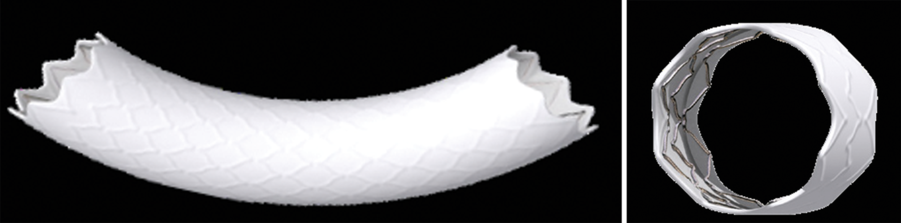
Stents (Figure 1)
The Bentley BeGraft Aortic stent received European Community approval in 2016. The stent is an open cell design made of Cobalt–Chromium alloy and covered with ~200 micrometers expanded-PolyTetraFluoroEthylene tubing. Although the stent is an open cell design, the Cobalt–Chromium alloy provides a good radial strength. The stent is pre-mounted on a high-pressure balloon and therefore cannot be delivered on a balloon-in-balloon system. The stent can cover a wide range of lesions, as it is available in 12–24 mm diameters in 2 mm increments and lengths of 19–59 mm. The 12 mm diameter stent can be delivered through a 9 Fr sheath, whilst the 14 and 16 mm diameter stents can be delivered through 11 Fr sheath. After post dilatation, the 12 and 14 mm diameter stent can be redilated to 20 mm and the 16 mm diameter stent to 24 mm. The nominal and burst pressure for the 12 mm diameter stent is 7 and 10 atm, respectively. The 14 mm diameter stent is 7 and 10 atm, and 16 mm diameter stent is 6 and 9 atm respectively.
Bentley BeGraft Aortic stent.

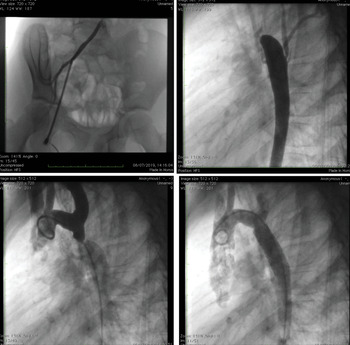
Left upper: Femoral artery angiography in anteroposterior view showing that a 9 Fr femoral introducer is completely filling the space of the femoral access; Right upper: angiography in the descending aorta in lateral view showing severely stenotic aorta with minimal retrograde flow into the proximal segment of the aortic arch; Left bottom: aortography in lateral view with a 5 Fr pigtail showing near-atretic aortic isthmus that is almost completely filled by the catheter; Right Bottom: Angiography in lateral view across the aortic arch after stent implantation.

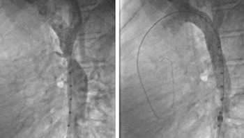
Left: aortography in lateral view showing severely stenosed aortic isthmus. A patent small patent ductus arteriosus can be seen; Right: final result after Bentley BeGraft implant.

Procedure (Figures 2 and 3)
All the procedures were performed in a standard biplane cardiac catheterisation laboratory under general anaesthesia and endo-tracheal intubation. The arterial access was obtained using ultrasound guidance with Seldinger technique. After obtaining the access, selective angiography was performed in the common femoral artery in 25–30° right anterior oblique or left anterior oblique projection to assess the appropriate level of puncture and the size of the common femoral artery. The angles for the initial aortography was based on 3D reconstruction of the CT using commercially available software. The patients received heparin at 100 International Units/Kg and received one dose of antibiotic during the procedure. A maximum dose of 5000 International Units of heparin was given in adults. The Activated Clotting Time was kept at 200–250 seconds. The coarctation was crossed from the femoral artery with 0.035” Terumo wire and a multipurpose catheter in all the patients except for one patient (Patient 3), in whom it was crossed from the right radial artery with 0.014” BMW coronary guidewire (Abbott, USA) and a 3 Fr Cobra catheter after creating a loop using 10 mm Goose-neck snare (Medtronic, Minneapolis, MN, USA). Angiography was performed in the left lateral and left anterior oblique views. Stent diameter was selected based on the diameter of transverse aorta or the aortic arch proximal to the coarctation site. The stent diameter did not exceed the diameter of the descending aorta at the level of the diaphragm. The chosen stent length was such that it covered completely the narrow segment. A 0.035” exchange length Amplatz super-stiff guide-wire was used for stent implantation and usually placed in the ascending aorta. A 9–12 Fr long Mullins sheath (Boston, USA) was advanced over the wire and positioned above the coarctation. Adequate positioning was checked by frequent hand or pump injections through the side arm of the long sheath. The sheath was withdrawn to expose the stent partially, allowing a more precise positioning, and implantation during its inflation. Then, the entire stent was exposed and inflated fully. Even in patients with almost atretic and tight stenosis, full inflation was performed. Then, the balloon was deflated and withdrawn through the sheath. The pressure gradient was measured from ascending to descending aorta at the end of the procedure. Angiogram was performed to rule out any vascular injury.
Repeat angiography was performed in the common femoral artery at the end of the procedure. Vascular access was closed by using a Perclose system (Abbott, USA). Antiplatelet dose of acetylsalicylic acid was given to all patients for 6 months.
The following data were obtained after reviewing the angiography and procedural data: age at the time of the procedure, weight and height, fluoroscopy and procedural time, baseline and final gradient across the coarctation, femoral artery size on cardiac tomography, angiography, and final sheath size used to deliver the stent, size and length of the initial stent implanted and percentage of shortening, complications, follow-up.
Results
During the period of study 5 Bentley BeGraft stents were implanted in five patients. The median age was 15 (range 8–30) years and the median weight was 63 (range 27.5–75) kg. The general profile and procedural data for these patients are shown in Table 1. The median size for common femoral artery on angiography was 4.3 (range 3.9–5.3) mm. Patient #2, #3, and #5 received stents opened to 12 mm diameters and patient #4 received a 14 mm diameter stent. This would suggest that the transverse arch in these patients were 12 and 14 mm in diameter, hypoplastic and small for the 15- and 16-year olds. However, these patients had reduction in gradient at the end of the procedure to <20 mmHg with no gradient noted across the arch. There for stent implantation in the transverse arch was not done.
Patients and procedural characteristics

Mullins long sheath size of between 9 and 12 Fr were used. All the stents were successfully deployed in the CoA. The median catheter-derived systolic pressure gradient decreased from 42 (range 29–52 mm of mercury) to 4 mm of mercury (range 0–10 mm of mercury) after stent implantation. The median diameter of the aorta at the site of corctation increased from 2.4 (range 1.2–4.4 mm) to 12 mm (range 12–16 mm) (Table 1). The stents opened fully and there were no differences across the lengths of the stent in terms of diameter. None of the patients had residual coarctation (gradient >20 mmHg) after stenting. None of the patients developed acute vascular injury or local access related complications. Median and range of fluoroscopy and procedural times were 15 minutes (range 10–27 minutes) and 91 minutes (77–170 minutes) respectively. The median percentage shortening of the stent was 21% (range 3–31%). During a median follow-up of 7 months (range 6–10 months), no major complications occurred. Patients were still hypertensive on only one single anti-hypertension therapy. Follow-up echocardiographic data showed no evidence of re-coarctation and absence of diastolic tailing.
Three patients underwent cardiac tomography scan showing a good ongoing result of stenting. Patient #2 experienced acute chest pain 1 week after the stent implantation. Clinical examination showed no residual gradient and normal biventricular function. Furthermore, a cardiac tomography scan showed a tiny clot at the origin of an intercostal artery almost 1 cm below the distal end of the aortic stent. The patient was treated with heparin and control cardiac tomography scan performed 3 days later showed complete resolution of the clot even though the chest pain persisted. Analgesics were given to control the symptoms. None of the patients underwent stent re-dilation during follow-up. All subjects were hypertensive at baseline and were normotensive at follow-up. Four out of five are still receiving one anti-hypertensive drug because the institutional approach is to keep anti-hypertensive treatment for 6 months after the procedure. However, two out of the four subjects reduced the amount and the number of anti-hypertensive agents.
Discussion
In this case series, we present the use of a novel expandable polytetrafluoroethylene covered stent in patients with near-aortic atresia and severe aortic coarctation. The stent was delivered through 9–12 Fr delivery sheath and was effective in reducing the gradient across the coarctation. Stent angioplasty has been used to treat coarctation in children and adults using either bare or covered stents. Reference Hussein and Walsh8 The bare metal stents offer the advantages of avoiding side branch occlusion and the possibility of parking the stent in alternative locations, in case of embolisation, without impairing the mesenteric and renal blood flow. However, they are not suitable for patients with near-atresia and severe coarctation. Reference Momenah, Khan, Qureshi and Hijazi7 Covered stents protect against aortic wall injury and therefore are used in patients, who may be more susceptible to this injury, such as those patients with near-aortic atresia and severe aortic coarctation. Reference Taggart, Minahan and Cabalka2 The currently available balloon-expandable covered stents include covered Cheatham–Platinum stent, Advanta V12 LD, and the Bentley BeGraft Aortic stent. Reference Taggart, Minahan and Cabalka2,Reference Hussein and Walsh8–Reference Bruckheimer, Birk, Santiago, Dagan, Esteves and Pedra10 . There is a large experience in the use of covered Cheatham–Platinum stents. Reference Taggart, Minahan and Cabalka2,Reference Meadows, Minahan, McElhinney, McEnaney and Ringel5,Reference Hussein and Walsh8 The covered Cheatham–Platinum stent is composed of 90% platinum and 10% iridium metal alloy, with the metal wire arranged in a “zig” pattern. Stents with eight “zigs” can be expanded up to 25 mm and those of 34–45 mm in length usually provide adequate covering of the coarctation lesion. The typical sheath size required is 3–4 Fr larger than the corresponding balloon. Reference Hussein and Walsh8 In fact, large introducers are needed from 11–12 Fr to 14–16 Fr depending on the size of the stent and balloon assembly. The COAST II trial (Coarctation of the Aorta Stent Trial) showed the effectiveness and safety of using covered Cheatham–Platinum stent to treat coarctation of aorta and prevent aortic wall injury; however, significant complications associated with access site were reported in 3% of patients, the main reason for this being the large sheaths required to implant Cheatham–Platinum stents. The Advanta V12 LD stent (Atrium Medical Inc., NH, USA) Reference Hayes, Podnar and Qureshi11 was a balloon-expandable laser-cut stainless steel open-cell stent with a covering made of expanded polytetrafluoroethylene. It was available in three lengths (29, 41, and 61 mm) and pre-mounted on balloons of 12-, 14-, and 16 mm diameters. The stent could be dilated up to a maximal diameter of 22 mm. Although Advanta V12 stent was delivered through 9–11 Fr sheaths, concerns about its radial strength were reported, as infolding and subsequent collapse was described in patients on follow-up. Reference Hayes, Podnar and Qureshi11,Reference Wan, Lee and Benson12 Furthermore, the Advanta stent was encapsulated from both the internal and external surface and this may have led to some degree of elastic recoil, that may prevent the stent from adhering to the vessel wall completely and may contribute to subsequent infolding or collapse of the stent and a need for re-dilation during follow-up. Reference Bruckheimer, Birk, Santiago, Dagan, Esteves and Pedra10,Reference Hayes, Podnar and Qureshi11 For these reasons, the stent is not used in clinical practice in this situation.
The main advantage of the Bentley BeGraft Aortic stent is the ability to deliver through smaller sheath size compared with covered Chetham–Platinum stent, whilst providing adequate radial strength. However, there are very few data published in the literature on the use of Bentley BeGraft stent. Nordemeyer et al reported on an 11-year-old patient, weighing 37 kg, with aneurysmal lesion associated with mild coarctation, in whom the Bentley BeGraft stent was used with good result. Reference Nordmeyer, Kramer, Berger and Schubert9 A larger series has been reported more recently by Promphan et al. Reference Wiegerinck, Marquering and Oldenburger14 They implanted the stent successfully in 12 patients. In their series, there were no patients with near-atretic aortic coarctation. In fact, the smallest coarctation diameter was in a 23-year-old patient, who had a 2 mm diameter size at the level of the aortic coarctation. In our series, we had two patients with near-atretic aortic coarctation and in one of the two, we were able to pass through the coarctation site only by using an antegrade approach and a coronary guidewire. Furthermore, we checked the size of the femoral artery and used the long sheath size as for the IFU in all patients except one. Promphan et al used long sheaths 1 Fr larger than those suggested in the indications for use and did not check the femoral arterial size. Our experience shows that small femoral arterial size may be present even in larger patients because of anatomical characteristics or spasm. We believe that it is safer to check in advance and chose the appropriate introducer sheath to avoid vascular injury. Finally, we fully inflated the stents in all patients, whilst Promphan et al adopted a staged approach in two patients. Because of the characteristics of the Bentley stent, we considered it safe to fully inflate it in a single procedure. However, Promphan et al experience shows that it is possible to redilate the stent during follow-up. This is of importance in growing children. In our series, stent foreshortening was ~25%, which is similar to covered Cheatham–Platinum stents foreshortening. As in other series, the results in our patients were maintained during follow-up. Finally, Preprocedural planning to implant the stent can be done using pre-registered computed tomography (CT) or magnetic resonance imaging (MRI) to create 3D image fusion with live fluoroscopy for live guidance of stent implantation. This technique is advantageous over 3D rotational angiography (3DRA) as it requires specific setup of the angiographic equipments and injection of relatively large volume of contrast and additional radiation exposure. Reference Goreczny, Dryzek, Moszura, Kuhne, Berger and Schubert13 To the best of our knowledge, they are no paediatric studies which compare accuracy of ultrasound versus tomography versus angiography to assess ilio-femoral arterial diameter. However adult data suggest smaller aortoiliac tract diameters measured on CT compared to projection measurements on angiographic images. Reference Wiegerinck, Marquering and Oldenburger14
Limitations
The limitations of our report are related to the small number of patients treated and the lack of data beyond short-term follow-up. Nevertheless, considering our experience, we believe that Bentley stents are useful tools for aortic coarctation, when femoral arteries are small and in patients with atretic aortic coarctation. In addition, balloon in Bentley BeGraft is rigid after deflation and removing it with covering it with a long sheath should be considered rather than pulling back.
Conclusions
Patients with severe aortic coarctation and near-atresia have higher risk of aortic wall injury and therefore the use of covered stents is recommended. Our case series, although limited by number of patients, demonstrated that the Bentley BeGraft Aortic stents provide additional advantage of having lower profile delivery sheath, whilst treating the lesions even when femoral artery access is small. Multicentre experience with adequate patient number is needed to demonstrate safety and efficacy.
Acknowledgements
None.
Financial support
This research received no specific grant from any funding agency, commercial, or not-for-profit sectors.
Conflicts of interest
None.