Introduction
Epistaxis is a common ENT emergency. Fortunately, most cases of epistaxis are self-limiting, and can be managed with simple first aid measures or nasal packing. However, surgical intervention may occasionally be required to manage severe, prolonged epistaxis that fails to settle. Surgical control may be achieved by septoplasty and nasal packing alone. However, ligation of the sphenopalatine artery, maxillary artery, anterior and posterior ethmoidal arteries, and rarely the external carotid artery, may be required.
Pregnancy results in increased vascularity of the nasal mucosa, which may prolong epistaxis. Granuloma gravidarum is a further uncommon complication of pregnancy. Although these can worsen epistaxis, the vast majority of cases can be managed with nasal cautery.
We report a case of a pregnant woman with severe, prolonged epistaxis, with no clear risk factors. The case highlights the complexity of managing such patients when conservative measures have failed.
Case report
A 31-year-old woman who was 24 weeks pregnant presented to a peripheral emergency department with a 1-day history of intermittent, profuse left-sided epistaxis. Except for a single self-limiting episode three months previously, the patient had no significant history of epistaxis. She had no personal or family history of bleeding tendencies, and was not taking any regular medications.
The bleeding was controlled by the insertion of a Merocel® nasal pack into the left nostril, and she was subsequently transferred to the ENT department at our unit.
She was tachycardic with a heart rate of 117 bpm, and her blood pressure was 100/69 mmHg. Her initial haemoglobin was 11.4 g/dl, with a platelet count of 229 × 109/l. Her clotting studies were within the normal range.
Within 4 hours of admission, her haemoglobin had dropped to 10.6 g/dl and she had a further bleed from the left nostril. This required replacement of a fully inserted Merocel nasal pack into the left nasal cavity, which was subsequently replaced with a Rapid Rhino® nasal balloon in order to achieve haemostasis. Despite this, her haemoglobin fell to 8.6 g/dl, necessitating a transfusion of 2 units of blood. She reported intermittent bleeding and haemoptysis during the night that settled to some extent with further inflation of the Rapid Rhino. She refused right nasal packing. She was also commenced on antibiotic prophylaxis with co-amoxiclav.
During her second day of admission, she became febrile (39.2°C), tachycardic (rate 152 bpm) and tachypnoeic (21 breaths per minute).
Repeat blood tests showed that her platelet count had fallen to 146 × 109/l and her haemoglobin remained persistently low at 9.1 g/dl, despite the recent transfusion. This was disproportionate to the volume of blood loss witnessed. She had a normal white cell count of 5.3 × 10⁹/l and an elevated C-reactive protein level of 121 mg/l. In addition, her arterial blood gas on air revealed paO2 levels of 9.1 mmHg.
The patient's clinical deterioration led to urgent reviews by both the obstetric and medical teams. The obstetricians recommended daily monitoring of fetal movements and fetal heart rate. A ventilation perfusion scan was performed to exclude a pulmonary embolus. The findings of a full septic screen were unremarkable. In order to prevent infection associated with prolonged nasal packing, co-amoxiclav was instead administered intravenously.
Due to continued profuse intermittent epistaxis, falling haemoglobin levels (8.6 g/dl) (despite blood transfusion), rapidly declining albumin levels and increased oxygen requirements, the patient was transferred to the high dependency unit and urgent surgical intervention was recommended.
She was taken to the operating theatre and underwent a nose examination under anaesthesia. Local anaesthetic infiltration and vasoconstrictor preparation (Moffat's solution) were not used due to the potential risk to the fetus. The nasal cavity was intermittently irrigated with normal saline in an attempt to improve visualisation.
Examination revealed a right septal deflection, and mucosal trauma to the left nasal cavity and a septal tear. No masses or granulation tissue were identified. A submucous resection was performed. A tear was noted on the left, but the right mucoperichondrial flap was intact. The left sphenopalatine artery was clearly identified and cauterised with bipolar diathermy. A head-down tilt and blood pressure challenge provoked no further bleeding. In light of the mucosal trauma, the nasal cavity was packed with ribbon gauze impregnated with bismuth iodoform paraffin paste. Blood loss was estimated at 150 ml, and the patient's intra-operative haemoglobin was recorded as 6.4 g/dl.
The patient was transferred to the intensive care unit where she remained intubated and ventilated for a further 24 hours. She was successfully extubated, and the nasal packs were removed 24 hours later. The patient suffered no further epistaxes.
She continued to have elevated oxygen requirements, and remained thrombocytopenic and anaemic despite the transfusion of a total of four units of blood during admission. Her albumin levels were recorded as 20 g/l. The patient developed gross perineal and lower limb oedema.
A systemic or metabolic condition was queried, specifically haemolysis, elevated liver enzymes, low platelets (‘HELLP’) syndrome. However, throughout admission the patient denied symptoms of blurred vision or epigastric pain, and remained normotensive. Whilst her albumin levels were low, her liver function tests were otherwise normal. A peripheral blood smear was unremarkable. Therefore, the obstetric team felt that this syndrome was unlikely. There was no evidence of fetal distress throughout this period. However, given the risk of preterm delivery, the patient was administered a course of dexamethasone. Her condition gradually improved and she was discharged 7 days following admission. It should be noted that coagulation factor assays did not reveal any deficiencies in any clotting factors.
The patient underwent an elective Caesarean section at 39 + 3 weeks and delivered a healthy baby girl weighing 3.23 kg. The patient was reviewed seven months post-operatively in the out-patient department. Her symptoms at that time included nasal obstruction and crusting secondary to a septal perforation. There was no cosmetic deformity and she had suffered no further epistaxes.
Discussion
Epistaxis is a common ENT emergency. It is estimated that 60 per cent of people will suffer at least 1 episode per lifetime, with 6 per cent of these seeking medical attention and 1.6 in 10 000 individuals being admitted to hospital.Reference Viehweg, Roberson and Hudson1
The prevalence of epistaxis in pregnant women is more than three times that in non-pregnant women.Reference Dugan-Kim, Connell, Stika, Wong and Gossett2 However, few cases of life-threatening epistaxis in pregnancy have been reported.Reference Howard3 The current case highlights the potential diagnostic and management difficulties that severe epistaxis in pregnancy can pose.
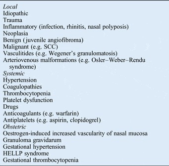
In the general population, epistaxis is precipitated by both local and systemic factors. The pregnant patient can develop several additional disorders that further predispose them to epistaxis. Table I summarises these aetiological factors. In the diagnostic investigation of our patient, we considered a number of causes of epistaxis in pregnancy (described below).
Local, systemic and obstetric causes of epistaxis

SCC = squamous cell carcinoma; HELLP = haemolysis, elevated liver enzymes, low platelets
Elevated oestrogen levels during pregnancy are thought to increase the vascularity of the nasal mucosa,Reference Sobol, Frenkiel, Nachtigal, Wiener and Teblum4 which may potentiate and prolong the bleeding.
Granuloma gravidarum is an uncommon, fast-growing benign fibrovascular tumour that occurs in 2–5 per cent of pregnancies.Reference Jones, Nguyen and Tabaee5–Reference Zarrinneshan, Zapanta and Wall8 These lesions typically involute after parturition, but are a potential source of profuse epistaxis during pregnancy. In our case, examination under anaesthesia did not reveal a granuloma gravidarum.
As with essential hypertension, hypertensive states during pregnancy can precipitate epistaxis. Gestational hypertension encompasses chronic hypertension and a transient rise in blood pressure (greater than 140/90 mmHg) in the latter half of pregnancy. In the current case, the patient was normotensive at both antenatal reviews and throughout her stay in hospital.
The development of thrombocytopenia raised the diagnosis of haemolysis, elevated liver enzymes, low platelets syndrome.Reference Garg, Nath, Bhalla and Kumar9, Reference Geary10 This syndrome is potentially fatal to both mother and fetus, and may necessitate emergency delivery. Although our patient was thrombocytopenic and hypoalbuminaemic, other liver function tests were within the normal ranges and a peripheral blood smear showed no evidence of haemolysis.
The most common cause of thrombocytopenia in pregnancy is gestational thrombocytopenia,Reference McCrae and Michelson11 which often occurs in the third trimester. The patient in the current study had a normal platelet count on admission, and the subsequent platelet drop was likely to be transfusion related.
The use of antibiotic prophylaxis is employed in cases of prolonged nasal packing. Fortunately, penicillin, cephalosporin and erythromycin can be used safely in pregnant women,Reference Crider, Cleves, Reefhuis, Berry, Hobbs and Hu12 hence the use of co-amoxiclav in this case.
Our management of epistaxis in this patient initially followed a conventional approach with the use of nasal packs. However, ongoing epistaxis and significant clinical deterioration necessitated urgent surgical intervention.
• Life-threatening epistaxis in pregnancy is rare
• Pregnant patients have additional risk factors for epistaxis compared with the general population
• Epistaxis is more common in pregnant patients versus non-pregnant patients
• This paper highlights the importance of multidisciplinary management for epistaxis in pregnancy
• It also highlights the risk to mother and fetus of using vasoconstrictor preparations
The surgical field and visualisation in endoscopic sinus surgery can be improved with the use of Moffat's solution (a variable mixture of adrenaline and cocaine), which produces a relatively bloodless field. However, cocaine can be systemically absorbed,Reference Wilkinson, Van Dyke, Jatlow, Barash and Byck13 and has been documented to cause ventricular tachycardia, myocardial infarction and death.Reference Chiu, Brecht, DasGupta and Mhoon14 It also has the potential to reduce uterine blood flow,Reference Foutz, Kotelko, Shnider, Thigpen, Rosen and Brookshire15 and its use may have devastating consequences for both mother and fetus. The recreational abuse of cocaine in the antenatal period is associated with intraventricular haemorrhage and maternal brain death.Reference Iriye, Asrat, Adashek and Carr16 Prenatal exposure also significantly increases the risk of premature labour, placental abruption, low birth weight and small for gestational age infants.Reference Gouin, Murphy and Shah17, Reference Chasnoff, Burns and Burns18 As a result, no topical preparation was used in this case, which hampered visualisation.
Conclusion
Life-threatening epistaxis in pregnancy is rare. It poses challenges from both a diagnostic and management perspective. It is important to consider the epistaxis risk factors that are specific to the pregnant patient. Moreover, multidisciplinary input from ENT surgeons, obstetricians and haematologists is integral in the management of complex epistaxis cases.
Acknowledgement
We would like to acknowledge the support of Vivienne Andrews, consultant haematologist (Medway Maritime Hospital).