Anomalous connection of the right superior pulmonary vein to the superior vena cava or the superior vena cava to the right atrium junction is the most frequent form of partial anomalous pulmonary venous connection.Reference Shahriari, Rodefeld, Turrentine and Brown1 Several surgical techniques have been described for repair of this type of anomaly, including one- or two-patch technique, Warden procedure, and various other modifications.Reference Gustafson2,Reference Pedro da Silva, da Fonseca da Silva, Melo and Lopes3 The objective of the present report is to describe a new surgical technique where the anomalous right superior pulmonary vein is connected to the left atrium by an extracardiac tunnel using pedicled autologous pericardium.
Case report
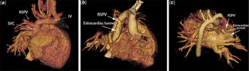
An 8-month-old boy presented with an initial diagnosis of perimembranous ventricular septal defect. He was mildly symptomatic and the physical examination showed III/VI holosystolic murmur in the left parasternal boarder. Preoperative echocardiography showed that besides an unrestrictive ventricular septal defect, there was also an anomalous connection between the right superior pulmonary vein and the superior vena cava. Computed tomography (CT) confirmed the diagnosis of partial anomalous pulmonary venous connection (Fig 1a). The patient was referred for surgical treatment after the parents provided informed consent.
( a ) Pre-operative computed tomography (CT). Anomalous connection between the right superior pulmonary vein (RSPV) and the superior vena cava (SVC). ( b and c ) Post-operative CT. ( b ) Anterior view and ( c ) posterior view of the extracardiac tunnel connecting the RSPV and the left atrium (LA). IV, Innominate vein.

A standard median sternotomy was performed and the thymus was sub-totally resected. The pericardial cavity was opened longitudinally on the left side of the aorta to preserve the pedicled autologous pericardium, and the heart was exposed. The superior vena cava was dissected out to the innominate vein, exposing the azygos vein and the anomalous right superior pulmonary vein. Attention is paid not to dissect the right superior pulmonary vein too much, like the sutureless technique used for total anomalous pulmonary venous connection. The connective tissue near the right superior pulmonary vein was preserved.
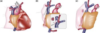
Cardiopulmonary bypass was established using ascending aortic and bicaval cannulation. The superior vena cava cannula was placed in the innominate vein (Fig 2a). The azygos vein was divided. The ascending aorta was cross-clamped and cardiac arrest was obtained. The fossa ovalis tissue was opened and the left atrial drainage tube was inserted. The anomalous right superior pulmonary vein was divided near the junction with the superior vena cava (Fig 2b). The superior vena cava was patched by a fresh autologous pericardium using 6-0 polypropylene suture. An incision was made in the left atrium toward the head at the junction with right lower pulmonary vein. An angled clamp through the incised fossa ovalis was used to guide the incision. The adventitia of the right superior pulmonary vein was sewed to the pericardium using interrupted 6-0 polypropylene stitches, leaving the orifice of right superior pulmonary vein open into the pericardium. Of note, the endothelium of right superior pulmonary vein was not caught by the suture to prevent potential inflammatory hyperplasia. The left atrium was also sewed to the pericardium at the site of the incision using interrupted 6-0 polypropylene stitches to prevent potential bleeding. The extracardiac conduit was created using 6-0 polypropylene suture in a running fashion (Fig 2c). The ventricular septal defect was closed in the usual manner using a Dacron patch. The incision in the fossa ovalis was closed after deairing. The patient was weaned from cardiopulmonary bypass.
( a ) Exposure of the anomalous right superior pulmonary vein (RSPV). Note that the pericardial cavity was opened longitudinally on the left side of the aorta to preserve the pedicled autologous pericardium (PAP). The dashed line indicates where to make the left atrium (LA) incision. ( b ) The anomalous RSPV was resected and the SVC was patched by a fresh pericardium. Note the incision made in the LA at the junction with right lower pulmonary vein. The LA was sewed to the PAP at the site of the incision. The adventitia of the RSPV was also sewed to the pericardium, leaving the orifice of RSPV open into the pericardium. ( c ) An extracardiac tunnel was created using PAP to connect the RSPV and the LA.

The patient had an uneventful recovery. The post-operative CT showed a satisfactory repair with unobstructed right superior pulmonary vein pathway to the left atrium (Fig 1b and c). The post-operative echocardiography showed an intact ventricular septum and laminal flow within the extracardiac tunnel, with no obstruction between right superior pulmonary vein and left atrium.
Discussion
The surgical technique for partial anomalous pulmonary venous connection varies according to the type of the anomalous connection. In the right partial anomalous pulmonary venous connection to the superior vena cava type, two different strategies have been most widely used. The first is the two-patch technique, which includes the use of a patch to baffle the blood from the anomalous pulmonary vein into the left atrium, with an additional patch to augment the superior vena cava incision.Reference Alsoufi, Cai, Van Arsdell, Williams, Caldarone and Coles4 However, an incision at the superior vena cava to the right atrium junction might be associated with higher incidence of sino-atrial node dysfunction.Reference DeLeon, Freeman, Ilbawi, Husayni, Quinones and Ow5 Our technique avoided this problem by not cutting or suturing near the sinus node area.
The second strategy, the Warden procedure, is a choice when the anomalous pulmonary vein connects to the superior vena cava at a higher position. The Warden procedure consists of dividing the superior vena cava right above the anomalous pulmonary vein, oversewing the lower superior vena cava end, connecting the upper superior vena cava end to the right atrial appendage, and placing an intra-atrial patch to direct the pulmonary venous drainage to the left atrium.Reference Gustafson2 This technique decreases the manipulation of the superior vena cava to the right atrium junction and thus avoids the sino-atrial node dysfunction. However, the major concern is the stenosis at the superior vena cava to the right atrium anastomosis.Reference Chandra, Gupta, Nath, Kazmi, Grover and Gupta6 In our case, construction of an extracardiac tunnel between right superior pulmonary vein and left atrium could avoid this sort of stenosis.
Another advantage of our surgical technique is the use of pedicled autologous pericardium to create the tunnel. Similar technique has been reported by Lugones et al for the surgical treatment of Scimitar Syndrome.Reference Lugones and García7 Compared with non-pedicled pericardium, pedicled autologous pericardium has preserved microvasculature and no evidence of calcification or shrinkage during the long-term follow-up.Reference Hibino, Shin’oka and Kurosawa8 Thus, the pedicled autologous pericardium might mimic vasculature tissue and have growth potential. A report by Masaki et alReference Masaki, Adachi, Kawamoto and Saiki9 found that a reconstructed right ventricular outlet tract conduit using pedicled autologous pericardium kept growing potential 14 years after the procedure. We believe in the repair of partial anomalous pulmonary venous connection, the use of pedicled autologous pericardium might also retain similar growing potential of the involved anatomic structure.
The surgical technique we have described provides a new choice for very young patients with right partial anomalous pulmonary venous connection to the superior vena cava. An extracardiac tunnel with pedicled autologous pericardium might keep growing potential, while avoid injury to the sino-atrial node or stenosis of superior vena cava or pulmonary veins. Further follow-up is necessary to elucidate the late results.
Financial Support
This research received no specific grant from any funding agency, commercial or not-for-profit sectors.
Conflicts of Interest
None.
Ethical Standards
The authors assert that all procedures contributing to this work comply with the ethical standards of the relevant national guidelines on human experimentation and with the Helsinki Declaration of 1975, as revised in 2008, and has been approved by the Institutional Committees.