Introduction
Some factors like financial difficulties, social isolation, and loss of independence are causes of chronic stress in older adults (Gaffey et al., Reference Gaffey, Bergeman, Clark and Wirth2016). In such conditions, the sustained increase of stress mediators (cortisol and catecholamines) may have long-term consequences as depressed mood, anxiety, and cognitive impairment. Higher morning cortisol levels and flatter diurnal cortisol slope were associated with poorer declarative memory and executive function in older people (Fiocco et al., Reference Fiocco, Wan, Weekes, Pim and Lupien2006; Beluche et al., Reference Beluche, Carriere, Ritchie and Ancelin2010). In addition, cortisol may have valence-specific effects on memory by facilitating the processing of emotionally arousing stimuli, especially the negative ones (Preuß et al., Reference Preuß, Schoofs and Wolf2009). Likewise, anxiety, which is a manifestation of chronic stress, has been related to lower memory performance (Curiel et al., Reference Curiel, Miller, Pollard, Kim and Kravitz2012) and memory bias for negative information (Herrera et al., Reference Herrera, Montorio and Cabrera2015). Nevertheless, some studies suggest that the relationship between both cortisol and anxiety and cognitive performance is curvilinear (Lupien et al., Reference Lupien2005; Bierman et al., Reference Bierman, Comijs, Rijmen, Jonker and Beekman2008). This pattern is reminiscent of the Yerkes–Dodson’ law, which states that certain degree of arousal is necessary to optimally perform cognitive tasks, but when arousal become too high or too low, performance decreases (Yerkes and Dodson, Reference Yerkes and Dodson1908).
Patients with Alzheimer's disease (AD) show high basal cortisol levels, which may represent not only a cause but also a result of hippocampal atrophy (Giubilei et al., Reference Giubilei2001; Gil-Bea et al., Reference Gil-Bea2010). In fact, experimental studies of CA3 lesions evidenced that hippocampus inhibits several aspects of the HPA axis functioning (Ulrich-Lay and Herman, Reference Ulrich-Lay and Herman2014). Thus, it has been proposed that hippocampal damage in AD leads to an increase in cortisol level that, in turn, may exacerbate AD pathology (Rothman and Mattson, Reference Rothman and Mattson2010).
Increased cortisol has been associated with poorer immediate memory in AD early stages (Souza-Talarico et al., Reference Souza-Talarico, Chaves, Lupien, Nitrini and Caramelli2010). Besides, some studies (Weiner et al., Reference Weiner, Vobach, Olsson, Svetlik and Risser1997) (Csernansky et al., Reference Csernansky2006), although not all (Swanwick et al., Reference Swanwick1998), found high cortisol level predicted faster cognitive decline. Consistently, a role of anxiety in clinical progression has been suggested (Bierman et al., Reference Bierman, Comijs, Jonker, Scheltens and Beekman2009; Mah et al., Reference Mah, Binns and Steffens2015).
AD patients show better memory for negative over positive information (Satler and Tomaz, Reference Satler and Tomaz2011; Gómez-Gallego and Gómez-García, Reference Gómez-Gallego and Gómez-García2017). This fact, that also been found in groups at risk of AD (mild cognitive impairment and late-onset depression) (Mah et al., Reference Mah, Anderson, Verhoeff and Pollock2017), is in contrast with the positivity effect present in healthy older adults. Although cortisol levels and anxiety have an influence on emotional memory, their effects in AD patients have not been studied yet. Therefore, the aim of this study is to examine the relation between both basal cortisol levels and state anxiety with memory for emotional information in patients with AD and in healthy controls. Based on previous studies, we hypothesized that participants’ cognitive status would moderate the relationship between both cortisol levels and anxiety and memory performance. In addition, cortisol and anxiety would be positively related to negative bias in memory.
Methods
Participants
Ninety-eight elderly participated in this study. Forty-six patients were recruited from several dementia day centers in the area of Murcia. All of them met criteria for “probable AD” (McKhann et al., Reference McKhann, Drachman, Folstein, Katzman, Price and Stadlan1984) and were in stage 4–5 according to the Global Deterioration Scale (Reisberg et al., Reference Reisberg, Ferris, de Leon and Crook1988). Controls (n = 52) were recruited from elderly participants who attended to older adult day centers in the same geographical area. They were selected to be comparable to the patients with respect to age, education, and gender. The exclusion criteria to participate in the study were (1) acute medical illness, (2) lifetime or current neurological or psychiatric disease (different from AD for patients), (3) medical condition that could be associated with changes in cortisol levels (e.g. Cushing syndrome, Addison syndrome), (4) use of medication that could affect cortisol levels (e.g. corticosteroids, hormones, narcotics).
Sociodemographic and clinical data of participants were obtained using semi-structured interviews with the participants and, in the case of patients, with a collateral source (generally a caregiver). All patients and controls were administered Mini-Mental State Examination (Folstein et al., Reference Folstein, Folstein and McHugh1975), Memory Alteration Test (Rami et al., Reference Rami, Bosch, Sanchez-Valle and Molinuevo2010), Trail Making Test Part B (Army Individual Test Battery, 1944), the 11-item Boston Naming Test (Nebreda et al., Reference Nebreda, García-Caballero, Asensio, Revilla, Rodriguez-Girondo and Mateos2011), Geriatric Depression Scale (GDS, Yesavage, Reference Yesavage1988), and the short form of Positive and Negative Affect Scale (Thompson, Reference Thompson2007). Patients were also administered Barthel Index (Mahoney and Barthel, Reference Mahoney and Barthel1965) and Functional Activities Questionnaire (Pfeffer et al., Reference Pfeffer, Kurosaki, Harrah, Chance and Filos1982).
Emotional memory assessment
In this study, pictures from the International Affective Picture System (Lang et al., Reference Lang, Bradley and Cuthbert2008) were used. This database contains high-resolution color photographs evoking a range of affective responses, accompanied by normative ratings of these images with respect to valence (or pleasure), arousal, and dominance (or control). Ratings of the three dimensions are based on a nine-point scale, where 1 represents the lowest (pleasure, arousal, or control) and 9 the highest. Since some cross-cultural differences have been reported in normative arousal ratings, Spanish normative valence and arousal ratings were used to select pictures (Moltó et al., Reference Moltó1999; Vila et al., Reference Vila2001; Moltó et al., Reference Moltó2013). Two sets of pictures were used: a study set (30 pictures) and a distractor set (30 pictures). Each set of pictures contained ten positive (pleasant) pictures, ten (neutral) pictures, and ten negative (unpleasant) pictures. We aimed to maximize the differences in valence between the pictures by selecting positive pictures with valence ratings higher than 6.5, negative pictures with valence rating lower than 3.5, and neutral pictures with valence ratings ranging from 4.5 to 5.5. Besides, we selected negative and positive pictures sets that were equated for arousal on the basis of normative ratings. Additionally, differences in the semantic content of the pictures between positive, negative, and neutral pictures were reduced by selecting, for each valence category, four pictures representing “people,” two pictures representing “animals,” and four pictures representing objects or landscapes. Both sets (study and distractor sets) were matched in terms of semantic content, mean arousal, and mean valence ratings (see Appendix for the IAPS codes as well as valence and arousal normative data of the pictures being used).
In the study phase, each picture was displayed on the computer screen for 6 seconds. Then, a fixation cross appeared for 6 seconds while participants identified the pictures with one or two words. Pictures were presented in a random order in both encoding and recognition phases. Besides, the order of the pictures differed across participants. They were given detailed instructions and were told to memorize the pictures (intentional encoding). The immediate free recall task started 1 minute after the presentation of the last slide. Participants had 5 minutes to describe verbally as many pictures as possible, in any order, from the ones they had been shown. The descriptions of the pictures were recorded and two independent raters, blind to study diagnosis, analyzed them and decided which pictures had been recognized. Agreement between judges was 95%, and discrepancies were discussed until a consensus was reached. Twenty minutes after the end of the free recall task, participants were given a yes–no recognition test for the pictures. This task was participant paced, although participants were instructed to respond as soon as they were sure of their answer. Between immediate free recall and delayed recognition tasks, the participants stayed in the same room, talking with the researchers about the activities they do in the centers and their current quality of life. We attempted to create an atmosphere of trust and respect that helps participants express their individual's feelings and thoughts, as recommended (Tappen et al., Reference Tappen, Williams-burgess, Edelstein and Touhy1997; McKeown et al., Reference McKeown, Clarke, Ingleton and Repper2010).
Saliva samples
Salivary cortisol samples were obtained using Salivette collection tubes (Sarstedt, Germany). Participants were told that, up to 2 hours before providing saliva, they only could drink water and they should not take any stimulant substance or smoke. Besides, they were instructed to keep the cotton swab in their mouth for 2 minutes, without chewing it. The samples were centrifuged at 3,000 rpm for 10 minutes, resulting in a clear supernatant of low viscosity that was stored at −20°C until assaying. The saliva cortisol levels were determined using a radioimmunoassay kit (Spectria Cortisol RIA (cat. Nu 06119) from Orion Diagnostica (Espoo, Finland). Assay sensitivity was 0.8 nmol/L, and the within- and inter-assay variation coefficients were all below 8%.
Procedure
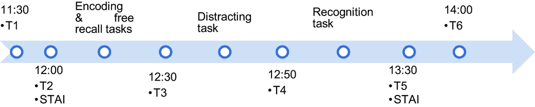
Cortisol was measured at baseline (T1 and T2), before (T3) and after the memory tasks (T4) and 30 minutes after finishing the session (T5) to assess potential changes due to the neuropsychological assessment (Figure 1). To reduce the impact of diurnal variations in cortisol levels, all testing was performed between 11:30 and 14:00 pm. All these measurements took place in the day centers. The short version of STAI was administered before and after memory assessment (Perpiñá-Galvañ et al., Reference Perpiñá-Galvañ, Richart-Martínez and Cabañero-Martínez2011).
Study design and experimental procedure including point in time of cortisol sampling.

Statistical analyses
The memory outcome variables were immediate recall score (computed as the percent of correct responses for each valence category), true recognition score (calculated as the percent of correct recognitions (Hits) minus the percent of false alarms), and measures of negative memory bias (calculated by subtracting the immediate recall scores and true recognition score for neutral pictures from the recall/recognition scores for negative pictures). To examine emotional enhancement effect, separate repeated-measure ANOVA tests were performed for immediate recall score and true recognition score, with valence as the within-participant factor and group as the between-participant factor. Greenhouse–Geisser corrected p values were used when the requirement of sphericity was not met. Planned comparisons were used to further analyze significant interactions. Bonferroni correction for multiple comparisons was used to correct p-values of post hoc tests for main effects of valence. Due to the low number of statistical tests performed in this step, this method allows an adequate control of type 1 error.
All the variables were checked for normality and homogeneity of variance with Kolmogorov–Smirnov test and Barlett test. Since cortisol samples were right skewed, values were natural log transformed. After transformation, all data were normally distributed. Differences between groups in normally distributed variables were analyzed using ANOVA test.
Selection of predictors and covariates
Changes in either cortisol levels or STAI scores were analyzed with repeated measures ANOVA. In the repeated-measures ANOVA with cortisol level as dependent variable, we did not find significant effects of the interaction group*time (F(4,96) = 1.15, p = 0.330) and time (F(4,96) = 1.76, p = 0.155). Since an association between average cortisol level and cognitive performance in mild cognitive impairment and healthy older people have been reported (Lupien et al., Reference Lupien1998; Wolf et al., Reference Wolf, Convit, Thorn and J de Leon2002), we used this metric as a measure of basal salivary cortisol. Analysis of the changes in STAI scores revealed significant effects of the interaction time*group (F(1,96) = 8.06, p = 0.006). Planned comparisons showed a reduction in STAI scores in the control group (t(51) = 3.46, p = 0.001), while no significant change in STAI scores was found in patients (t(45) = −1.26, p = 0.213). The main effect of the group was not significant (Patients: 8.65 ± 2.73, controls: 8.01 ± 3.24, F(1,96) = 1.05, p = 0.308). Because significant differences between groups were found in pre-task STAI scores (patients: 7.61 ± 3.74, controls: 9.19 ± 2.97, F(1,96) = 5.42), we used average STAI scores as a predictor.
Since gender, age, education (years of education), and GDS are variables with a potential effect on memory performance, their associations with memory scores were tested. Negative significant correlations were found between age and both immediate and delayed total recognition scores (r = −0.33 and −0.41 p < 0.001 for both contrasts). Positive significant correlations were found between education and total immediate recall scores (r = 0.31, p = 0.020), negative bias in immediate recall task (r = 0.19, p = 0.050), and delayed total recognition scores (r = 0.51, p < 0.001). GDS was negatively correlated with total immediate recall (r = −0.28, p = 0.005), negative bias immediate recall (r = −0.18, p = 0.050), and delayed total recognition scores (r = −0.25, p = 0.010). Men had higher negative bias scores in immediate task and higher delayed recognition total scores than women had (F(1,97) = 5.97, p = 0.016, and F(1,97) = 4.82, p = 0.030, respectively). Based on these data, age, gender, education, and GDS were included as covariates in the analyses.
Design of the models
In order to determine if cognitive status moderated the relation between stress markers and memory performance, group was added as a moderator, according to the suggested (Aiken et al., Reference Aiken, West and Reno1991). Besides, we investigated the existence of curvilinear relationships between total memory scores and both stress markers by adding to the regression analyses the square of cortisol level (cortisol2) and the square of STAI scores (STAI2).
The design of the analyses to predict negative bias was as follow: Step 1 – we entered covariates (gender, age, educative level, GDS); Step 2 – we added group; Step 3 – we included the predictor; Step 4 – we entered the interaction term (predictor*group). In the models predicting total scores, two further steps were included: Step 5 – cortisol2 or STAI2 and Step 6 – cortisol2*group or STAI2*group. All predictors were standardized prior to entry into the regression model to reduce multicollinearity.
When an interaction term was significant, separate analyses were performed in each group (patients and controls). The design of these analyses was Step 1 – covariates (gender, age, educative level, GDS); Step 2 – predictor (cortisol or STAI). Only if quadratic interaction was significant, we added a further step: cortisol2 or STAI2. All the statistical analyses were performed with SPSS Statistics 20.0. The criterion for statistical significance was 0.05.
Results
The sample consisted of 52 healthy older adults and 46 AD patients (31 with mild dementia (Global Deterioration Scale stage 4) and 15 with moderate dementia (Global Deterioration Scale stage 5). Groups did not significantly differ in mean age (patients: 78.3 ± 4.8; controls: 76.7 ± 7.5, F(1,97) = 3.62, p = 0.06), gender (patients: 32 women; controls: 36 women, (χ 2 = 0.367, p = 0.544) and education (patients: 9.6 ± 4.9 years; controls: 10.4 ± 5.8 years, F(1,97) = 0.49, p = 0.486).
As expected, significant differences were found in MMSE scores (patients = 18.8 ± 4.9; controls = 28.6 ± 1.5, F(1,97) = 183.46, p < 0.001), Memory Alteration Test (patients: 20.0 ± 11.9, controls: 46.6 ± 3.3, F(1,97) = 218,76, p < 0.001), Trail Making Test part B (patients: 210.4 ± 72.1 seconds, controls: 87.5 ± 68.5 seconds, F(1,97) = 191.82, p < 0.001), and 11-item Boston Naming Test (patients: 3.1 ± 0.6, controls: 7.7 ± 0.4, F(1,97) = 170.66, p < 0.001). Nevertheless, no significant difference was found in GDS scores (patients: 7.4 ± 1.8; controls: 6.8 ± 2.2, F(1,97) = 2.24, p = 0.138), positive PANAS subscale (controls: 15.95 ± 3.61, patients: 14.94 ± 3.10, F(1,91) = 3.23, p = 0.075), and negative PANAS subscale (patients: 10.33 ± 3.11), controls: 9.0 ± 3.48), F(1,91) = 0.29, p = 0.594).
Patients showed severe impairment of instrumental activities of daily living (ADL) as shown Functional activities Questionnaire scores (mean = 18.35, SD = 6.01) and mild-to moderate impairment of basic ADL as shown Barthel index scores (mean = 61.08, SD =15.84). Concerning the use of psychotropic medication, 39 patients were taking cholinesterase inhibitors, 23 benzodiazepines, and 17 antipsychotic drugs. No control participant was taking psychotropic drugs.
Stress markers and memory outcomes
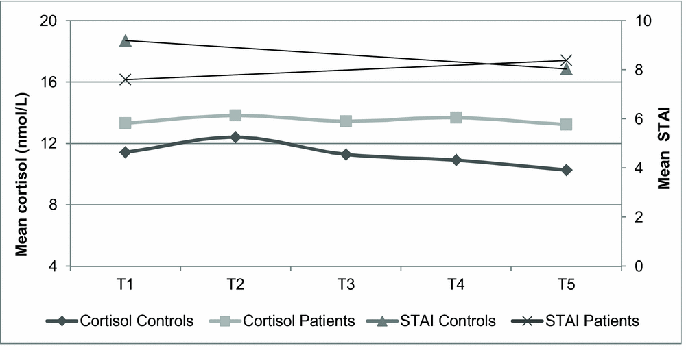
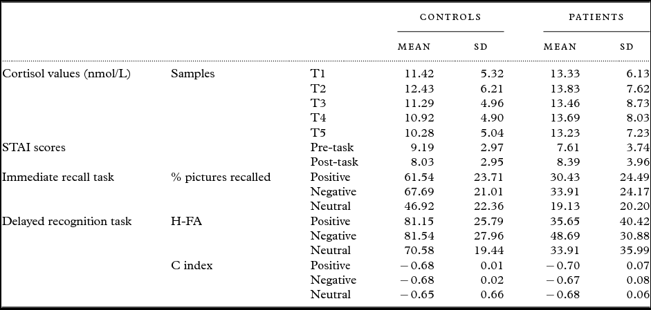
The mean cortisol values at different time points and mean STAI scores at baseline and post-task time are shown in Figure 2. The descriptive statistics of cortisol values, STAI scores and memory scores in immediate and delayed recognition tasks are shown in Table 1. As described in Methods, there was no significant difference in average cortisol levels or average STAI scores between patients and controls (p > 0.05 in both contrasts).
Changes in mean cortisol level and STAI scores over the experiment.

Descriptive statistics of the cortisol level, STAI scores, and memory scores for picture valence

In repeated-measures, ANOVA with immediate recall scores as dependent variable, interaction group*valence was not found to be significant (F(2,96) = 0.76, p = 0.47). However, there was a significant effect of group, with patients’ scores being lower than controls’ F(1,96) = 72.84, p < 0.001). Besides, the main effect of the valence was significant (F(2,96) = 28.16, p < 0.001). The recall of pictures was significantly lower for neutral than for pleasant and unpleasant ones (Bonferroni-adjusted p-values < 0.001 in both cases).
The results of the repeated measures ANOVA with total recognition scores as dependent variable showed a significant interaction group*valence (F(2,96) = 3.40, p = 0.041) as well as significant effects of group (F(1,96) = 50.10, p < 0.001) and valence (F(2,96) = 12.24, p < 0.001). In planned comparisons, we observed that controls had higher true recognition score for positive and negative pictures than for neutrals (t(51) = 4.10, p < 0.001 and t(51) = 3.72, p = 0.001, respectively), while patients had higher true recognition score for negative pictures than for positive and neutral ones (t(45) = 2.88, p = 0.006 and t(45) = 4.81, p < 0.001).
Relation between AD status, cortisol, and memory
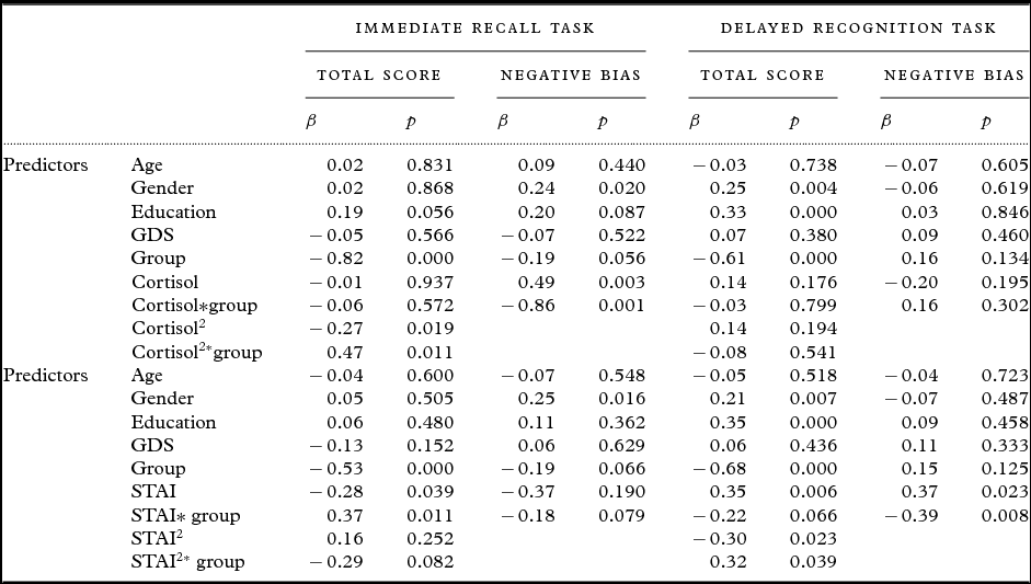
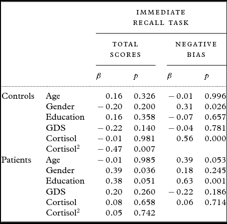
In the regression model to predict immediate recall scores (Table 2), the quadratic interaction term was significant (β = 0.47, p = 0.011). As shown in Table 3, an inverted U-shaped effect of cortisol was observed in controls (β = −0.47, p = 0.007), while no significant curvilinear or linear effects were found in patients (p > 0.1 for both contrasts). The results of the regression model to predict negative bias in immediate recall showed a significant interaction group*cortisol (β = −0.86, p = 0.001) (Table 2). In controls, cortisol had a significant effect (β = 0.56, p < 0.001) as opposed to patients (p > 0.1) (Table 3).
Models to predict memory scores from cortisol levels and from STAI scores

Regression models to predict immediate recall scores from cortisol level in patients and in controls

In the analyses to predict delayed total scores and negative bias (Table 2), neither the interaction terms nor the main effects of cortisol were significant (p > 0.1 for all the contrasts).
Relation between AD status, STAI, and memory
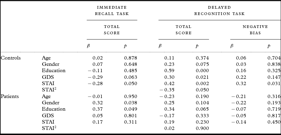
In the regression model with immediate recall scores as dependent variable, we found a significant linear interaction term (β = 0.37, p = 0.011) (Table 2). In controls, we found a negative association between STAI and memory scores (β = −0.28, p = 0.050) while in patients, such relation was not significant (β = 0.17, p = 0.311) (Table 4). As to negative bias, neither the interaction term nor the main effect were significant (β = −0.18, p = 0.079, β = −0.37, p = 0.190, respectively) (Table 2).
Summary of the models to predict memory scores from STAI scores in patients and in controls

As to total recognition scores is concerned, there was a significant quadratic interaction effect (β = 0.32, p = 0.039) (Table 2). In controls, a curvilinear effect was found (β = −0.35, p = 0.050), while in patients no significant main effects were found (p > 0.1 for both terms) (Table 4). In the regression model predicting negative bias, we found the linear interaction STAI*group be significant (β = −0.39, p = 0.008) (Table 2). In controls, STAI score was positively related to bias (β = 0.32, p = 0.030) while in patients this predictor had no significant effect (β = −0.14, p = 0.450) (Table 4).
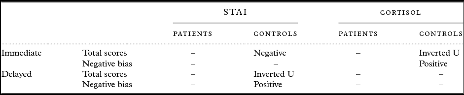
Table 5 shows a summary of the relationships between predictor variables (cortisol and STAI) and memory outcomes variables in patients and controls.
Summary of the relationships between predictor variables (basal cortisol and STAI) and memory outcomes variables (total scores and negative bias) in patients and controls

Discussion
In this study, we investigated the relationships between both cortisol and anxiety levels and memory for emotional information as well as the moderating role of cognitive impairment. Our finding supports the notion that emotional memory is preserved in AD patients (Mah et al., Reference Mah, Anderson, Verhoeff and Pollock2017). The results of the immediate recall task showed a better performance for arousing pictures than for neutrals by both groups. For the delayed recognition task, the pattern of results showed that patients discriminate better the negative information than positive or neutral one. This negative bias has been previously reported (Fleming et al., Reference Fleming, SH, Doo, Maguire and Potkin2003; Schultz et al., Reference Schultz, De Castro and Bertolucci2009) and is not explained by patients’ negative mood (Gómez-Gallego and Gómez-García, Reference Gómez-Gallego and Gómez-García2017). However, we cannot discard the possibility that transient anxiety or mood changes in patients, following questions about their quality of life, could have been affected their performance in the delayed memory task. One alternative interpretation of these results is that AD patients had a failure in remembering positive information over neutral one. In fact, AD patients classified positive pictures as neutral in a higher percent than healthy controls did (Gomez-Gallego and Gomez-Garcia, Reference Gómez-Gallego and Gómez-García2017), which may lead to a worse memory of these pictures.
Although patients’ mean cortisol levels at different times were slightly higher than controls’ though such difference did not reach statistical significance. Moreover, in both groups, cortisol values were within the reference range (Kirschbaum and Hellhammer, Reference Kirschbaum, Hellhammer and Fink2000). Although neuropsychological testing may be a mild stressor for some participants (Neupert et al., Reference Neupert, Miller and Lachman2006; Rosnick et al., Reference Rosnick, Rawson, Butters and Lenze2013), no significant change in cortisol level was found in either group. Conversely, anxiety decreased in healthy controls.
As previously reported in healthy older adults (Lupien et al., Reference Lupien2005), an inverted U-shaped association between cortisol and immediate recall was found, with the best performance for moderate levels. Besides, cortisol levels were associated with better memory for negative information. However, no relationship was found between cortisol and immediate recall in AD patients.
Since high cortisol levels have a great impact on the functioning of hippocampal neurons, an effect on short-term memory is expected (Krugers et al., Reference Krugers, Lucassen, Karst and Joëls2010). In contrast, we did not find a significant relationship between cortisol and short-term memory scores. Because cortisol samples were collected at early afternoon (when hormone levels are stable and low) and stress was not induced, little effects were expected. In addition, the performance of the recognition task is influenced by familiarity processes, which require neocortical activation, and may be less related to cortisol levels than recollection-based memory tasks, which are hippocampal dependent (Souza-Talarico et al., Reference Souza-Talarico, Chaves, Lupien, Nitrini and Caramelli2010; McCullough et al., Reference McCullough, Ritchey, Ranganath and Yonelinasa2016). Furthermore, the delay between encoding and recognition tasks might have not been long enough to separate the effects of cortisol on retrieval, from those on consolidation processes. Some evidence exists that stress-induced cortisol levels during consolidation phase improve memory performance, while such increase during retrieval worsens performance (Wolf, Reference Wolf2009). One possible explanation for the lack of association between cortisol and short-term memory may relay in that specific effects on different memory processes can be counteracted. Similar findings were reported by Ackermann et al. (Reference Ackermann, Hartmann, Papassotiropoulos, Quervain and Rasch2012). Additionally, the association between cortisol and memory may depend on the time of the day in which cortisol samples were taken. Stress induced in the morning, when cortisol is at their peak, has been negatively related to memory performance opposed to stress provoked in the afternoon that may not affect memory functioning (Maheu et al., Reference Maheu, Collicutt, Kornik, Moszkowski and Lupien2005).
Coordinated functional interactions between the HPA axis and the noradrenergic systems participate in the production of the anxiety response (Charney and Drevets, Reference Charney, Drevets, Kenneth, Charney, Coyle and Charles2002). The impairment of attentional processes (inhibition and shift) due to anxiety may affect immediate recall performance (Eysenck et al., Reference Eysenck, Derakshan, Santos and Calvo2007). Consistent with this, worse immediate recall score was observed in relation to higher anxiety levels in controls. However, the relationship between anxiety and short-term memory is found to be curvilinear. As predicted, anxiety increases the memory for negative information. In AD patients, anxiety levels were not related to either memory performance or bias.
Overall, our results in healthy elderly support the notion that anxiety and cortisol levels are related to better memory for negative information (Herrera et al., Reference Herrera, Montorio and Cabrera2015). Instead, memory performance increases with stress to an optimal point, above or below which memory decreases. The different association patterns between both anxiety and cortisol and short-term memory may be partly due to the type of memory task. The performance of a recognition task of visual information seems to be more related to the arousal response to stress than to cortisol levels (Ackermann et al., Reference Ackermann, Schwabe, Scho, Schönfeld, Ackermann and Schwabe2014).
In the other hand, in patients, the association between immediate and short-term visual memory and stress is not evident. In AD, an increase in the expression of mineralocorticoid receptors in frontal cortex has been shown to occur in relation to the cognitive impairment (Gil-Bea et al., Reference Gil-Bea2010). This adaptive change might cause that patients in moderate stages have lower sensitivity to stress hormones effects (ter Heegde et al., Reference ter Heegde, De Rijk and Vinkers2015). Likewise, as the disease progresses, a greater HPA axis dysfunction and atrophy of amygdala (Csernansky et al., Reference Csernansky2006; Poulin et al., 2012) may affect the memory enhancement effect of emotional items. Previous literature suggests that patients with damage in left amygdala show greater and more specific deficits in emotional memory than those with damage in right amygdala (Buchanan et al., Reference Buchanan, Denburg, Tranel and Adolphs2001). Heterogeneity in the AD pathology may explain why some studies reported no emotional effect in AD patients (Abrisqueta-Gomez et al., Reference Abrisqueta-Gomez, Bueno, Oliveira and Bertolucci2002; Chainay et al., Reference Chainay, Sava, Michael, Landré, Versace and Krolak-Salmon2014), while others found preservation of this effect (Perrin et al., Reference Perrin, Henaff, Padovan, Faillenot, Merville and Krolak-Salmon2012; Gómez-Gallego and Gómez-García, Reference Gómez-Gallego and Gómez-García2017).
Some limitations should be considered in interpreting the current findings. First, the transversal design of the study prevents us from establishing causal associations. Second, although afternoon cortisol is a good indicator of basal cortisol levels and was found to predict disease progression (Weiner et al., Reference Weiner, Vobach, Olsson, Svetlik and Risser1997), it does not reflect either the reactivity of HPA axis or the circadian fluctuations. In addition, our results on the relationship between stress markers and memory of emotional information could not be generalizable to verbal emotional stimuli, though pictures selected contained familiar stimuli that were verbally identified. Although a depression scale was included as covariate in this study and no participant was depressed, we cannot completely exclude a potential effect of transient mood changes on our results (Holland et al., Reference Holland2014). Futures studies should include patients in early stages of AD to examine the moderating role of depression in the association between stress and memory. Similarly, based on recent studies on the effect of cholinesterase inhibitors on emotional memory (Jiang et al., Reference Jiang2016), we think it would be advisable to replicate the present findings in a larger sample that allows adjustment for these drugs.
Conflict of interest
None.
Description of authors’ roles
María Gómez-Gallego designed the study, collected and analyzed the data, interpreted the results, and wrote the paper. Juan Gómez-Garcia collected data, supervised the development of work, helped to analyze data, and draft the paper.
Ethical standards
This study was approved by the Ethics Committee of the Catholic University San Antonio. Previously to participate in it, the participants or their legal representatives gave their written, informed consent.
Acknowledgments
We would like to thank patients and older adults who have participated in this study for their active participation as well as the centers staff for the excellent cooperation.