1. Introduction
The Demospongiae, considered part of the most basal evolutionary lineage in the Metazoa (Larroux et al. Reference Larroux, Fahey, Liubicich, Hinman, Gauthier, Gongora, Green, Wörheide, Leys and Degnan2006), is the most speciose of the classes within the phylum Porifera (Degnan et al. Reference Degnan, Adamska, Richards, Larroux, Leininger, Bergum, Calcino, Taylor, Nakanishi, Degnan and Wanninger2015) and has received particular attention in taxonomic and cladistic studies (e.g. Botting, Muir & Lin, Reference Botting, Muir and Lin2013; Botting, Cárdenas & Peel, Reference Botting, Cárdenas and Peel2015). Recently, Ehrlich et al. (Reference Ehrlich, Rigby, Botting, Tsurkan, Werner, Schwille, Petrášek, Pisera, Simon, Sivkov, Vyalikh, Molodtsov, Kurek, Kammer, Hunoldt, Born, Stawski, Steinhof, Bazhenov and Geisler2013) proposed that the Vauxiidae, exhibiting an apparently reticulate, aspiculate fibrous skeleton (Rigby & Collins, Reference Rigby and Collins2004), were likely to be the most basal definitive demosponge group known because recent molecular work often places the ‘keratosans’ at the base of demosponge phylogeny (Erpenbeck &Wörheide, Reference Erpenbeck, Wörheide, Zhang and Shear2007). Previously described vauxiid sponges are all of roughly similar age (Cambrian Series 3), mainly from the Burgess Shale of British Columbia, Canada (Walcott, Reference Walcott1920; Rigby, Reference Rigby1980, Reference Rigby1986; Rigby & Collins, Reference Rigby and Collins2004), and the Spence Shale and Wheeler Formation of Utah, USA (Rigby, Reference Rigby1980; Robison, Babcock & Gunther, Reference Robison, Babcock and Gunther2015). Related but poorly known material has also been reported from North Greenland (Peel & Ineson, Reference Peel and Ineson2011), and Botting (Reference Botting, Clarkson, Harper, Taylor and Anderson2007) briefly described and illustrated a Silurian vauxiid.
Strata of South China show evidence that the Porifera experienced an adaptive radiation near the beginning of the Cambrian Period (Xiao et al. Reference Xiao, Hu, Yuan, Parsley and Cao2005; Yang et al. Reference Yang, Zhao, Wang and Wang2005 a, Reference Yang, Zhao, Wu, Zheng and Zhu2014). Sponges continued to flourish through the Cambrian Period, becoming common components of the marine biota, and have maintained important positions in marine ecosystems through the remainder of Phanerozoic time. In the Cambrian Period, non-archaeocyathan poriferans are both abundant and diverse in Konservat-Lagerstätten of North America (Walcott, Reference Walcott1920; Rigby, Reference Rigby1986; Robison, Reference Robison, Simonetta and Conway Morris1991; Rigby & Collins, Reference Rigby and Collins2004; Robison, Babcock & Gunther, Reference Robison, Babcock and Gunther2015) and China (Steiner et al. Reference Steiner, Mehl, Reitner and Erdtmann1993; Xiao et al. Reference Xiao, Hu, Yuan, Parsley and Cao2005; Yang et al. Reference Yang, Zhao, Wang and Wang2005 a,b, Reference Yang, Sun, Zhao, Peng and Zhen2012, Reference Yang, Zhao, Wu, Zheng and Zhu2014; Wu, Zhu & Steiner, Reference Wu, Zhu and Steiner2014), and archaeocyaths are abundant and diverse in shelf carbonate deposits from a wide range of palaeolatitudes (e.g. Wood, Reference Wood1999; Zhuravlev & Riding, Reference Zhuravlev and Riding2001). In this paper, a new genus and species of vauxiid sponge, Angulosuspongia sinensis gen. et sp. nov., is described from the middle and upper part of the Kaili Formation (Cambrian Series 3, Stage 5) at Jianhe, Guizhou, South China. These vauxiid remains are the first to be described in detail from China, as well as the first reported from outside Laurentia, and represent only the second genus attributed to the family. The new material shows apparent spicular structures preserved in relief that are interpreted as spicules here, and are possibly composed of silica according to results of Energy Dispersive X-ray Spectroscopy (EDS), although further work needs to be done in this regard. Its age is close to but still slightly older than the Burgess Shale Biota, and it appears to be a primitive relative to other members of the Vauxiidae. This new material not only extends the geographic distribution of vauxiids, but also helps to fill a chronostratigraphical gap between the North Greenland and North American material, and provides additional evidence for understanding the evolutionary history of the Demospongiae.
2. Geologic setting
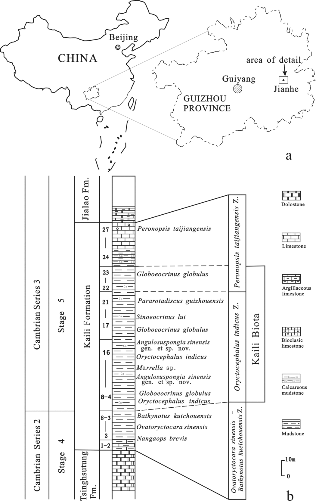
Fossils reported here were collected from the middle and upper part of the Kaili Formation (Cambrian, provisional Series 3, provisional Stage 5) in the Miaobanpo section quarry at Balang village, Jianhe County, Guizhou Province, South China (Fig. 1). The section is located c. 1.2 km northeast of the Wuliu–Zengjiayan section, which is under consideration as a potential stratotype for the Series 3/Stage 5 GSSP (Zhao et al. Reference Zhao, Yuan, Guo, Peng, Yin, Yang, Wang and Sun2014). In the Wuliu–Zengjiayan section, the Kaili Formation overlies the Tsinghsutung Formation, and is overlain by the Jialao Formation. It contains representatives of some 120 animal genera distributed among at least 10 phyla (Zhao et al. Reference Zhao, Zhu, Babcock and Peng2011), plus a small number of algae and bacteria. Overall, the Kaili Biota ranks as the third-most diverse Burgess Shale-type biota known, and its age is close to but still slightly older than the Burgess Shale Biota (Zhao et al. Reference Zhao, Zhu, Babcock, Yuan, Parsley, Peng, Yang and Wang2005, Reference Zhao, Zhu, Babcock and Peng2011).
Location and stratigraphy of the Miaobanpo section. (a) Map showing the location of the Miaobanpo section. (b) Stratigraphic column of the Miaobanpo section showing the level that produced specimens of Angulosuspongia sinensis gen. et sp. nov.

The Kaili Formation in the Miaobanpo section is a 156 m thick (Zhao et al. Reference Zhao, Zhu, Babcock, Yuan, Parsley, Peng, Yang and Wang2005) succession of fine-grained siliciclastic rocks with minor carbonates (Gaines et al. Reference Gaines, Mering, Zhao and Peng2011; Fig. 1). The base of the formation is not exposed because a fault occurs between it and the underlying Tsinghsutung Formation. The Miaobanpo section is the most productive site for exceptionally preserved fossils of the Kaili Biota. Notably, the section has yielded many specimens of the nonbiomineralized worm Ottoia, as well as numerous remains of nonbiomineralized arthropods such as Marrella, Naraoia and Anomalocaris. Three trilobite biozones are recognized in the Kaili Formation. In ascending order, these are the Bathynotus kueichouensis – Ovatoryctocara sinensis Assemblage Zone, Oryctocephalus indicus Zone and Peronopsis taijiangensis Zone (Zhao et al. Reference Zhao, Yuan, Guo, Peng, Yin, Yang, Wang and Sun2014, Reference Zhao, Yuan, Peng, Yang and Esteve2015). Available biostratigraphic evidence shows that the vauxiid sponge-yielding horizon belongs to the Oryctocephalus indicus Zone. Specimens were collected from the lower and middle parts of the zone.
3. Material and methods
Sixty-two specimens of vauxiid sponge remains were examined in this study. They are reposited in the Guizhou Research Centre for Palaeontology, Guizhou University, Guiyang, China (GRCP, GU). Some specimens (Figs 2–4) were imaged using a Canon EOS Rebel T3i Digital SLR camera with MP-E 65 mm macro lens. Others were imaged by Y. Fang using a LEO1530VP Scanning Electron Microscope (SEM) equipped with an Energy Dispersive X-ray Spectrometer (EDS), located in the State Key Laboratory of Palaeobiology and Stratigraphy, Nanjing Institute of Geology and Palaeontology, the Chinese Academy of Sciences. One sample, GTBM16-1192 was gold-coated, whereas all others imaged under the SEM were left uncoated.
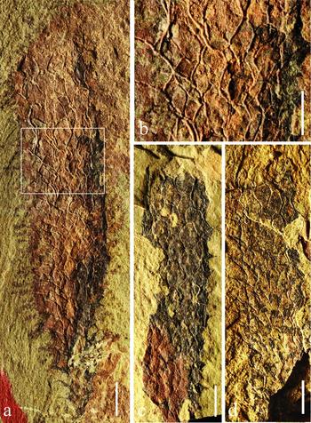
Angulosuspongia sinensis gen. et sp. nov. (a) Complete adult individual, holotype, GTBM17-765a. (b) Close-up of the rectangle area in (a). (c) Nearly complete adult individual, GTBM16-109b. (d) Adult individual without the base, GTBM17-1077b. All scale bars are 2 mm, except (b) 1 mm. All specimens photographed dry in reflected light.

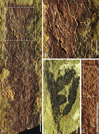
Angulosuspongia sinensis gen. et sp. nov. (a) Nearly complete adult individual, GTBM22-1816. (b) Close-up of the rectangle area in (a). (c) Nearly complete adult individual, paratype, GTBM9-3-3613. (d) Nearly complete juvenile, GTBM16-1192. All scale bars are 2 mm, except (b) 1 mm. All specimens photographed dry in reflected light.

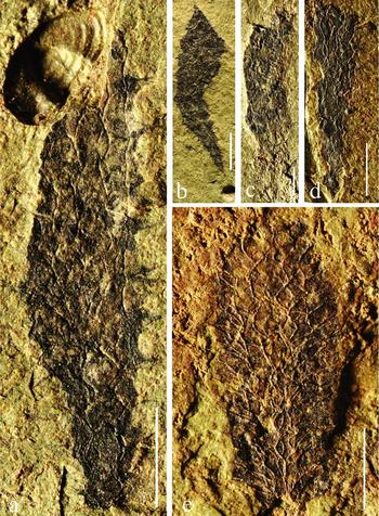
Angulosuspongia sinensis gen. et sp. nov. (a) Nearly complete juvenile, GTBM9-2-4792b. (b) Nearly complete juvenile, GTBM17-1169. (c) Nearly complete juvenile, GTBM9-4-2581. (d) Nearly complete juvenile, GTBM9-2-816b. (e) Nearly complete juvenile, GTBM22-1-18. All scale bars are 2 mm. All specimens photographed dry in reflected light.

4. Systematic palaeontology
Class DEMOSPONGEA Sollas, Reference Sollas1875
Order VERONGIDA Bergquist, Reference Bergquist1978
Family Vauxiidae Walcott, Reference Walcott1920
Emended diagnosis. Unbranched to branched thin-walled conical-tubular sponges, double-layered skeleton: endosomal layer of fused cored fibres or spicules united to produce a net with polygonal or hexagonal openings; dermal layer thin, irregular or regular reticular supported by radial fibres or spicules, respectively.
Remarks. At present, the family Vauxiidae is monogeneric, embracing only Vauxia, and body fossil remains are known mainly from strata of Cambrian Series 3, ranging from Stage 5 (provisional) to the Drumian Stage in Laurentia. The systematic position of the family has been discussed a number of times since the time of Walcott (Reference Walcott1920), who classified these sponges as hexactinellids, noting that the specimens preserved in the Burgess Shale appeared to have the original siliceous matter of the spicules removed and replaced. Others who likewise classified vauxiids in the Hexactinellida include de Laubenfels (Reference de Laubenfels and Moore1955) and Finks (Reference Finks1960). On the basis of skeletal symmetry, Rigby (Reference Rigby1980) reassigned the Vauxiidae to the order Lithistida of the Demospongiae. Rigby (Reference Rigby1986) and Rigby & Collins (Reference Rigby and Collins2004) rejected the interpretation that vauxiid spicules were originally biomineralized, and proposed instead that they were composed of keratose fibres of spongin, prompting classification of the Vauxiidae in the Order Verongida (Demospongiae). Ehrlich et al. (Reference Ehrlich, Rigby, Botting, Tsurkan, Werner, Schwille, Petrášek, Pisera, Simon, Sivkov, Vyalikh, Molodtsov, Kurek, Kammer, Hunoldt, Born, Stawski, Steinhof, Bazhenov and Geisler2013), who identified chitin in the skeletal network of Vauxia gracilenta specimens from the Burgess Shale, confirmed alignment of this group with the ‘Keratosa’ (Demospongiae).
Sponge fossils from calcareous mudstones of the Kaili Formation (Cambrian Stage 5), Jianhe area, Guizhou, South China, are characterized by rigid skeletons of fused spicules, and represent a new genus and species of vauxiid sponges. Botting, Muir & Lin (Reference Botting, Muir and Lin2013) also identified the presence of monaxon spicules within Vauxia bellula. Superficially, classification of vauxiids in the Lithistida seems reasonable because lithistid sponges are united by the common possession of an interlocking siliceous desma skeleton (see Kelly-Borges & Pomponi, Reference Kelly-Borges and Pomponi1994; Winder, Pomponi & Wright, Reference Winder, Pomponi and Wright2011) that incorporates spicules of tetraxial, monaxial, polyaxial and anaxial forms (Kelly, Reference Kelly2007). However, because vauxiid sponges were reported by Rigby (Reference Rigby1986) that had an aspiculate construction and because of the possibility that spongin skeletons evolved at least twice in the Demospongiae (Maldonado, Reference Maldonado2009), we regard it as best to retain the Family Vauxiidae within the Order Verongida.
Angulosuspongia gen. nov.
Type species. Angulosuspongia sinensis gen. et sp. nov.
Etymology. ‘angulosus’ – [Gr.], angular
Diagnosis. Unbranched to branched thin-walled, conical to tubular sponges, characterized by a double-layered skeleton and angular margin along the flattened cone. Both endosomal and dermal skeletons composed of fused spicules united to produce open nets with polygonal or hexagonal openings. Openings often more regular and larger in upper part of the sponge body than lower part. Individual skeletal components formed by tetraxial-based spicules with one ray aborted or missing, and also having monaxon spicules.
Discussion. The specimens discussed here resemble Vauxia with regular reticulation of the endosomal layer. However, distinct differences are present in terms of the skeletal components and dermal skeletal morphology. In particular, the new genus is mainly composed of siliceous spicules and these spicules are united to form a rigid and regular net with hexagonal or polygonal openings in the dermal layer. These openings are almost totally distinct from the relatively coarse network of the endosomal layer. Vauxia is composed of fused-core fibres and missing siliceous spicules, and its dermal skeleton is thin and irregular, supported by radial fibres, and sometimes has an almost spider web or multi-veined pattern, as in leaves, which is different from Angulosuspongia gen. nov. In terms of overall shape and skeleton construction, the new material is most similar to Vauxia magna Rigby, Reference Rigby1980 from the Spence Shale (Cambrian Stage 5) of Utah. The new material, however, differs most notably in having a less consistent shape for the openings. Vauxia gracilenta Walcott, Reference Walcott1920, from the Burgess Shale (Cambrian Stage 5) of British Columbia and the Spence Shale of Utah, is similar in size but commonly has more branches. It also has a pronounced vertically lineated net, composed of a spicular skeleton arranged more nearly at right angles to one another, which is different from the open hexagonal pattern in the Jianhe material. Vauxia densa Walcott, Reference Walcott1920, from the Burgess Shale, is also a simple obconical to branched sponge, but it has vertical rod-like structures and a more regular skeletal structure than specimens discussed here. Vauxia bellula Walcott, Reference Walcott1920, from the Burgess Shale and the Wheeler Formation (Drumian Stage), is unbranched and relatively large, with a smooth surface and regular hexagonal openings, without any angular margin along the flattened cone. The skeletal openings in V. bellula are more consistently hexagonal than is the case in the new genus.
Angulosuspongia sinensis gen. et sp. nov.
Figures 2, 3, 4, 5
2003 Angulosuspongia conia and Angulosuspongia brevitubula; Yang, Zhao & Wu, p. 288, fig. 3 (nom. nud.)
2006 Angulosuspongia conia; Tai & Deng, p. 33 (nom. nud.)
2011 Gen. et sp. uncertain; Zhao et al. pp. 72–3, figs 49–51
Etymology. From Sina, Latin for China, the origin of the new species.
Holotype. Unbranched sponge, GTBM17-765a,b.
Paratype. Branched sponge, GTBM9-3-3613.
Material. A total of 62 specimens preserved in calcareous mudstone, including juvenile and adult individuals, among which 45 specimens are nearly complete, and eight specimens are known from both parts and counterparts. All collected from the Miaobanpo section of the Kaili Formation, Balang village, near Jianhe County, and reposited at GRCP, GU.
Diagnosis. As for genus.
Description. Unbranched to branched, thin-walled, conical to tubular sponges; body of medium height, the tallest individual being 28 mm (Fig. 2a); bodies straight, only one specimen bent (Fig. 4b); adult body at least 15 mm in height, reaching a maximum width at one-third of height from the base (Fig. 2a) and becoming tubular above that level (Figs 2, 3a–c); juveniles less than 15 mm in height (Figs 3d, 4), with a conical shape. A dark, organic-stained film outlines the specimens.
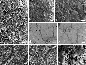
One of the best-preserved specimens, the holotype, GTBM17-765a (Fig. 2a), is an unbranched conical-cylindrical sponge with thin walls, c. 28 mm high. It expands from a flattened width at the fairly sharp base of 0.5 mm to a maximum width of c. 6 mm at one-third of the distance from the base. A simple spongocoel penetrates completely through the sponge and has a thin wall made of two distinct layers. The characteristic endosomal and dermal nets are composed of fused spicules outlining packed quadrangular, pentagonal and hexagonal openings. In the lower part of the endosomal layer, distinct individual spicules have three rays subparallel with the surface that diverge at c. 120–130°. However, these individual spicules are speculated to be tetraxial-based with one ray aborted or missing because the central axes of small nodes (Figs 2b, 5a–c) or a hollow hole (Fig. 5f–h) are observed. Tetraxial-based spicules are commonly slightly curved to wavy, tips fused or cemented to the ends of the side rays, or to the upper side of one of them, and produce a rhomboidal or hexagonal pattern that is 0.9–1.2 mm wide and c. 1.2–1.4 mm high at the upper part of the sponge. Downwards, the skeletal nets become slightly and gradually narrower. One ray of the three surface rays is laterally projected and extends up to c. 1.25 mm out beyond the general margin of the sponge. These extensions give the sponge an almost prickly appearance marked by spines. The dermal layer is composed of regular hexagonal or polygonal openings formed by tetraxial-based spicules, which is almost totally distinct and regular from the relatively coarse network of the endosomal layer. Both dermal and endosomal layers produce a reticulated-appearing wall in a staggered pattern.
SEM images of skeletons of Angulosuspongia sinensis gen. et sp. nov. (GTBM16-1192). (a–c, f–h) Secondary electron (SE) images showing individual spicules with three rays subparallel with the surface and diverging at c. 120–130°; and the central axes of small nodes in (a–c) or a hollow hole in (f–h). (d, e) Backscattered electron (BSE) images showing continuous skeletal structure formed by fused spicules. Scale bars 300 μm in (a–e), 60 μm in (f, g), 4 μm in (h).

The paratype, GTBM9-3-3613, is a branched sponge (Fig. 3c). The sponge body is somewhat like a saguaro cactus and composed of a main branch c. 19 mm tall and three side branches. All three branches extend out from one side of the main branch. The first branch is 12 mm long, a little curved, and initiates c. 3.6 mm above the sponge base. It expands upwards from a basal diameter of 0.8 mm to a maximum width of c. 2 mm, appearing at 6 mm in length. The oscular is pointing upwards and its margin is gently rounded to nearly flat. The second branch is very short, c. 2 mm upwards from the first one, only 2.8 mm long and 2.5 mm wide, and is inclined at a 45° angle relative to the main branch. The third branch is straight, c. 6.5 mm long and 2 mm wide, and oriented parallel to the second branch. Two distinct layers are visible in the skeleton. The endosomal and dermal layers are a characteristic reticulated net of fused spicules that surround openings. In addition to tetraxial-based spicules, long monaxon spicules are observed.
The other specimens show the same general characteristics of the holotype and paratype; well-formed hexagonal nets were preserved in adults, such as the whole specimen GTBM16-109b, and the upper part of GTBM22-1816 and GTBM17-1077b.
Discussion. Yang, Zhao & Wu (Reference Yang, Zhao and Wu2003) first mentioned Angulosuspongia conia and Angulosuspongia brevitubula in listing sponges of the Kaili Formation at the Miaobanpo section, but did not provide formal descriptions or figures. Subsequently, Angulosuspongia was listed as a new genus by Yang et al. (Reference Yang, Zhu, Zhao and Wang2005 b), again without description or figures. In 2006, the genus was figured poorly as Angulostuspongia [sic] conia in a booklet by Tai and Deng. All of these names fail to meet mandatory requirements of the Rules of Zoological Nomenclature and should, therefore, be regarded as nomen nudum. As a result, these names were not included in the Kaili Biota as described by Zhao et al. (Reference Zhao, Zhu, Babcock and Peng2011). In view of its morphology and geographical significance, a new name, Angulosuspongia sinensis, is established here.
5. Palaeobiogeographic significance
The vauxiid remains from South China are the first reported from outside Laurentia, indicating a cosmopolitan distribution of vauxiid sponges with two distinct biogeographic provinces. The distribution of vauxiids was apparently restricted to low palaeolatitude regions near the equator when we plot their occurrences on the Cambrian palaeogeographic reconstruction of McKerrow, Scotese & Brasier (Reference McKerrow, Scotese and Brasier1992). This pattern is consistent with algal biogeography in early and middle Cambrian time (Yang et al. Reference Yang, Mao, Zhang and Jiang2002), and also with that described for other groups, such as trilobites and echinoderms (Zamora, Gozalo & Liñán, Reference Zamora, Gozalo and LiÑÁn2009), and could be interpreted as climatic control on the geographic distribution of vauxiid sponges. Studies suggest that demosponges have short-dispersal potential in both shallow waters (Uriz & Turon, Reference Uriz and Turon2012) and deep-sea settings (Cárdenas & Rapp, Reference Cárdenas and Rapp2015). Detailed examination reveals the Chinese material to belong to a different genus than Laurentian vauxiid sponges. This difference may be a result of vicariance, because a study of living sponges did not support species distinction based solely on phenotypic criteria (Wörheide et al. Reference Wörheide, Degnan, Hopper and Reitner2000) and that morphologically different sponges in different geographically distinct areas were most likely geographic subspecies (Wörheide, Reference Wörheide1998). Furthermore, vicariance patterns in olenellid trilobites were also successfully applied to determine the compatibility of palaeogeographical models with palaeobiogeographical patterns matching palaeomagnetically derived models for the Neoproterozoic and early Cambrian (Meert & Lieberman, Reference Meert and Lieberman2004). However, this view contradicts a notion suggested by Botting & Peel (Reference Botting and Peel2016) that the differences between Laurentian and Chinese assemblages owe more to taxonomic overturn than to geographical separation. Such an issue also exists in the research on recent sponges, which shows it is still an open, unaddressed issue. Future work could certainly shed light on the assessment of relationships between phylogeography and taxonomy with the discovery of more new material.
Acknowledgements
This research was supported by the National Basic Research Programme of China (2013CB835002), the National Natural Science Foundation of China (grant numbers 41362002, 41330101), State Key Laboratory of Palaeobiology and Stratigraphy (Nanjing Institute of Geology and Palaeontology, CAS) (No.153113), Guizhou science and technology plan (Gui. Sci. Z. [2014] 4003) and Study Abroad Scholarship of China. Special thanks are due to Feng Liu and Zhefu Liu, Balang Village, Jianhe County, for assistance in fieldwork; to Yan Fang, Nanjing Institute of Geology and Palaeontology (NIGP), for assistance with SEM microphotography analyses; to J. P. Botting and an anonymous reviewer for providing critical comments; and to Steve LoDuca of Eastern Michigan University, for improving the English writing of the manuscript.