Introduction
The genus Pleurogenoides was erected by Travassos (Reference Travassos1921) to receive P. tener as its type-species which was described by Looss (Reference Looss1898) as Prosotocus tener. The genus comprises more than 26 valid species reported from frogs in different parts of the world. As many as eight species have been reported from Indian frogs: P. gastroporus (Luhe, 1901), P. sphaericus Klein, 1905, P. sitapurii Srivastava, 1934, P. orientalis (Srivastava, 1934), P. bufonis Kaw, 1953, P. ovatus Rao, 1977, P. jamshedpurensis Hasnain and Sahay, 1987 and P. ranchiensis Dan and Hasnain, 1991.
The life cycles of only five species of Pleurogenoides have been established (Mathias, Reference Mathias1924; Neuhaus, Reference Neuhaus1941; Buttner, 1951; Okabe & Shibue, Reference Okabe and Shibue1951; Shibue, Reference Shibue1953; Macy, Reference Macy1964; Madhavi et al., Reference Madhavi, Dhanumkumari and Ratnakumari1987; Janardanan & Prasadan, Reference Janardanan and Prasadan1991). In addition, reports are available on metacercariae from crabs, dragonfly nymphs, damselflies and other insect larvae (Dissanaike & Fernando, Reference Dissanaike and Fernando1960; Muraleedharan & Pande, Reference Muraleedharan and Pande1967; Prakash & Pande, Reference Prakash and Pande1969; Krasnolobova, Reference Krasnolobova1970; Vojtkova, Reference Vojtkova1970; Janardanan et al., Reference Janardanan, Ramanandan and Usha1987; Dhanumkumari, Reference Dhanumkumari2000).
During an explorative study on the digenean fauna of frogs in Kerala, we discovered a species of Pleurogenoides in the intestine of the Indian bullfrog Hoplobatrachus tigerinus (Daudin) and the skipper frog Euphlyctis cyanophlyctis (Schneider) collected from irrigation canals and paddy fields at Malampuzha in Palakkad district, Kerala. Further studies proved that it is a new species and is named P. malampuzhensis sp.nov. Examination of snail hosts from the same habitat where infected frogs were caught revealed that the bithynid snail Digoniostoma pulchella (Benson) was infected with a virgulate xiphidiocercaria during the monsoon months (June–September). Encysting metacercariae were found in muscle tissues of dragonfly nymphs belonging to the families Gomphidae and Libellulidae. These metacercariae, when fed to H. tigerinus, developed into adults of the new species in the intestine. The life-cycle stages, from cercaria to egg-producing adult, were established successfully in the laboratory. This forms the third report on the life cycle of an Indian species of Pleurogenoides.
Materials and methods
Collection and examination of frogs and snails
The Indian bullfrog Hoplobatrachus tigerinus (Daudin) and the skipper frog Euphlyctis cyanophlyctis (Schneider), collected from irrigation canals and paddy fields at Malampuzha in Palakkad district from December 2008 to October 2009, were brought to the laboratory and examined for digenetic trematodes. The recovered flukes were studied live using a phase-contrast microscope with or without supravital staining with neutral red. Those used for permanent preparations were killed in hot distilled water, fixed in ethyl alcohol and stained with alum carmine following the procedure outlined by Cantwell (Reference Cantwell and Clark1981).
The bithynid snail Digoniostoma pulchella (Benson) was collected from the same habitat where infected frogs were caught. Living snails were transferred to the laboratory in plastic containers with ‘well-water’ and then maintained in groups of 5–10 in beakers similarly containing ‘well-water’. Snails that shed cercariae were isolated and kept individually in separate beakers. The water in the beakers was checked for cercariae. Cercariae emerging from infected snails were studied live with or without vital staining using a phase-contrast microscope. Genital primordia were observed in lactoacetic carmine-stained cercariae. Measurements (in μm) were made on heat-killed specimens. A few infected snails were later crushed and examined for intramolluscan stages of development. Sporocysts recovered from the hepatopancreas of infected snails were studied after supravital staining with neutral red.
Experimental infections
In order to establish metacercarial infection experimentally, dragonfly nymphs of the family Gomphidae obtained from a tank in the Calicut University Campus and which proved to be free of natural infections with Pleurogenoides sp. metacercariae, were exposed to newly emerged cercariae. Nymphs thus exposed were maintained in aquarium tanks and sacrificed at intervals to follow the course of development of metacercariae. Excysted metacercariae were fixed in ethyl alcohol, stained with alum carmine and prepared as whole mounts.
Hoplobatrachus tigerinus only was used in experimental infections. Frogs used in experiments were collected from infection-free ponds in and around Calicut University Campus and kept in the laboratory; their faecal matter was carefully analysed for any eggs of Pleurogenoides for a continuous period of 3 months. Infection-free frogs, force-fed with infective metacercariae, were dissected at intervals and examined for trematodes.
Descriptions of larval and adult stages are based on the measurements of 15 specimens. Measurements are given in micrometres, as the range followed by the mean in parentheses. Figures were drawn with the aid of a camera-lucida, and details were added free-hand from observations made on live specimens.
Results
Adult worms
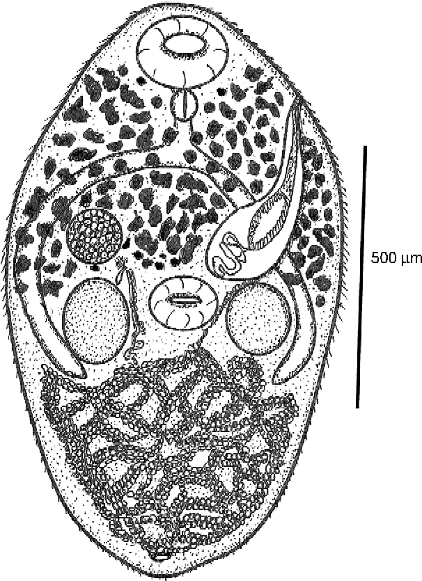
Natural infections with adult flukes were found in 65 of 998 (6.5%) H. tigerinus and 45 of 687 (6.6%) E. cyanophlyctis. The intensity of infection in these two hosts varied from 5 to 9 (8 ± 1.6) and 6 to 8 (7 ± 0.8), respectively. The following description is based on mature adults (fig. 1) recovered from naturally infected frogs.
Adult worm of Pleurogenoides malampuzhensis sp. nov.

Description
Body elongate-oval, 780–1404 (1071) long, 603–820 (734) wide. Forebody 390–700 (525) long, 47–58% (52%) of body length. Hindbody 390–643 (512) long. Tegument spinose, spines densely distributed to level of ventral sucker and sparsely behind it. Oral sucker sub-terminal, round, 132–234 (215). Ventral sucker post-equatorial, round, 100–172 (135). Sucker ratio 1:0.43–0.75 (0.65). Mouth sub-terminal; pre-pharynx short, 8–13 (9); pharynx subglobular, 49–78 (60). Oral sucker/pharynx ratio, 1:0.33–0.44 (0.38). Oesophagus 58–78 (66) long. Caeca narrow, 454–625 (525) × 45–75 (68), extend a short distance beyond posterior margin of testes, curve inwardly and terminate blindly 485–549 (514) from posterior end of body.
Testes two, ovoid, symmetrical, postero-lateral to ventral sucker. Right testis 125–281 (212) × 109–218 (170), left testis 109–265 (204) × 110–265 (178). Cirrus sac claviform, 412–624 (518) × 109–187 (160), extends from anterior region of ventral sucker to left body margin and opens through genital pore at the level of the pharynx. Cirrus sac encloses bipartite seminal vesicle, well-developed pars prostatica, long and narrow ejaculatory duct and unspined cirrus.
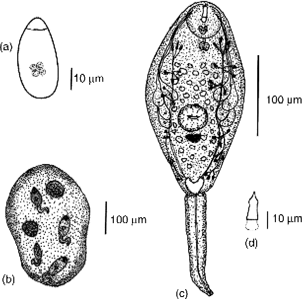
Ovary ovoid, intercaecal, dextral, anterior to ventral sucker; 94–123 (114) × 72–156 (103) in size. Ootype complex post-ovarian. Seminal receptacle round, situated behind ovary. Mehlis' gland well developed. Laurer's canal absent. Vitellaria follicular, follicles extending from mid-level of oral sucker to mid-level of ventral sucker, confluent in forebody. Uterine coils occupy most of body posterior to testes and contain many eggs. Metraterm inconspicuous, opens at the side of male genital pore. Excretory bladder V-shaped, 312–566 (498) × 78–106 (104); excretory pore sub-terminal. Eggs (fig. 2a) pale brown, ovoid, operculate, 40–56 (50) × 15–26 (23).
(a) Egg, (b) sporocyst, (c) cercaria and (d) stylet of cercaria of Pleurogenoides malampuzhensis sp. nov.
Sporocysts and cercariae
Sporocysts (fig. 2b) ovoid, 312–437 × 210–280 in size, containing 3–4 developing cercariae (fig. 2c) and a few germ balls at various stages of development.
Natural infections with cercariae were found in 25 of 1500 (1.66%) snails collected. Cercariae emerged during the daytime, with peak emergence from 08.00 to 11.00 h, and exhibited positive phototaxis.
Description
Virgulate xiphidiocercaria. Body elongate–oval, 109–231 (189) × 83–125 (106), spinose, contains large number of refractile golden yellow globules. Tail shorter than body, 116–149 (128) × 20–33 (25), aspinose, attached to body ventrally. Oral sucker sub-terminal, round, 38–48 (42). Ventral sucker post-equatorial, round, 18–28 (24). Stylet lancet-shaped (fig. 2d), 22–25 (23) long, 4–6 (5) wide, inserted in antero-dorsal region of oral sucker. Stylet with pointed tip, moderately developed shoulder region at its anterior fourth and round base. Stylet walls reinforced except at base. Virgula organ bilobed, at the posterior half of oral sucker.
Mouth ventral, sub-terminal. Pre-pharynx short. Pharynx globular, muscular, 10–13 (11) in size. Post-pharyngeal system not observed. Penetration glands four pairs, situated antero-lateral and lateral to ventral sucker; each gland with round nucleus and finely granular contents; ducts of penetration glands open individually at the sides of stylet. Genital primordium represented by a mass of cells lying posterior to ventral sucker. Excretory bladder V-shaped, thin-walled. Primary collecting vessels run from antero-lateral margins of bladder, to the level of ventral sucker where they divide into anterior and posterior collecting tubules. Flame cell formula: 2[(3+3+3)+(3+3+3)] = 36. Caudal excretory duct originates from posterior margin of excretory bladder, runs through tail and opens at its tip.
Metacercariae
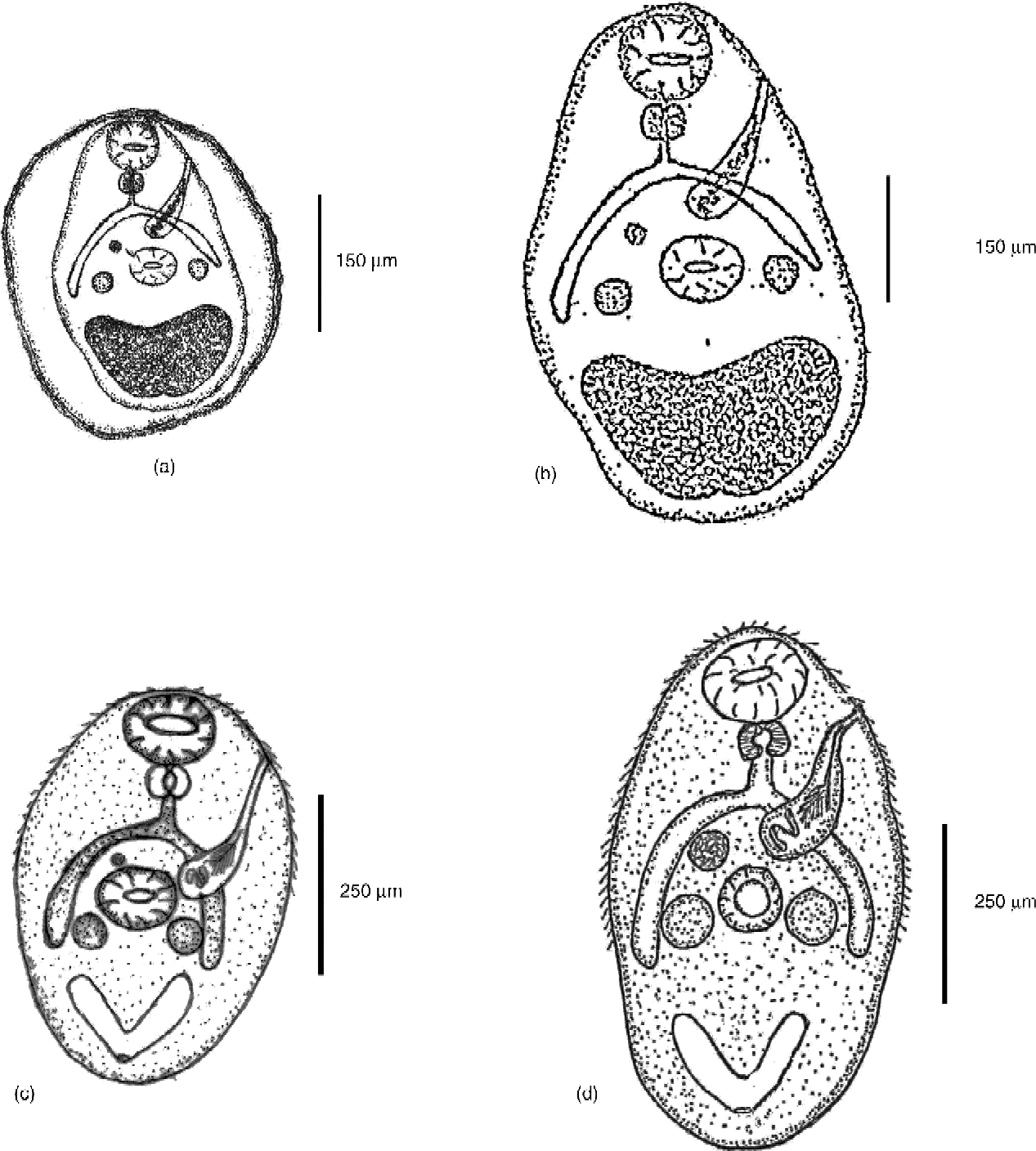
Natural infections with metacercariae (fig. 3a and b) were found in dragonfly nymphs of the families Gomphidae (13 of 59) and Libellulidae (17 of 61) collected from the same habitat where infected snails were present. The intensity of infection was 12–15 (13 ± 1.3) in Gomphidae and 14–18 (15 ± 2.1) in Libellulidae. Metacercariae were found encysted in the muscle tissues of dragonfly nymphs.
(a) Encysted and (b) excysted metacercariae, together with (c) 5-day-old and (d) 15-day-old immature worms of Pleurogenoides malampuzhensis sp. nov.
Experimental infection
Growth and development of metacercariae were observed in experimentally infected, clean dragonfly nymphs of the family Gomphidae. Cercariae actively penetrated tegument of the nymphs using their stylet, and free cercarial bodies were observed in the haemocoel 2–3&th;h after exposure. Metacercariae enclosed in a thin, delicate cyst wall were observed after 24 h. Cysts were round, 166–198 (176) in diameter. Stylet could be seen in the oral sucker. On the tenth day post-infection, cysts enlarged in size, measured 221–255 (237). Cyst wall became double-layered and stylet seemed to disappear. Excysted metacercaria ovoid, spinose, 230–355 (257) × 200–278 (250). Oral sucker and ventral sucker increased in diameter; oral sucker 72–78 (76) and ventral sucker 44–49 (46). Pharynx globular, muscular, 23–33 (28). Caeca 59–110 (86) long, extend beyond developing testes. Excretory bladder V- shaped, 100–178 (151) in size. Metacercariae recovered after 22 days post-exposure were found to be infective to the definitive host. These metacercariae were identical to those obtained from natural infections. The following description is based on the observations made on 22-day-old experimentally infected metacercariae.
Description
Cysts white, ovoid, 300–456 (381) × 314–410 (351) in size. Cyst wall two-layered, outer one thin and transparent, inner thick and opaque. Excysted metacercariae ovoid, 455–495 (478) × 235–334 (292) in size. Tegument spinose, spines prominent in forebody. Oral sucker round, 83–116 (101) in diameter. Ventral sucker round, post-equatorial, 62–108 (78) in diameter. Mouth sub-terminal, leads to short pre-pharynx. Pharynx globular, muscular, measured 21–36 (28). Oesophagus 25–34 (32) long. Caeca extend laterally a short distance posterior to developing testes; 132–155 (143) × 31–42 (36) in size. Developing testes two, round, post-equatorial, 20–34 (25) in diameter. Cirrus sac claviform, extending from anterior region of ventral sucker to left body margin and opens through genital pore at the level of the pharynx; 86–120 (98) × 15–20 (17) in size. Ovary single, rudimentary, pre-equatorial, near right testis. Excretory bladder V-shaped, filled with excretory concretions, occupied the posterior third of body; 14–164 (146) × 25–45(32) in size. Excretory pore sub-terminal.
Adult worms from experimental infections
Development of adult flukes was observed in experimentally infected H. tigerinus. On the first day post-infection, excysted metacercariae were found in the intestine of the frog. Five-day-old immature flukes (fig. 3c) increased in size, 462–562 (511) × 245–285 (262). Oral sucker round, 116–145 (132). Ventral sucker round, 76–125 (115). Prepharynx 10–13 (12) long. Pharynx globular, muscular, 40–63 (51) × 34–40 (37) in size. Oeosphagus 34–40 (37) long. Caeca 231–276 (265) long, extend laterally a short distance posterior to developing testes. Developing testes two, left testis 111–124 (120), right testis 100–122 (118). Developing ovary round, anterior to ventral sucker, 54–62 (58) in diameter. Cirrus sac claviform, opens at left body margin at the level of pharynx; 254–270 (258) × 66–77 (74) in size. Excretory bladder V-shaped, without excretory concretions, 116–126 (119) × 55–69 (60) in size. Fifteen-day-old adults (fig. 3d) increased in size, 613–755 (684) × 308–493 (423). Oral sucker 132–145 (143). Ventral sucker 87–116 (100). Pharynx globular, muscular, 54–77 (67) × 54–70 (67). Oeosphagus 34–40 (37) long. Caeca 308–397 (357) long. Testes two, ovoid, symmetrical; left testis 131–156 (142) × 100–145 (122), right testis 132–152 (138) × 133–154 (144). Cirrus sac well-developed, encloses seminal receptacle and pars prostatica and opens through genital pore at left body margin at the level of pharynx; 264–270 (268) × 66–77 (76) in size. Ovary round, 62–69 (68). Excretory bladder V-shaped, 126–154 (138) × 59–77 (69). At 20 days post-infection, flukes are fully mature, with their uterine coils filled with many eggs. The experimentally developed flukes were identical to those recovered from natural infections.
Taxonomic summary
Genus. Pleurogenoides Travassos, Reference Travassos1921.
Species. Pleurogenoides malampuzhensis sp. nov.
Type host. Hoplobatrachus tigerinus (Daudin); Ranidae.
Additional host. Euphlyctis cyanophlyctis (Schneider); Ranidae.
Site. Intestine.
Type locality. Malampuzha, Palakkad district, Kerala, India.
Snail host. Digoniostoma pulchella (Benson); Bithyniidae.
Second intermediate host. Dragonfly nymphs; Gomphidae and Libellulidae.
Holotype. Deposited in the Department of Zoology, University of Calicut, Kerala, India. No. Z./Par./Dig. 2009 1a.
Paratypes. Z./Par./Dig. 2009 1b–d.
Date of collection. 25 January 2009.
Etymology. Named after the type locality.
Discussion
The genus Pleurogenoides Travassos, Reference Travassos1921 belongs to the subfamily Pleurogenetinae Looss, 1901 of the family Pleurogenidae Looss, 1899. The present Pleurogenoides species has a sub-terminal oral sucker, symmetrical testes at the level of the ventral sucker, intercaecal ovary, follicular vitellaria extending from the mid-level of the oral sucker to the mid-level of the ventral sucker and genital pore at the level of the pharynx, in addition to other characters. Based on these characters, the present fluke comes close to P. gastroporus (Luhe, 1901) and P. orientalis (Srivastava, 1934) among the 26 recognized species of Pleurogenoides. The remaining 24 species are distinctly different in one or more of the above characters.
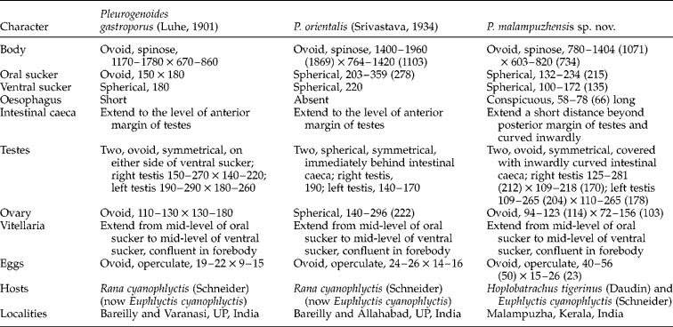
A comparative study of characters of P. gastroporus, P. orientalis and the present fluke (table 1) revealed that while in body size the present form is smaller than the other two, it has eggs which are double or more in size when compared with those of the others. Besides, the caeca of the new species extend a short distance beyond the posterior margin of testes and are curved inwardly, while in the other two species they end near the anterior margin of the testes, making the new species distinctly different. Pleurogenoides gastroporus also differs in the size of the suckers, with the oral sucker being smaller than the ventral sucker, whereas in P. orientalis and in the present species the reverse is true. The absence of an oesophagus and the presence of a large spherical ovary differentiates P. orientalis from the new form. It is evident that the present species differs significantly from all the known species of Pleurogenoides, and is, therefore, reported here as a new species and named P. malampuzhensis sp. nov., after the type locality.
A comparison of the characteristics (in μ_rm;m) of Pleurogenoides gastroporus (Luhe, 1901), P. orientalis (Srivastava, 1934) and P. malampuzhensis sp. nov.
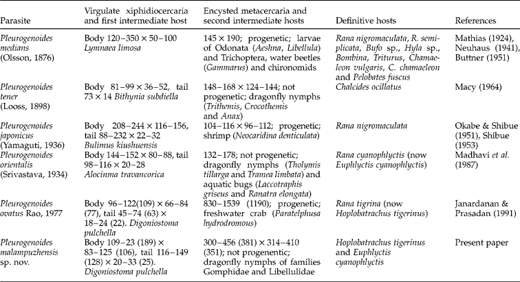
Information on life cycles of the genus Pleurogenoides is limited to five species: P. medians, P. japonicus, P. tener, P. orientalis and P. ovatus. All of them exhibited a three-host life-cycle pattern. Pleurogenoides malampuzhensis sp. nov. also has a typical three-host life cycle, differing only with regard to hosts involved and the structural details of different stages in the life cycle. A summary of selected characteristics and life cycles of five species of Pleurogenoides and P. malampuzhensis sp. nov. is presented in table 2.
A summary of seleted characteristics and life cycles of five species of Pleurogenoides and P. malampuzhensis sp. nov.
The snail hosts used by P. medians, P. japonicus, P. tener, P. orientalis and P. ovatus are Lymnaea limosa (Linnaeus), Bulimus kiushuensis Hirase, Bithynia subdiella Bourguignat, Alocinma travancorica (Benson) and Digoniostoma pulchella (Benson), respectively. The virgulate xiphidiocercaria of P. malampuzhensis sp. nov. recovered from D. pulchella during the present investigation differs in morphological features and dimensions from the cercariae of five species of Pleurogenoides whose life cycles have been established, but its characters agree fully with the description of Cercariae indicae LXIX given by Murty (Reference Murty1976) from Alocinma travancorica (Benson) collected at Gopalapatnam, Kakinada, and, therefore, both are identical.
The metacercaria of P. japonicus is reported from shrimps. Pleurogenoides medians, P. tener, P. orientalis and the present species use aquatic insects as second intermediate hosts, in contrast to the use of freshwater crabs as second intermediate hosts by P. sitaprii and P. ovatus. Metacercariae of Pleurogenoides spp. undergo considerable growth and development inside the second intermediate hosts. Metacercariae of P. medians, P. japonicus, P. sitapurii and P. ovatus exhibited the phenomenon of progenesis, whereas those of P. tener and P. orientalis are not progenetic. In the present species, also, the metacercaria is not progenetic. The present metacercaria is distinctly different from the metacercariae of other Pleurogenoides spp. in morphological features and dimensions.
The life-cycle stages of P. malampuzhensis sp. nov., from cercaria to egg-producing adult, took 42 days or more to complete: 22 days to develop an infective metacercaria in the second intermediate host and a 20-day pre-patent period in the final host.
Acknowledgements
The authors express their sincere thanks to Dr David I. Gibson (Natural History Museum, London) for providing us with the valuable literature. We also express our sincere thanks to the Head of the Department of Zoology, University of Calicut for the facilities provided. One of us (R.B.) is grateful to the University of Calicut for the financial support in the form of a Research Fellowship.