Introduction
Infertility has become a global health concern affecting approximately 190 million people, with nearly half of the cases are due to male infertility (Refs Reference Agarwal1–Reference Minhas3). Non-obstructive azoospermia (NOA), the most severe type of male infertility, is a condition in which spermatozoa are absent in the ejaculate due to spermatogenic dysfunction in the testes. NOA affects approximately 1% of men worldwide (Refs Reference Li4–Reference Caroppo and Colpi6). Although several underlying causes are known, including chromosomal abnormalities, Y-chromosome microdeletions, cryptorchidism, hypogonadism, viral or bacterial orchitis, chemotherapy/radiation therapy, varicocele, etc., the exact aetiology of most NOA cases remains unclear, which is therefore referred to as idiopathic NOA (INOA) (Refs Reference Kang7, Reference Cannarella8). Exogenous hormone therapy, such as supplementing gonadotropins in hypogonadotropic hypogonadism and bromocriptine in hyperprolactinaemia, is typically effective in some NOA cases with hormonal imbalances (Refs Reference Kang7, Reference Tao9, Reference Alkandari and Zini10). However, no appropriate pharmacological therapies have been proven effective in most patients with NOA. A series of approaches has attempted to retrieve sperm from the testis of these patients, and microdissection testicular sperm extraction (micro-TESE) is deemed the most promising approach (Refs Reference Schlegel11, Reference Hibi12).
Although micro-TESE has contributed to sperm retrieval, yielding satisfactory outcomes of intracytoplasmic sperm injection (ICSI) in some patients with INOA (Refs Reference Corona13–Reference Ravizzini16), its low surgical success rate and associated iatrogenic testicular trauma are undeniable drawbacks to this invasive approach (Refs Reference Sertkaya17–Reference Donoso20). Therefore, accurate preoperative predictive models to estimate the probability of surgically retrieving sperm using micro-TESE are of great significance. Such models allow clinicians to tailor personalised clinical practices for patients with NOA, predicting the success rate of micro-TESE (Ref. Reference Caroppo and Colpi6). Several candidate predictors have been proposed to evaluate success rates; for instance, the NOA aetiology is a key predictor for successful sperm retrieval (SSR). Sperm can be retrieved in more than half of patients with a cryptorchidism history (Refs Reference Ozan21–Reference Vernaeve24). SSR rates vary from 20 to 66% in NOA patients with Klinefelter syndrome (Refs Reference Caroppo and Colpi6, Reference Ramasamy25–Reference Chen28). However, no sperm can be retrieved by micro-TESE in patients with complete AZFa or AZFb deletions, although better chances of SSR are present in patients with AZFc deletions (Refs Reference Caroppo and Colpi6, Reference Yuen29–Reference Majzoub31). For patients with INOA, the SSR varies from 20 to 50.1% (Refs Reference Arafa32–Reference Ozkanli36). Testicular histopathology strongly correlates with SSR following micro-TESE in patients with INOA. Patients with hypospermatogenesis have significantly higher SSR rates than those with maturation arrest and Sertoli cell-only syndrome (Refs Reference Caroppo and Colpi6, Reference Das37–Reference Caroppo42). Although it is perhaps the best predictor for micro-TESE outcomes, a primary limitation of testicular histopathology should be considered as results can only be obtained by invasive operation of the testis. Therefore, it is critical to explore appropriate non-invasive molecular biomarkers to preoperatively predict the outcomes of sperm recovery, which might play an important role in assessing the trade-off between potential risks and benefits.
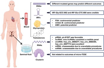
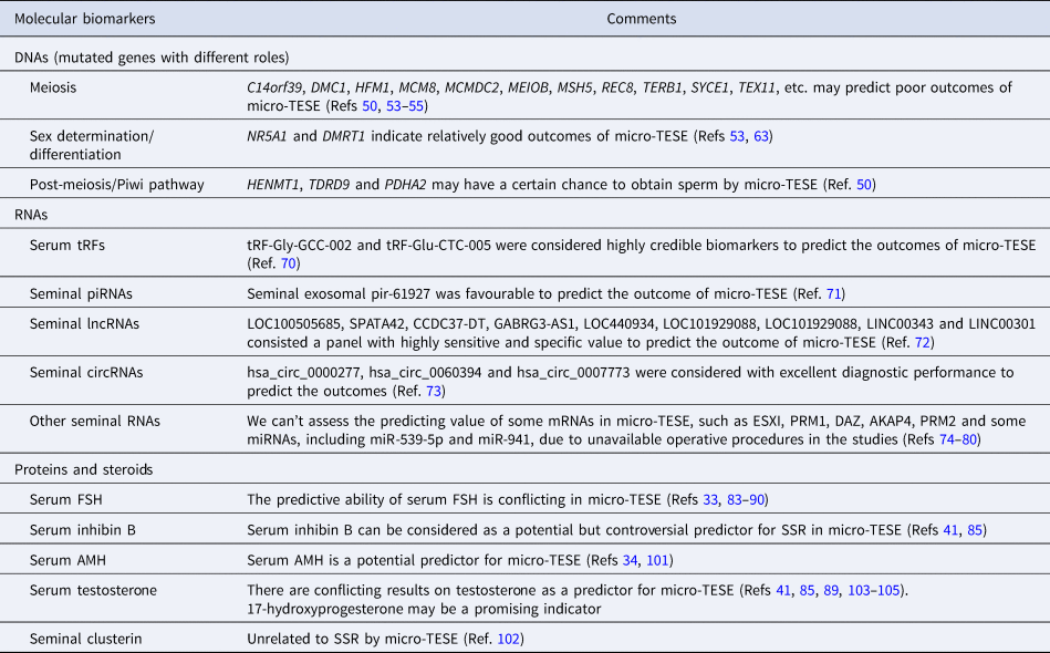
In recent years, an increasing number of predictors has been evaluated. In this review, we assessed these preoperative non-invasive molecular biomarkers, such as DNAs, RNAs, protein and steroid levels, from different tissues and their predictive values for SSR by micro-TESE in patients with INOA. The predictors are summarised in Table 1 and Figure 1.
Overview of preoperative molecular biomarkers for predicting the outcomes of micro-TESE in INOA patients (by Figdraw, www.figdraw.com).

Overview of preoperative molecular biomarkers for predicting the outcomes of micro-TESE in INOA patients

DNAs
In animal models, more than 600 genes have been reportedly associated with reduced fertility or infertility (Ref. Reference Matzuk and Lamb43). More than 400 genes have been confirmed to be specifically expressed in human testicular tissues (Ref. Reference Uhlen44). These results suggest that genetic factors and single genetic mutations may be important causes of NOA. In recent years, several studies identified a series of pathogenic genes involved in the impairment of spermatogenesis, leading to NOA, such as TEX11 (OMIM: 300311) (Refs Reference Yatsenko45, Reference Sha46), ADAD2 (OMIM: 619532) (Ref. Reference Krausz47), CCDC155 (OMIM: 618125) (Ref. Reference Fakhro48), DMRT1 (OMIM: 602424) (Refs Reference Krausz47, Reference Lima49), TDRD9 (OMIM: 617963) (Ref. Reference Kherraf50) and TEX14 (OMIM: 605792) (Refs Reference Kim51, Reference Gershoni52). In parallel, several researchers explored the association between precise genetic diagnosis and SSR by micro-TESE in patients with INOA, aiming to propose genetic predictors for SSR prior to surgery (Refs Reference Kherraf50, Reference An53–Reference Tang55).
Kherraf et al. conducted a genetic study enrolling 96 unrelated patients with INOA. Of these, 22 patients were diagnosed with genetic defects, and all underwent micro-TESE (Ref. Reference Kherraf50). A significant phenomenon was noted indicating that no sperm can be retrieved in patients with NOA linked to underlying genetic disorders involving meiotic genes, such as C14orf39 (OMIM: 617307), DMC1 (OMIM: 602721), HFM1 (OMIM: 615684), MCM8 (OMIM: 608187), MCMDC2 (OMIM: 617545), MEIOB (OMIM: 617670), MSH5 (OMIM: 603382), REC8 (OMIM: 608193) and TERB1 (OMIM: 617332). Spermatozoa were successfully retrieved in two out of five patients with mutations in genes involved in the Piwi pathway, such as HENMT1 (OMIM: 612178), TDRD9 (OMIM: 617963) and TDRKH (OMIM: 609501); a patient with a mutated HENMT1 gene and another with a mutated TDRD9 gene. Additionally, regarding genes involved in post-meiotic maturation, such as DDX25 (OMIM: 607663) and PDHA2 (OMIM: 179061), one patient with mutated PDHA2 had positive micro-TESE (Ref. Reference Kherraf50). Consistent results have been reported by several other studies. In a study by Ann et al., six patients with possible genetic disorders involving four genes, namely NR5A1 (OMIM: 184757), TEX11 (OMIM: 300311), TEX14 (OMIM: 605792) and KLHL10 (OMIM: 608778), underwent micro-TESE (Ref. Reference An53). No spermatozoa were retrieved following micro-TESE in two patients harbouring TEX11 mutations. Although very few spermatozoa were retrieved in another patient with mutated TEX14, spermatozoa were insufficient for ICSI. These results validated the conclusion of Kherraf et al. that genetic disorders involving meiotic genes predict poor micro-TESE outcomes. Of six patients, only one with a mutated NR5A1 gene, encoding a transcription factor involved in steroidogenesis and sexual differentiation in a dose-dependent manner (Refs Reference Fabbri-Scallet56–Reference Chen58), produced viable spermatozoa through micro-TESE. In addition, two patients with mutated KLHL10 unsuccessfully underwent micro-TESE (Ref. Reference An53). However, it is worth discussing whether KLHL10 variants were causative factors for spermatogenetic failure in these two patients with NOA, as KLHL10 mutations were reportedly linked with oligozoospermia (Ref. Reference Yatsenko59). Therefore, KLHL10 variants may not be deemed suitable genetic biomarkers to predict SSR in micro-TESE.
Furthermore, Feng et al. reported a NOA case with a homozygous mutation in SYCE1 (OMIM: 611486) (Ref. Reference Feng54), which encodes an essential component of the synaptonemal complex and plays a key role in first meiosis (Refs Reference Maor-Sagie60–Reference Bolcun-Filas62). No mature sperms were retrieved using micro-TESE (Ref. Reference Feng54). Our team also identified two homozygous variants in the meiotic gene HFM1 in two unrelated patients with INOA. Although these patients underwent micro-TESE, no spermatozoa were retrieved in the testes (Ref. Reference Tang55). In another study, we found that SSR was achieved by micro-TESE in a patient with INOA bearing a variant of the DMRT1 (OMIM: 602424) gene (Ref. Reference Tang63), which encodes a transcription factor involved in male sex determination and differentiation by controlling testis development and male germ cell proliferation (Ref. Reference Macdonald64).
In general, no significant evidence supports the value of gene variants in predicting the outcomes of micro-TESE in patients with NOA. Nevertheless, according to available information, gene mutations involved in meiosis may predict poor outcomes of micro-TESE. Conversely, mutations in genes associated with sex determination/differentiation may indicate relatively good outcomes of micro-TESE. Mutated genes involved in the Piwi pathway/post-meiotic maturation may indicate a certain chance of achieving spermatozoa retrieval by micro-TESE from the testis of patients with NOA. As current evidence was obtained from several small cohorts with several cases, further studies with larger cohorts are essential to explore the association between gene mutations and outcomes of micro-TESE. Additionally, even the association between gene mutations and ICSI outcomes warrants further investigation. Based on well-documented evidence, we may develop an appropriate panel to detect gene variants before micro-TESE to predict the likelihood of SSR in the near future.
RNAs
In addition to the limited evidence of DNA information from blood preoperatively predicting SSR outcomes in patients with INOA, some studies have also focused on RNA information in serum or seminal plasma as molecular biomarkers to estimate the presence of residual spermatozoa retrievable by micro-TESE.
Due to the stability of exosomes in bodily fluids, exosome-carrying biomolecules have received increasing attention as non-invasive predictors of various diseases (Refs Reference Nishiwada65–Reference Zheng67). Given the critical roles of exosomes in the progress of spermatogenesis, such as germ cell differentiation and Sertoli cell activities (Refs Reference Jamin68, Reference Ahmed69), a few studies have focused on the predictive value of exosome-carrying RNAs on the outcomes of micro-TESE. In a study conducted by Zhang et al., 30 patients with INOA underwent micro-TESE (12 with SSR and 18 with failed sperm retrieval). The significant differential expression of two circulating plasma exosomal transfer RNA-derived fragments (tRFs), tRF-Gly-GCC-002 and tRF-Glu-CTC-005, was identified. Moreover, the receiver operating characteristic area under the curve analysis presented these two tRFs as highly credible biomarkers for predicting SSR by micro-TESE in patients with INOA (Ref. Reference Zhang70). Exosome-carrying RNAs in seminal plasma have also been used to establish predictive models of the presence of testicular spermatozoa by micro-TESE in patients with INOA (Refs Reference Chen71, Reference Xie72). In a study by Chen et al., 10 NOA patients with SSR and 10 NOA patients who unsuccessfully underwent micro-TESE were enrolled as the training cohort to develop prediction models using exosome-carrying piRNAs in the seminal plasma. The resulting model, based on pir-61927 expression, could promisingly predict the outcome of micro-TESE (Ref. Reference Chen71). In addition to piRNAs, long noncoding RNAs (lncRNAs) in seminal plasma-derived exosomes have also been indicated as sensitive and specific predictive markers of the outcomes of micro-TESE in patients with NOA. Thirty patients with NOA, including 18 and 12 patients with successful and unsuccessful SSR by micro-TESE, respectively, were selected to develop a panel based on seminal plasma-derived exosome-carrying lncRNAs to predict the presence of sperm in the testis. Nine differentially expressed testis-specific lncRNAs from seminal plasma exosomes, including LOC100505685, SPATA42, CCDC37-DT, GABRG3-AS1, LOC440934, LOC101929088, LOC101929088, LINC00343 and LINC00301, yielded a highly sensitive and specific panel for predicting the outcome of micro-TESE, whose predictive accuracy was 95.238% when the score exceeded the cut-off value of 0.532 in the panel (Ref. Reference Xie72). Nevertheless, it is worth noting that the two models above were developed for the general population of patients with NOA, and whether these predictors are sufficiently accurate in patients with INOA remains unclear. Further studies with larger sample sizes in patients with INOA are needed to verify the accuracy of these predictive tools.
The RNAs predicting the outcomes of micro-TESE in the aforementioned studies were all exosome-derived. RNAs directly extracted from seminal plasma were also considered applicable molecular biomarkers to predict the outcome of micro-TESE in patients with INOA. Ji et al. selected 52 patients with INOA who underwent micro-TESE, including 20 and 32 successful and failed cases, respectively, to detect the differentially expressed circRNAs in seminal plasma and explore their predictive value on the outcome of micro-TESE. Three circRNAs, namely hsa_circ_0000277, hsa_circ_0060394 and hsa_circ_0007773, were considered to have excellent diagnostic performance in predicting micro-TESE outcomes (Ref. Reference Ji73).
Several other studies have also investigated the predictive value of some RNAs from seminal plasma on the outcomes of sperm retrieval, such as ESXI-mRNA, PRM1-mRNA, DAZ-mRNA, AKAP4-mRNA, PRM2-mRNA and some specific miRNAs, including miR-539-5p and miR-941 (Refs Reference Bonaparte74–Reference Barcelo80). However, as information on operative procedures (conventional testicular sperm extraction or micro-TESE) was unavailable, we could not assess exact predicting performance for micro-TESE outcomes in patients with INOA.
Proteins and steroid hormone
A wide variety of proteins exert diverse biological functions throughout life, including spermatogenesis. Therefore, spermatogenesis-related proteins have recently gained a spotlight as to whether they can predict the presence of residual spermatozoa in the testes of patients. In particular, serum glycoprotein hormones, including follicle-stimulating hormone (FSH), inhibin B and anti-Müllerian hormone (AMH), have been extensively researched as predictors of micro-TESE outcomes. Despite some controversy, these glycoprotein hormones seemingly have predictive value.
FSH
FSH, produced by the pituitary gland, has received the most attention due to its critical role in spermatogenesis. Many studies have focused on the predictive value of this glycoprotein hormone on the presence of spermatozoa in testes. Although FSH levels in serum are considered significantly valuable for indicating SSR rates in conventional TESE (Refs Reference Mitchell81, Reference Boitrelle82), their predictive abilities are conflicting in micro-TESE.
Some studies have concluded that higher FSH levels indicate better micro-TESE outcomes. Liu et al. observed that FSH levels were significantly higher in the spermatozoa-positive group than in the negative group using micro-TESE. A positive correlation was observed between FSH levels and SSR in NOA patients undergoing micro-TESE, as opposed to patients going through testicular sperm aspiration (Ref. Reference Liu83). Modarresi et al. also observed that high FSH levels indicated increased SSR by micro-TESE in NOA patients with presumed Sertoli cell-only syndrome. The authors established a prediction model based on the 15.25 mIU/ml FSH cut-off level. When the FSH values exceeded 15.25 mIU/ml, the SSR rate was 28.9%, higher than 11.8% in the group with FSH values lower than the cut-off (Ref. Reference Modarresi84). Beyond the population of patients with NOA enrolled in the aforementioned studies, Zhang et al. also suggested that increased FSH levels correlated with better micro-TESE outcomes, representing an independent predictor in men with INOA. The SRR rate was 32.6% in the subgroup with FSH values greater than 24.8 mIU/ml, significantly higher than 15.8 and 14.3% in the subgroups with FSH values within 12.4–24.8 mIU/ml and less than 12.4 mIU/ml, respectively (Ref. Reference Zhang33).
On the contrary, a negative correlation between FSH level and SSR rates by micro-TESE in NOA patients was supported by Tsujimura et al. Their study revealed decreased FSH levels in the successful SSR group compared with the failed group in a cohort of 100 patients with NOA (Ref. Reference Tsujimura85). Moreover, some studies have focused on the association between FSH levels and SSR during salvage micro-TESE. In a study by Yücel et al., a lower mean FSH level was observed in the successful salvage micro-TESE group than in the unsuccessful group (Ref. Reference Yucel86). Nonetheless, Xu et al. reported that FSH level was not an independent factor in predicting spermatozoa retrieval (Ref. Reference Xu41). Some studies also hold the view that FSH levels cannot be used as valuable predictors or independent factors for sperm retrieval in the first micro-TESE (Refs Reference Cetinkaya87–Reference Ramasamy90).
FSH levels may only be related to the number of germ cells in the testis. Although it is the main indicator of overall spermatogenetic function, it may not be sufficient to reflect the presence of few residual spermatozoa in the testis (Refs Reference Tsujimura91–Reference Bernie93). Indeed, it is not uncommon for NOA patients with histological results of maturation arrest to present normal FSH levels but experience poor micro-TESE outcomes (Ref. Reference Hung94).
Inhibin B
Inhibin B is a glycoprotein hormone produced by the Sertoli cells. As a negative regulatory factor of FSH in the hypothalamus–pituitary–gonad axis, its serum level was also reported as a potential but controversial predictor of micro-TESE outcomes. In the study by Tsujimura et al., the mean level of inhibin B was significantly higher in the SSR group than in the failed group following micro-TESE. Hence, its level could serve as a predictive factor for micro-TESE outcomes (Ref. Reference Tsujimura85). For salvage micro-TESE, inhibin B level was not considered an independent factor for predicting spermatozoa retrieval (Ref. Reference Xu41). Although a few studies with controversial conclusions have focused on the predictive value of inhibin B on the outcomes of micro-TESE, the contentious conclusions of its predictive value in conventional TESE have been presented in most studies (Refs Reference Bohring95–Reference Meachem98).
AMH
Similar to inhibin B, AMH is a glycoprotein hormone secreted by Sertoli cells in the seminiferous tubule. Although major physiological functions remain unclear in adult males, its persistence in serum and seminal plasma has drawn researchers' attention to its predictive value of spermatozoa retrieval both in conventional TESE and micro-TESE (Refs Reference Alfano34, Reference Mitchell99–Reference Aboukhshaba101). Aboukhshaba et al. noted that preoperative serum AMH levels were lower in the positive spermatozoa retrieval group than in the negative group. The optimal AMH cut-off value was 0.133 ng/ml with a sensitivity of 0.91 and a specificity of 0.29; hence, AMH can be used as a predictor of micro-TESE outcomes (Ref. Reference Aboukhshaba101). A similar conclusion was presented by Alfano et al. According to them, circulating AMH levels (cut-off value 4.62 ng/ml), as well as the AMH-to-total testosterone ratio (cut-off value 1.02), were independent predictors of micro-TESE outcomes (Ref. Reference Alfano34). Nonetheless, further studies with larger sample sizes are required to confirm predictive accuracy.
Seminal clusterin
To the best of our knowledge, only one study has focused on proteins in the seminal plasma as non-invasive biomarkers to predict the outcomes of micro-TESE in patients with NOA. For instance, Fukuda et al. assessed the impact of seminal clusterin levels on spermatogenesis in patients with NOA. Seminal clusterin concentrations were measured in 28 patients, and micro-TESE was performed. However, seminal clusterin concentrations were not independently related to sperm retrieval by micro-TESE (Ref. Reference Fukuda102). Further studies should be conducted, such as proteomic studies on seminal plasma, to explore the predictive values of proteins on micro-TESE outcomes in patients with INOA.
Testosterone
Testosterone, which is mainly produced in the testicles, plays an important role in the development and maintenance of male sexual characteristics and spermatogenesis. However, different studies yielded conflicting results on whether it can serve as a predictor of micro-TESE in patients with NOA. In a review by Caroppo et al., a cohort of 2029 NOA patients undergoing micro-TESE was described. An increased SSR was found in the group with normal testosterone levels compared with the abnormal testosterone-level group (Ref. Reference Caroppo and Colpi103). In a study conducted by Cetinkaya et al., 191 patients with NOA underwent micro-TESE, and total testosterone level was shown to be an independent predictive factor for SSR. The sensitivity was 52.2%, and the specificity was 60%, with an optimal cut-off value of 400 ng/ml (Ref. Reference Cetinkaya87). Comparable results were reported by Tsujimura et al. (Refs Reference Xu41, Reference Tsujimura85). Nonetheless, conflicting conclusions have been reported by other studies. Reifsnyder et al. conducted a retrospective study and reported that baseline testosterone levels were unrelated to SSR in micro-TESE (Ref. Reference Reifsnyder104). Enatsu et al. also reported that serum testosterone levels were not independently related to successful micro-TESE (Ref. Reference Enatsu89). Althakafi et al. also reported no significant differences in testosterone levels between the successful and failed micro-TESE groups (Ref. Reference Althakafi105).
Considering the disputable results on the predictive status of circulating total testosterone for micro-TESE outcomes, intratesticular testosterone may serve as a better indicator (Ref. Reference Caroppo and Colpi6). However, to bypass the invasive measurement of intratesticular testosterone, serum 17-hydroxyprogesterone level could reliably be used as a proxy for intratesticular testosterone level (Ref. Reference Lima106). Further studies on the predictive value of serum 17-hydroxyprogesterone levels on micro-TESE outcomes in patients with INOA are warranted.
Conclusion and perspectives
Collectively, micro-TESE has become an effective and essential procedure for the managing patients with INOA. It is beneficial to both clinicians and patients to perform preoperative evaluations of the possibility of SSR to assess the trade-off between benefits and risks of micro-TESE. We reviewed reports involving molecular biomarkers as non-invasive predictors for micro-TESE in patients with INOA, aiming to provide a global insight into this topic. Regarding genes that predict micro-TESE outcomes in INOA patients (according to various gene variants involved in different biological roles), it seems promising that these genes not only provide accurate clues about the aetiology of INOA but also predict the outcomes of micro-TESE. However, we must realise that genetic causes can only account for a small proportion of INOA cases, and available evidence remains limited. Large-scale studies should be conducted to enhance the genetic predictive efficacy. RNAs in the serum and seminal fluid are prospective indicators for predicting micro-TESE outcomes. The main limitation is that the small number of studies was insufficient to provide conclusive evidence and valid recommendations. Although proteins and steroids, especially various hormones, have been receiving relatively more attention, inconsistent and conflicting results limit their clinical application to a large extent. Some new hormone biomarkers, as well as other proteins and steroids from serum or seminal fluid, need to be developed to further assess their value in predicting the outcomes of micro-TESE in patients with INOA. Predictive models based on artificial neural networks, including a series of molecular biomarkers, may increase prediction accuracy.
Acknowledgement
This study was supported by the National Natural Science Foundation of China (82101681 and 82101944) and National Key R&D Program of China (No. 2021YFC2700901).
Conflict of interest
None.