INTRODUCTION
Disease is a significant impediment to teleost aquaculture worldwide (Shaw and Kent, Reference Shaw, Kent, Wittner and Weiss1999). A major contributor to disease-associated losses is microsporidians, some of which colonize edible fish musculature and render it unsightly and unacceptable for consumption. Among microsporidians parasitizing fish muscles, members of the genus Pleistophora occupy an important position (Canning and Nicholas, Reference Canning and Nicholas1980). Pleistophora hyphessobryconis, the causative agent of neon tetra disease, is one of the most detrimental microsporidians in ornamental fish, particularly those originating from the Amazon River watershed. The host range of this microsporidian is relatively wide and includes a number of fish species from the families Characidae, Cyprinidae, Cyprinodontidae, Poecilidae, and Cichlidae (Lom and Corliss, Reference Lom and Corliss1967; Lom, Reference Lom2002).
As with other Pleistophora spp. infections, P. hyphessobryconis infections begin when mature spores inject sporoplasms into the cytoplasm of the host cell through a polar filament. Once in the host cell, the sporoplasms undergo merogony and sporogony, processes by which meronts develop into sporonts, sporoblasts and in turn, mature spores. Once spores completely fill the host cell, the sporophorous vesicle ruptures and releases mature spores that can infect surrounding host cells, be transported to other sites within the host, or excreted in feces or urine to infect new hosts (Canning and Nicholas, Reference Canning and Nicholas1980).
Most of the P. hyphessobryconis infections are limited to the musculature where the parasite causes liquefactive necrosis that leads to muscular disruption (Canning and Nicholas, Reference Canning and Nicholas1980; Dyková and Lom, Reference Dyková and Lom1980; Shaw and Kent, Reference Shaw, Kent, Wittner and Weiss1999); however, in severe infections, spores can spread to visceral organs, including the kidney, spleen, ovaries, intestine and mesenteries (Dyková and Lom, Reference Dyková and Lom1980; Sanders et al. Reference Sanders, Watral and Kent2012). Recently, Sanders et al. (Reference Sanders, Lawrence, Nichols, Brubaker, Peterson, Murray and Kent2010) observed P. hyphessobryconis infections in laboratory raised zebrafish (Danio rerio) from three different research facilities, a matter that raised questions about the origin of the parasite since these fish had been kept in captivity for multiple generations (Piron, Reference Piron1978). The authors suspected that food may have been the source of infection.
Although the disease caused by P. hyphessobryconis is named after an Amazonian fish, the neon tetra (Paracheirodon innesi), this microsporidian has never been reported from an Amazonian fish species in their native habitat. Additionally, the infection has never been reported from any aquaculture facility in the Amazonian River watershed despite the exponential expansion in aquaculture in this region (Food and Agriculture Organization of the United Nations, 2015). Among the promising aquaculture species of South America are pimelodid hybrids (Family Pimelodidae, Order: Siluriformes) which have profitable growth characteristics in addition to their palatability (Hashimoto et al. Reference Hashimoto, Senhorini, Foresti and Porto-Foresti2012). As aquaculture of these hybrids expands, concerns regarding potential economic losses due to infections with local opportunistic pathogens have been raised; therefore, multiple disease surveys were initiated (de Pádua et al. Reference de Pádua, Ishikawa, Ventura, Jerônimo, Martins and Tavares2013; Ventura et al. Reference Ventura, Jerônimo, Gonçalves, Tamporoski, Martins and Ishikawa2013).
Herein, we describe a microsporidian infecting the skeletal muscle of the pimelodid hybrid jundiara (Leiarius marmoratus × Pseudoplatystoma reticulatum). Based on morphological and molecular data, as well as histological changes associated with the infection, we demonstrate that the observed parasite is P. hyphessobryconis. This study is the first report of a P. hyphessobryconis infection in a non-ornamental fish living in the Amazon region.
MATERIALS AND METHODS
Specimen collection
A lot of farmed hybrid jundiara raised in Sorriso, Mato Grosso, Brazil, was rejected at the slaughter facility. The fish had softened areas of musculature along its sides that often bulged. The affected fillets exhibited multiple opaque foci that were scattered throughout the musculature (Fig. 1). Affected fish came from a single pond (about 5 ha) and were without noticeable morbidity or mortality.
Photographs of dorsal aspect of the body (a) and skeletal muscle (b and c) of hybrid jundiara infected with Pleistophora hyphessobryconis.

Histopathology
Affected muscle tissues with the whitish areas were fixed in 10% neutral buffered formalin and were dehydrated in a graded series of ethanol, embedded in paraffin, cut into 3–4 µm thick serial sections, and submitted to Mayer's hematoxylin and eosin stain (Luna, Reference Luna1968), Gomori's trichrome stain (Gomori, Reference Gomori1950), Grocott's methenamine silver stain (Luna, Reference Luna1968), Periodic acid–Schiff reaction (Lillie, Reference Lillie1965) and Ziehl–Neelsen stain (Luna, Reference Luna1968).
Transmission electron microscopy
Samples of affected muscle tissues were fixed in 2% glutaraldehyde and 2% paraformaldehyde in sodium cacodylate-buffered solution. Ultrastructural studies were performed on representative, heavily infected skeletal muscle tissue samples. The samples were then dehydrated in a graded series of ethanol, and embedded in Epon 812. Semi-thin sections were stained with methylene blue, and ultra-thin sections from selected areas were stained with 2% (w/v) uranyl acetate in 50% ethanol followed by Reynold's lead citrate, and observed using a Zeiss EM 109 transmission electron microscope (Jena, Germany) at an accelerating voltage of 80 kV.
DNA isolation, amplification and sequencing
Genomic DNA from infected tissue from a single fish preserved in 70% ethanol was extracted using the PowerSoil DNA extraction kit (MOBIO, Carlsbad, CA) according to the manufacturer's instructions. Polymerase chain reaction (PCR) amplification of microsporidian rDNA (complete 16S small subunit, complete internal transcribed spacer and partial 23S large subunit) was performed using microsporidian primers V1f (forward) 5′-CACCAGGTTGATTCTGCCTGAC-30 (Vossbrinck and Woese, Reference Vossbrinck and Woese1986) and 580r (reverse) 5′-GGTCCGTGTTTCAAGACGG-3′ as detailed in Baker et al. (Reference Baker, Vossbrinck, Didier, Maddox and Shadduck1995). A negative control containing no DNA was included in the PCR reaction. The resulting PCR product was visualized by agarose gel electrophoresis to confirm only a single fragment was amplified, cloned using a TOPO TA Cloning Kit® (Invitrogen, Carlsbad, CA) following the manufacturer's protocol, cultured on Luria-Bertani agar plates (Fisher Scientific Inc., Pittsburgh, PA) containing 50 µg mL−1 Kanamycin as directed by the manufacturer's protocol, and the purified plasmid DNA from a single clone was sequenced using the M13f (5′-GTT TTC CCA GTC ACG AC-3′), M13r (5′-CAG GAAACA GCT ATG ACC-3′) the amplification primers, and primers described by Ghosh and Weiss (Reference Ghosh and Weiss2009). The resulting sequence (1908 bp) was deposited in GenBank (Accession #: KM458272).
Sequence and phylogenetic analysis
The gene sequence was submitted for Basic Local Alignment Search Tool (BLAST) (Altschul et al. Reference Altschul, Gish, Miller, Myers and Lipman1990) analysis and highly similar matches were included in the dataset for phylogenetic analysis. Although the amplified sequence from hybrid jundiara microsporidian spanned the 16S small subunit, internal transcribed spacer, and 23S large subunit regions, inferred phylogenic assignment of the sequence was based on nearly complete 16S rRNA gene sequences of a total of 30 other microsporidians that were and similar to the amplified sequence. Sequences were aligned with ClustalW as implemented in MEGA 6·0 (Tamura et al. Reference Tamura, Peterson, Peterson, Stecher, Nei and Kumar2011) using default settings. The length of final alignment was 1237 nucleotide positions. Estimation of pairwise genetic distances among sequences was also performed in MEGA 5·0 using p-distance as a measure of genetic distance.
Bayesian inference phylogenetic construction was performed with MrBayes v 3·1·2 (Huelsenbeck and Ronquist, Reference Huelsenbeck and Ronquist2001) using the general time-reversible model (Rodriguez et al. Reference Rodriguez, Oliver, Marin and Medina1990) with γ distributed rates and invariant sites (GTR + I + G) as selected by the program jModelTest (Darriba et al. Reference Darriba, Taboada, Doallo and Posada2012). Bayesian analysis included four Monte Carlo Markov chains for 1 000 000 generations with one tree retained every 100th generation. After discarding the burn-in samples (first 25% of samples), the remaining data were used to generate a 50% majority-consensus tree. To test the robustness of the tree topology, the resulting tree was used as the initial tree for maximum likelihood analysis (1000 replications) as implemented in MEGA 6·0 using the selected model, Nearest-Neighbor-Interchange as the heuristic method, and the strongest branch swap filter.
RESULTS
Light microscopic examination
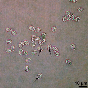
Wet mount preparations of affected muscles revealed the presence of spores that measured 4·0 × 6·0 µm. Spores were generally pyriform, slightly acute in the anterior portion and blunt in the posterior portion. Most of the spores were translucent and had a refractile wall (Fig. 2).
Spores of Pleistophora hyphessobryconis in a wet mount preparation of infected hybrid jundiara muscle. Note prominent posterior vacuole (arrow).

Histopathology
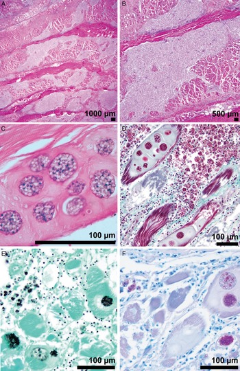
Affected hybrid jundiara exhibited massive infections in skeletal muscle fibres. In the grossly pale and soft areas, fish abdominal musculature was extensively separated and often replaced by marked inflammatory cell infiltrate that was composed primarily of phagocytic cells containing microsporidial spores. Many of the myofibers that remained in markedly inflamed foci had the sarcoplasm distended by one or more cystic stages with either different stages of merogonial proliferation or, more commonly, spores (Fig. 3a–f). Occasionally, the accompanying inflammation extended into parasitized myofibers (Fig. 3b) and the overlying subcutaneous adipose tissue. Some remaining myofibers had variable degrees of degeneration characterized by sarcoplasmic fragmentation, vacuolization and hyalinization with loss of cross-striations (Fig. 3c). The cystic stages were commonly surrounded by pale eosinophilic homogeneous material. The various parasitic stages in the skeletal muscle stained bright red with Trichrome stain (Fig. 3d). A proportion of the encysted and free parasitic stages also stained black with Grocott's methenamine silver (Fig. 3e), weakly pink to purple with Periodic acid-Schiff (not shown) and weakly pink with the Ziehl–Neelsen stain (Fig. 3f). No abnormalities or xenomas were present in the skin.
Histological characteristics of Pleistophora hyphessobryconis infecting the skeletal muscle of hybrid jundiara (a–f). Inflammation extending into parasitized myofibers (a and b); myofibers with variable degrees of degeneration characterized by sarcoplasmic fragmentation, vacuolization and hyalinization with loss of cross-striations (c). Various parasitic stages in the skeletal muscle stained bright red with Trichrome stain (d), black with Grocott's methenamine silver (e), and weakly pink with the Ziehl–Neelsen stain (f).

Ultrastructural observations
By transmission electron microscopy (TEM), multinucleate meronts appeared as rounded cells surrounded by a simple plasma membrane (Fig. 4a). As the microsporidian entered sporogony, the outer membrane surrounding sporogonial plasmodia thickened (Fig. 4b). Within the same sporophorous vesicle (~17 µm in diameter), synchronous development of sporoblasts was observed (Fig. 4c). As the sporoblasts developed into mature monomorphic spores, they became more regular in appearance and the posterior vacuole was discernable (Fig. 4d).
Ultrastructural features of the different developmental stages of Pleistophora hyphessobryconis parasitizing hybrid jundiara (a–f). Developing meront with multiple nuclei (N) (a); microsporidia undergoing early stages of sporogony and thickening of the outer membrane surrounding sporogonial plasmodia (b); sporophorous vesicle with synchronous development of sporoblasts (Sb) (c); developing spore with a discernable posterior vacoule (*) (d).

The spore wall of mature spores was thick (~130 nm) and composed of two layers: an outer zone with an eletrondense wavy aspect, and an inner zone composed of hyaline substance (Fig. 5a). A prominent posterior vacuole was observed (Fig. 5a–d). The polar filament had a external membrane and a central electron dense mass and coiled several times around the posterior vacuole (Fig. 5b). The polaroplast at the apical region of mature spores appeared as alternating layers of electron dense and electron lucent densities (Fig. 5c and d).
Ultrastructure of microsporidian spores of Pleistophora hyphessobryconis in hybrid jundiara. (a) Immature spore delimited by an undulating Ex, which is interrupted at the AD, multiple packed membranes forming the PP membrane, the PF and the posterior vacuole (*); (b) developing spore with a posterior vacuole (*) and coiled PF on each side of the parasite (arrow); (c) mature spore with a prominent posterior vacuole (*); (d) higher magnification of (c) showing the bilaminate spore wall, exospore and endospore, (between black arrows) and the polaroplast (white arrow) occupying the anterior portion of the spore. Abbreviations: AD, anchoring disc; Ex, exospore; PF, polar filament; PP, polaroplast.

Phylogenetic analysis
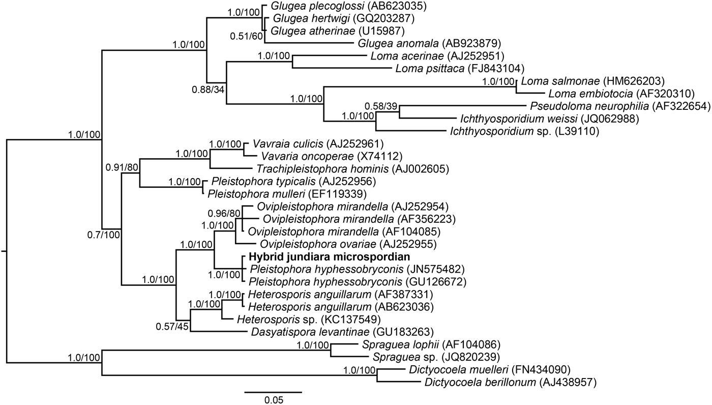
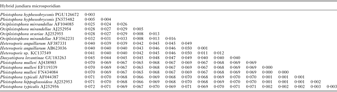
Pairwise analysis of genetic distances (Table 1) showed that the sequence from infected hybrid jundiara is most similar to the two P. hyphessobryconis sequences from the neon tetra (Paracheirodon innesi, PGU126672, 99·7%) and the tiger barb (Puntius tetrazona, JN575482, 99·5%). The remaining microsporidian sequences in the dataset are less than 97·6% similar to the sequence from infected hybrid jundiara. The resulting tree of phylogenetic inference showed the sequence obtained from infected hybrid jundiara positioned within a clade containing P. hyphessobryconis sequences (Fig. 6). Both posterior probabilities of branching points based on Bayesian inference and maximum-likelihood bootstrap values indicated that the node support of the clade containing the hybrid jundiara microsporidian taxon was 1·0 and 100%, respectively.
Dendogram (50% majority-rule consensus) of nearly complete small subunit ribosomal gene sequences showing the phylogenetic position of the microsporidian infecting hybrid jundiara within the species Pleistophora hyphessobryconis. Numbers at the nodes are Bayesian posterior probabilities/maximum-likelihood bootstrap values.

Pairwise genetic distances between a microsporidian infecting hybrid jundiara in a Brazilian aquaculture facility and similar microsporidian based on nearly full length 16S small subunit rDNA sequences

DISCUSSION
A comparison of the microsporidian observed in this study with previously described species of non-xenoma-forming microsporidia known to infect fish musculature revealed a number of morphological differences. While all of the life stages (i.e. meronts, sporonts and sporophorous vesicles with sporoblasts and spores) of members of the genus Heterosporis are contained within a sporophorocyst (Lom and Nilsen, Reference Lom and Nilsen2003), those of the observed microsporidian are not contained within a sporophorocyst. Similarly, members of the genus Kabatana have spores that are not contained within a sporophorous vesicle (McGourty et al. Reference McGourty, Kinziger, Hendrickson, Goldsmith, Casal and Azevedo2007; Barber, Reference Barber, Davies, Ironside, Forsgren and Amundsen2008; Casal et al. Reference Casal, Matos, Teles-Grilo and Azevedo2010), whereas the observed microsporidian was contained within a sporophorous vesicle. In the same context, the vesicle wall surrounding the younger plasmodium members of the genus Ovipleistophora are surrounded by a membranous labyrinth with a hairy appearance (Pekkarinen, Reference Pekkarinen1996; Pekkarinen et al. Reference Pekkarinen, Lom and Nilsen2002), while that of the observed microsporidian was simple. Finally, the only described Dasyatispora species (Dasyatispora levantinae) was reported to be uni-nucleate throughout developmental cycle and produce spindle-shaped subcutaneous swellings that develop into massive, elongated, tumor-like protuberances (Diamant, Reference Diamant, Goren, Yokeş, Galil, Klopman, Huchon, Szitenberg and Karhan2010), while the observed microsporidian is multinucleate during morogeny and causes massive infections in muscle cells with large areas of muscular atrophy.
The microsporidian infecting hybrid jundiara of this study clearly belongs to the genus Pleistophora based on the reported characteristics of this genus such as round merogonial plasmodia bound by a simple membrane; large number of spores produced within each sporophorous vesicle; and microsporidia infecting specifically muscular tissue without xenoma formation (Canning and Nicholas, Reference Canning and Nicholas1980). Moreover, the observed spores were similar in size and shape to those reported for P. hyphessobryconis isolated from the zebrafish and tiger barb in that (1) developing meronts were multinucleated, (2) mature spores measuring 4·0 × 6·0 µm2 had a wrinkled bi-layer spore wall and (3) polar filament had an external membrane and a central electron dense mass (Sanders et al. Reference Sanders, Lawrence, Nichols, Brubaker, Peterson, Murray and Kent2010; Li et al. Reference Li, Chang, Wang, Liu, Liang and Wu2012). Additionally, results of the phylogenetic analysis confirm the observed microsporidian is P. hyphessobryconis. Therefore, based on its light and ultrastructural morphology, developmental stages, genetic sequence, host and location in the host, we conclude that the observed microsporidian is indeed P. hyphessobryconis.
Pleistophora hyphessobryconis causes lethal infections primarily of freshwater aquarium fish (Thieme, Reference Thieme1954). Since the original description by Schäperclaus (Reference Schäperclaus1941), P. hyphessobryconis has been reported to cause massive infections in the muscles of ~20 species of aquarium fish. Herein, we found, for the first time, P. hyphessobryconis infection in hybrid jundiara, a commercially important fish species kept under farming conditions. The histologic alterations in the hybrid jundiara were similar to those described in other Pleistophora infections of aquarium fish (Dyková and Lom, Reference Dyková and Lom1980; Sanders et al. Reference Sanders, Lawrence, Nichols, Brubaker, Peterson, Murray and Kent2010; Li et al. Reference Li, Chang, Wang, Liu, Liang and Wu2012).
Due to the unsightly appearance of infected muscle tissue in fish hosts, P. hyphessobryconis infection can greatly reduce the salability of fish products (Grabda, Reference Grabda1978). Additionally, concurrent bacterial infections including Mycobacterium marinum and Flavobactreium columnare have been reported in cardinal (Paracheirodon axelrodi) (Novotný and Dvořák, Reference Novotný and Dvořák2006) and neon tetras (P. innesi) (Michel et al. Reference Michel, Messiaen and Bernardet2002), respectively. These reports provide evidence to suggest P. hyphessobryconis can compromise the immune system of hosts thereby making hosts susceptible to opportunistic infections. It is well recognized that losses due to opportunistic pathogens are a common problem in the aquaculture industry (Austin and Austin, Reference Austin and Austin2007). It is also possible that farmed cross-breed fish such as hybrid jundiara have a compromised immune system and are more susceptible to infection by P. hyphessobryconis compared with non-hybrid fish, a matter that requires further investigation. Furthermore, P. hyphessobryconis is suspected to be easily transmitted among aquaculture facilities (Sanders et al. Reference Sanders, Watral and Kent2012). Collectively, results of the current study and previous reports of P. hyphessobryconis infections in other fish species suggest the observed microsporidian can be a significant pathogen in commercially exploited fish, especially where the muscle quality is adversely affected (Nigrelli, Reference Nigrelli1946). The spread of such a pathogen in an aquaculture setting may therefore have devastating effects on production and result in economic losses.
Prior to the current study, only two studies have provided both morphological and molecular descriptions of P. hyphessobryconis isolates, both of which describe infection in ornamental fish (Lom and Corliss, Reference Lom and Corliss1967; Li et al. Reference Li, Chang, Wang, Liu, Liang and Wu2012). This study is the first report of a P. hyphessobryconis infection in a pimelodid. Additionally, this is the first report of a P. hyphessobryconis infection in an Amazonian fish species in its native region. Given the potential for P. hyphessobryconis infections to cause severe infections and long-term subclinical chronic infections in zebrafish used as a model in research facilities, it has been suggested that P. hyphessobryconis be added to the list of monitored laboratory pathogens and that a specific PCR test to screen fish and their progeny for P. hyphessobryconis infection be developed (Sanders et al. Reference Sanders, Watral and Kent2012). The information generated in the current study greatly adds to our understanding of both the host range and phylogeny of P. hyphessobryconis.
ACKNOWLEDGEMENTS
The authors thank two anonymous reviewers for their helpful, critical evaluation of this manuscript.
FINANCIAL SUPPORT
Funding for this study was generously provided by the United States Department of Agriculture - Animal and Plant Health Inspection Service (Grant#: 10-9100-1293-GR awarded to M. F.).