Introduction
Although scientists have known about the presence of microbes in the tonsils of swine for more than 100 years, little is understood about the interactions among the microbes at this site or between the host and the resident microbes. The tonsil of the soft palate (tonsilla veli palatini) is the largest (5 cm long by 3 cm wide in adult pigs) and arguably the most important tonsil in the pig. It is a complex and heterogeneous collection of host cells and microbial agents. The tonsil of the soft palate is not only involved in immune surveillance, but paradoxically, it is also the site of colonization and entry of many bacterial and viral pathogens including those that cause septicaemia, respiratory tract and even gastrointestinal tract disease. In this review, we briefly discuss key features of the tonsil of the soft palate, culture-dependent studies that have been done to characterize bacteria and other microbes at this site, and more recent culture-independent analyses of bacterial communities (loosely referred to here as microbiome) in healthy and diseased pigs. Strictly speaking, a microbiome is the totality of microbes (bacteria, viruses, protozoa, fungi, and protists), their genetic elements, and environmental interactions in a particular environment. In the absence of appropriate tools and databases, many ‘microbiome’ studies have focused on bacteria. Studies of the tonsil of the soft palate of swine are no exception.
The tonsil of the soft palate
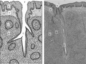
Mammalian tonsils are secondary lymphoid organs in the oral cavity that are part of the mucosa-associated lymphoid tissue (MALT) (Casteleyn et al. , Reference Casteleyn, Breugelmans, Simoens and Van den Broeck2011a). Their position at the beginning of the digestive and respiratory tracts allows for immune surveillance of materials entering the body through the mouth or nares and provides a first line of defense against foreign antigens (Casteleyn et al. , Reference Casteleyn, Simoens and Van den Broeck2011b). Although they carry out similar functions, there is considerable variation in the number, size, organization, and histological properties of pharyngeal tonsils in different species (Casteleyn et al. , Reference Casteleyn, Breugelmans, Simoens and Van den Broeck2011a). Pigs have five types of tonsil: lingual, paraepiglottic, pharyngeal, tubal and the tonsil of the soft palate (often erroneously called palatine) (Casteleyn et al. , Reference Casteleyn, Breugelmans, Simoens and Van den Broeck2011a, Reference Casteleyn, Simoens and Van den Broeck2011b). The tonsil of the soft palate contains numerous fossules that open into tonsillar crypts (Belz and Heath, Reference Belz and Heath1996). These crypts run deep into the tonsil, branching extensively then terminating blindly in the underlying lymphoid tissue (Belz and Heath, Reference Belz and Heath1996; Horter et al. , Reference Horter, Yoon and Zimmerman2003; Fig. 1).
Composite diagram (a) and hematoxylin and eosin staining (b) of a cross-section through a region of the porcine tonsil of the soft palate. The fossule (F) opens into the tonsillar crypt (C). The surface of the soft palate and the crypt neck are lined with a thick layer of non-keratinized stratified squamous epithelium (E) with underlying connective tissue (T). Lymphoid follicles (L) are interspersed in the parenchyma and surrounded by diffuse lymphoid tissues (D). (Tonsil illustration by Rebecca Palmer).

The surface of the tonsil of the soft palate is covered with a thick layer of non-keratinized stratified squamous epithelium as are the openings of the crypts of the tonsil, while the crypts themselves are lined with a thinner layer of lymphoepithelium. Within the tonsil there is considerable variation in the morphology and number of layers of the epithelial cells and in the concentration of M cells and goblet cells (Belz and Heath, Reference Belz and Heath1996; Horter et al. , Reference Horter, Yoon and Zimmerman2003). Subpopulations of professional phagocytes are also reported to be present in the tonsil of the soft palate including macrophages, neutrophils (Belz and Heath, Reference Belz and Heath1996), and dendritic cells (Jamin et al. , Reference Jamin, Gorin, Le Potier and Kuntz-Simon2006). Although the precise identities of the subpopulations are not clear, and no doubt vary with age and environment, the lymphocyte population of the tonsil of the soft palate in pigs is thought to comprise approximately 75% T-lymphocytes and 25% B-lymphocytes (reviewed in Horter et al. , Reference Horter, Yoon and Zimmerman2003).
The epithelial cells that line the surface and crypts of the tonsil of the soft palate are believed to provide a physical barrier against the entry of pathogens into the host and no doubt, provide multiple receptors for microbial attachment. However, some organisms are able to penetrate this line of defense, perhaps by means of the M cells (Belz and Heath, Reference Belz and Heath1996). Mucus and possibly complement components and antimicrobial peptides may play a role in controlling microbes in the tonsil of the soft palate in swine (Horter et al. , Reference Horter, Yoon and Zimmerman2003). At least at this site in humans, antigen that is not effectively cleared by the innate immune response is subject to the adaptive immune response perhaps following contact with the M cells and transport to the parafollicular area where it is sampled by antigen-presenting cells such as macrophages or dendritic cells (Brandtzaeg et al. , Reference Brandtzaeg, Baekkevold, Farstad, Jahnsen, Johansen, Nilsen and Yamanaka1999; Jahnsen and Brandtzaeg, Reference Jahnsen and Brandtzaeg1999; Brandtzaeg, Reference Brandtzaeg2003). In addition, T cells are thought to play an important regulatory role in adaptive immunity in the tonsil of the soft palate, as well as at the systemic level through cytokine production resulting in the activation and direction of lymphocytes, antigen-presenting cells, and neutrophils (Sallusto et al. , Reference Sallusto, Lanzavecchia and Mackay1998; Zuckermann, Reference Zuckermann1999); however, the details of these processes and how they might shape the composition of the microbiome remain to be elucidated.
Despite the tonsil's role in immunity, it is well known as a portal of entry for microbes as well as a site of replication, colonization, and infection. Bacteria persist mainly in the tonsillar crypts, while viruses are more often localized to the lymphoepithelium and the lymphoid follicles (Horter et al. , Reference Horter, Yoon and Zimmerman2003). Within the crypts, bacteria are often found as communities, and it may be postulated that they pool resources in order to persist in the inhospitable host environment. These communities (which might also be thought of as biofilms) could be the site of acquisition of antibiotic resistance or virulence genes though that remains to be demonstrated. To date, there are no data to indicate whether the morphologically different epithelial cells of the tonsil of the soft palate express similar receptors, but it would not be unreasonable to assume that there are many similar, but not identical environments in the tonsil that can select for specific species, and in turn, lead to the development of different microbial communities.
Culture-dependent analysis of tonsillar microbiota
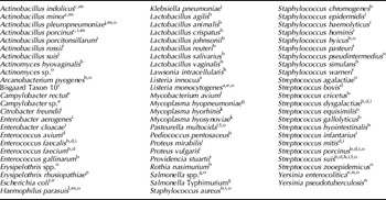
Over the past 30 years there have been 100's of reports of the presence of bacteria in tonsil of the soft palate of swine. To date, the culture of more than 70 species belonging to 14 families has been described (Table 1). Initially, this work was done using classical culture and identification methods; in later studies culture was supplemented with species-specific or even pathotype-specific PCR tests. In these studies, much of the emphasis was placed on the identification of primary pathogens of swine and zoonotic agents. In the case of organisms such as Mycoplasma hyopneumoniae (Marois et al. , Reference Marois, Le Carrou, Kobisch and Gautier-Bouchardon2007) or Salmonella enterica subspecies Typhimurium (Lomonaco et al. , Reference Lomonaco, Decastelli, Bianchi, Nucera, Grassi, Sperone and Civera2009) the pathogenic potential of these agents is reasonably clear. Then again, bacteria such as Actinobacillus minor (Moller et al. , Reference Moller, Andersen, Christensen and Kilian1993; Chiers et al. , Reference Chiers, Haesebrouck, Mateusen, Van Overbeke and Ducatelle2001) and Actinobacillus porcitonsillarum (Tonpitak et al. , Reference Tonpitak, Rohde and Gerlach2007) are almost certainly benign commensals. However, with very many members of the microbiome of the soft palate, the virulence potential can vary greatly depending on the immune status of the host, as well as the serotype or specific genotype of the organism. For example, Streptococcus suis serotype 2 isolates are swine pathogens with zoonotic potential, whereas other serotypes of S. suis have little zoonotic potential and some, little if any ability to cause disease in swine (Staats et al. , Reference Staats, Feder, Okwumabua and Chengappa1997; Zhang et al. , Reference Zhang, Ning, Zhang, Song, Qiu, Gao and Fan2009). Similarly, some serotypes/strains of Haemophilus parasuis can behave as primary pathogens, while others seem to be benign normal microbiota (Devriese et al. , Reference Devriese, Hommez, Pot and Haesebrouck1994; Zhang et al. , Reference Zhang, Ning, Zhang, Song, Qiu, Gao and Fan2009; Ngo et al. , Reference Ngo, Tran, Tran, Nguyen, Campbell, Pham, Huynh, Nguyen, Bryant, Tran, Farrar and Schultsz2011). It is also difficult to evaluate the significance of species such as Escherichia coli that are common environmental isolates. Moreover, with most of these strains, it is not clear whether they are true members of the autochthonous microbiota or transitory allochthonous microbes that are present in large numbers in the environment.
Bacterial species cultured from the tonsil of the soft palate in swine

aAutio et al. (Reference Autio, Markkula, Hellstrom, Niskanen, Lunden and Korkeala2004), bBaele et al. (Reference Baele, Chiers, Devriese, Smith, Wisselink, Vaneechoutte and Haesebrouck2001), cChiers et al. (Reference Chiers, Haesebrouck, Mateusen, Van Overbeke and Ducatelle2001), dDevriese et al. (Reference Devriese, Hommez, Pot and Haesebrouck1994), eFredriksson-Ahomaa et al. (Reference Fredriksson-Ahomaa, Wacheck, Koenig, Stolle and Stephan2009), fEllsworth et al. (Reference Ellsworth, Kirkbride and Johnson1980), gLomonaco et al. (Reference Lomonaco, Decastelli, Bianchi, Nucera, Grassi, Sperone and Civera2009), hJensen et al. (Reference Jensen, Møller, Lindecrona and Jorsal2000), iLowe et al. (Reference Lowe, Marsh, Isaacs-Cosgrove, Kirkwood, Kiupel and Mulks2011), jMacInnes et al. (Reference MacInnes, Gottschalk, Lone, Metcalf, Ojha, Rosendal, Watson and Friendship2008), kMakhanon et al. (Reference Makhanon, Tummaruk, Thongkamkoon, Thanawongnuwech and Prapasarakul2012), lMarois et al. (Reference Marois, Cariolet, Morvan and Kobisch2008), mMoller et al. (Reference Moller, Andersen, Christensen and Kilian1993), nOrtiz Martinez et al. (Reference Ortiz Martinez, Mylona, Drake, Fredriksson-Ahomaa, Korkeala and Corry2010), oO'Sullivan et al. (Reference O'Sullivan, Friendship, Blackwell, Pearl, McEwen, Carman, Slavic and Dewey2011), pTakahashi et al. (Reference Takahashi, Sunama, Satra, Cholsindhu, Kongthon, Jitnupong, Yamamoto, Kijima and Furuuchi1999), qMarois et al. (Reference Marois, Le Carrou, Kobisch and Gautier-Bouchardon2007) ; h,j: culture-independent methods used, but organisms cultured in other studies; q: experimental infection.
In addition to studies that focused on specific organisms, several culture-based analyses have been done that more broadly describe bacterial communities in the tonsil of the soft palate in swine. For example, Baele et al. (Reference Baele, Chiers, Devriese, Smith, Wisselink, Vaneechoutte and Haesebrouck2001) characterized the Gram-positive nasal and tonsillar microbiota of piglets from four different farms before 2 weeks of age and after weaning at 6 weeks of age. Both aerobic and anaerobic cultures were done; species identification was carried out by tDNA intergenic spacer PCR (tDNA-PCR) analysis with a subset further characterized by the sequencing of 16S rRNA genes. In addition to six unknown species, these researchers reported the presence of 25 different species, including, for the first time, Rothia nasimurium, Streptococcus gallolyticus, and Pediococcus pentosaceus (Table 1). In this study, S. suis, Streptococcus dysgalactiae, Staphylocccus hyicus, Staphylococcus aureus, Arcanobacterium pyogenes, and Actinomyces hyovaginalis were isolated from the greatest number of animals both before and after weaning. S. aureus and several species of Lactobacillus became more prevalent after weaning. Enterococcus spp. were also detected in low numbers in a few animals, mostly after weaning. Although enterococci are known to be members of the normal microbiota of the gastrointestinal tract of pigs, their presence in the upper respiratory tract may just be incidental. The findings of Baele et al. (Reference Baele, Chiers, Devriese, Smith, Wisselink, Vaneechoutte and Haesebrouck2001) are generally consistent with the study by Devriese et al. (Reference Devriese, Hommez, Pot and Haesebrouck1994) ; however, molecular tools for species identification were not available when this earlier work was done.
O'Sullivan et al. (Reference O'Sullivan, Friendship, Blackwell, Pearl, McEwen, Carman, Slavic and Dewey2011) also used culture to assess the bacterial burden of tonsils of the soft palate in samples collected over a 7-month period from 395 animals originating from 264 farms. In this study, the presence of commensal organisms and opportunistic pathogens in normal carcasses and carcasses diverted to the hold rail were evaluated at slaughter. Streptococcal species (most notably S. suis) were detected most frequently; A. pyogenes and Pasteurella multocida were also common residents of the tonsils of both groups. Salmonella spp. and Yersinia enterocolitica were detected infrequently (0.5 and 1.8%, respectively). These researchers found that tonsils collected from abnormal carcasses were more likely to be positive for S. hyicus, Streptococcus porcinus, and S. suis, but less likely to be positive for S. aureus. Except S. suis and Listeria monocytogenes, carcasses diverted to the hold rail did not have increased odds of being positive for zoonotic pathogens. Despite the analysis of this relatively large sample, there were no clear differences between microbes associated with normal and abnormal carcasses at slaughter; the failure to see an association may have been due in part to the many reasons for carcasses to be diverted to the hold rail and the many differences among the farms sampled.
In a recent work by Lowe et al. (Reference Lowe, Marsh, Isaacs-Cosgrove, Kirkwood, Kiupel and Mulks2011), both culture-dependent and -independent methods were used to characterize the core microbiome present in the tonsils of healthy pigs. In this study, four 18- to 20-week-old pigs from a high health status herd with no history of respiratory disease and four similar animals from a healthy herd with a history of respiratory disease were evaluated. The herds were similar farrow-to-finish operations that weaned at 21–24 days of age and used all-in all-out procedures when filling grow–finish rooms. Herd 1 contained Hampshire–Yorkshire cross-bred pigs that received no vaccinations or in-feed antibiotics. Hampshire–Cambrough cross-bred pigs were sampled in Herd 2. Animals in this herd were given Tylan in-feed; in addition, they were vaccinated against PCV-2 and received Pulmotil (a tilmicosin in-feed Premix), until 8 weeks of age. For culture, tonsil homogenates were plated on five different media and variously incubated aerobically with and without CO2. In addition to unknowns, a total of 26 species that are members of five different families belonging to two different phyla (Firmicutes and Proteobacteria) were identified by culture. On average, 1.5×10⁷ CFU/g tissue were obtained on rich media, while fewer colonies were seen on selective media (1.0×10⁶ CFU/g). Similar to the findings of Baele et al. (Reference Baele, Chiers, Devriese, Smith, Wisselink, Vaneechoutte and Haesebrouck2001), ~27.7% of the 253 isolates were streptococci or enterococci, 20.6% were staphylococci, while the remaining isolates were Gram-negatives belonging to the families Enterobacteriaceae (~20%) or Pasteurellaceae (~20%) (Table 1). P. multocida, S. suis, S. dysgalactiae, S. aureus, Staphylococcus epidermidis, and E. coli were detected in more than half of the pigs in both herds. Several species of Actinobacillus, Streptococcus, and Staphylococcus, as well as a number of different Enterobacteriaceae were isolated in small numbers; several isolates of Gram-positive bacilli, genotypically related to Bacillus spp. or Actinomyces spp. were detected, but these organisms could not be classified to species. Only one species appeared to be herd specific. Klebsiella pneumoniae was recovered from most animals tested in Herd 1, but was not recovered from any of the animals sampled in Herd 2. Lowe et al. (Reference Lowe, Marsh, Isaacs-Cosgrove, Kirkwood, Kiupel and Mulks2011) also reported that Gram staining revealed considerable variation in the colonization of individual crypts; some crypts containing both Gram-positive and Gram-negative cells in equal proportions, while in others, either Gram-positive or Gram-negative cells were most prevalent.
Culture independent methods
16S sequence libraries
DNAs from the same tonsil samples described above were also studied by Lowe et al. (Reference Lowe, Marsh, Isaacs-Cosgrove, Kirkwood, Kiupel and Mulks2011) using ‘clone libraries’ containing virtually full-length 16S genes. A total of 831 16S rRNA sequences were analyzed; 682 produced using the bacterial domain universal 63 forward primer and 149 generated using the bacterial domain universal 27 forward primer. Rarefaction plots for individual pigs revealed detectable variation among animals and indicated that there was some unsampled diversity. Further, analysis of a composite rarefaction plot of the two herds suggested that Herd 2 was marginally more complex than Herd 1. These conclusions were further supported by the calculated diversity indices and Chao 1 estimations of richness at the herd and individual animal levels with an estimated 67 unique species in Herd 1 and 88 in Herd 2.
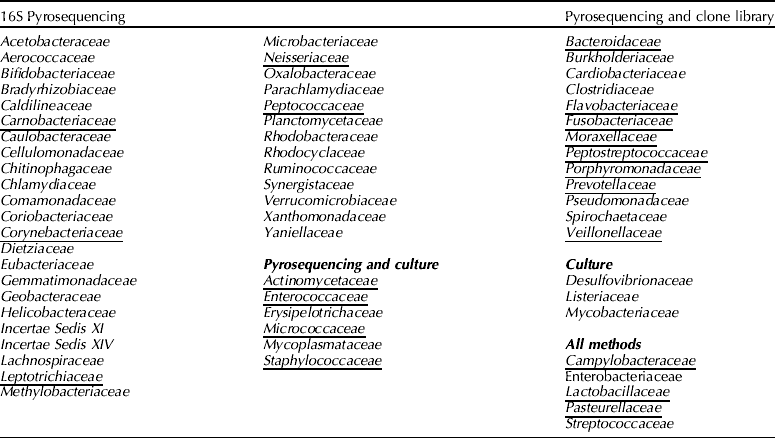
A total of 43 species that are members of 19 families belonging to five phyla (Bacteroidetes, Firmicutes, Fusobacteria, Proteobacteria, and Spirochaetes) were identified from the analysis of the 16S rRNA libraries (Table 2). Many of the same members of Enterobacteriaceae and Pasteurellaceae were observed by both methods, but in contrast to their culture results, very few Gram-positive cocci were identified (Table 2). Conversely, other genera were observed using this culture-independent method that were not obtained by culture. Such a disparity was not unanticipated, in part because anaerobic culture were not done and the tonsillar crypts are thought to be low oxygen environments. However, the paucity of Gram positives in the 16S rRNA clone libraries, even with the use of a second universal forward primer, was unexpected. These results underline the fact that with any method of microbiome analysis there are biases, and with some species, culture is more sensitive than molecular methods.
Bacterial families identified in the tonsil of the soft palate by 16S rRNA gene pyrosequencing (Lowe et al. , Reference Lowe, Marsh, Isaacs-Cosgrove, Kirkwood, Kiupel and Mulks2012), in clone libraries (Lowe et al. Reference Lowe, Marsh, Isaacs-Cosgrove, Kirkwood, Kiupel and Mulks2011), and/or by culture (Table 1)
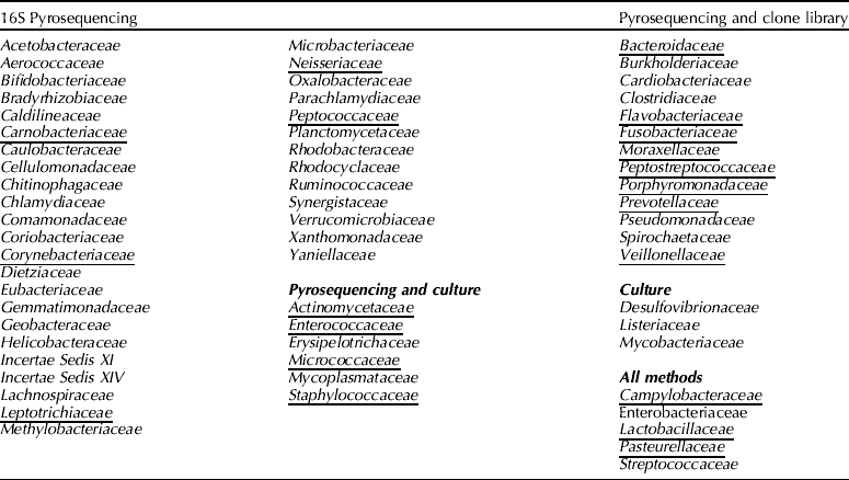
Families found in all pigs in 16S pyrosequencing studies are indicated in bold. Underlined families have also been reported in human tonsil (Kasenõmm et al. , Reference Kasenõmm, Kull and Mikelsaar2002; Aas et al. , Reference Aas, Paster, Stokes, Olsen and Dewhirst2005). Note that the analysis of the clone library data was done at an earlier time than the 16S rRNA data so it is possible that the data presented underestimate the number of families identified by both methods.
Seven genera (Actinobacillus, Haemophilus, Pasteurella, Porphyromonas, Fusobacterium, Bacteroides, and Prevotella) were frequently found in both herds; however, differences in prevalence were noted. For example, 59% of the species in Herd 1, but only 32% in Herd 2 were members of the family Pasteurellaceae. At the genus level, Actinobacillus spp. were more prevalent in Herd 1 versus Herd 2 (49% versus 14% of clones recovered), while Pasteurella spp. were relatively more abundant in Herd 2 (14% versus 5%). Other notable differences between the two herds included a relative predominance of Porphyromonas (13% versus 7%), Bacteroides (13% versus 4%), Dysgonomonas (3% versus 0%), Parabacteroides (8% versus 0%) and unidentified OTUs (13% versus 7%) in Herd 2 and the presence of greater numbers of Fusobacterium (8% versus 3%) in Herd 1. Five additional genera (Campylobacter, Morganella, Proteus, Providencia, and Tannerella) were found in at least one pig from each herd. On the other hand, a number of genera were unique to Herd 1 (Bisgaard Taxon 10, Clostridium, Enterobacter, Filifactor, Klebsiella, Lactobacillus, Moraxella, Planococcus, Pseudomonas, and Suttonella) or to Herd 2 (Acinetobacter, Bergeyella, Burkholderia, Chryseobacterium, Dysgonomonas, Parabacteroides, Peptostreptococcus, and Veillonella). Although most of these unique species represented only a very small proportion of the total microbiome their presence is consistent with the notion that there can be many herd-specific differences, a finding similar to that seen in other studies of swine microbiome (Mulder et al. , Reference Mulder, Schmidt, Stokes, Lewis, Bailey, Aminov, Prosser, Gill, Pluske, Mayer, Musk and Kelly2009). Biological and other experiments are needed; however, to determine the significance of such ‘minor’ constituents and to identify controlling factors.
In the study of Lowe et al. (Reference Lowe, Marsh, Isaacs-Cosgrove, Kirkwood, Kiupel and Mulks2011) herd-specific differences were also seen in a phylogenetic analysis of the sequences of 305 clones belonging to the family Pasteurellaceae and 30 belonging to the family Enterobacteriaceae. In this analysis, 74% of the Pasteurellaceae were identified as A. minor, Actinobacillus porcinus, or P. multocida. With the exception of Pasteurella aerogenes and Bisgaard Taxon 10 (found only in Herd 1) and Pasteurella mairii (found only in Herd 2) most Pasteurellaceae species were present in both herds. Clones of several species (A. minor, A. porcitonsillarum, Actinobacillus indolicus, and H. parasuis) had 97% similarity to each other, but could be grouped into clades and assigned to individual species based on the greatest similarity to the reference strains. Single clades of H. parasuis, A. indolicus, and A. porcitonsillarum were identified with representatives in both herds, whereas four distinct clades were identified as A. minor and three clades of A. porcinus were variously distributed in the two herds. A number of herd-specific species was also seen among members of the family Enterobacteriaceae. For example, Proteus mirabilis and Providencia rustigianii were detected only in Herd 1, whereas Proteus vulgaris was only seen in Herd 2.
In their study, Lowe et al. (Reference Lowe, Marsh, Isaacs-Cosgrove, Kirkwood, Kiupel and Mulks2011) also found that members of the order Bacteroidales including representatives of the families Porphyromonadaceae, Bacteroidaceae, and Prevotellaceae were the second most dominant group comprising 30% of the clones sequenced. Using a 94% cut-off, analysis of sequences belonging to the family Porphyromonadaceae revealed members of the genera Parabacteroides, Dysgonomonas, Tannerella and Porphyromonas; however, more than 80% of Porphyromonadaceae clones belonged to new OTUs at the species level. Of the six clades of Porphyromonas spp. identified, including three clades of new OTUs, three were found in both herds, whereas Porphyromonas gulae was found only in Herd 1, and Porphyromonas catoniae and a new OTU were found only in Herd 2. In addition, Parabacteroides and Dysgonomonas were identified only in Herd 2, whereas Tannerella forsythensis was present in both herds.
Similar patterns of distribution were seen with clones identified as belonging to the families Bacteroidaceae and Prevotellaceae. The vast majority (83%) of Bacteroides spp. and 66% of Prevotellaceae spp. were identified as new OTUs. Given that there have been few reports of anaerobic culture of swine tonsil, this finding is not surprising. As with the Pasteurellaceae and Porphyromonadaceae, there were clades containing clones from both herds as well as some from only one herd.
Pyrosequencing
In a second study, Lowe et al. (Reference Lowe, Marsh, Isaacs-Cosgrove, Kirkwood, Kiupel and Mulks2012) used 454-FLX pyrosequencing to investigate the ‘core microbiome’ of the eight pigs from the two herds described above and samples from an additional four animals obtained from the first herd 2 years after the first samples were collected. In contrast to time 1 (2007), the animals sampled in Herd 1 at time 2 (2009) were purebred Yorkshire (versus Hampshire–Yorkshire), had received Tylan in-feed, and were vaccinated against PCV-2. An additional four samples were also collected from Herd 2 at time 2 using tonsil brushes. One forward and four different reverse primers were used to amplify the V4 region (bases ~576–682) of the 16S rRNA genes. Data analysis was performed using Ribosomal Database Project (RDP) pipeline at Michigan State University and after processing, high-quality sequences were taxonomically identified using the Classifier tool at a 60% confidence level. Using a 97% cut-off for species identification, the number of OTUs detected per sample ranged from 57 to 730. Herd 2 had the greatest animal to animal variability, while pigs from Herd 1, regardless of sampling time or method, showed less variation. Roughly twice as many sequences were obtained from Herd 2 as Herd 1 (99,894 versus 54,932); the number of detected OTUs and Chao 1-estimated OTUs in Herd 2 were ~3- and 4-fold greater than those of Herd 1, respectively. Shannon and Simpson's diversity indices also suggested that Herd 2 was more diverse than Herd 1.
In the Lowe et al. (Reference Lowe, Marsh, Isaacs-Cosgrove, Kirkwood, Kiupel and Mulks2012) study, more than 200,000 reads were obtained. Although there was considerable variation from group to group (from 15,929 to 99,894) an average of >97% of the reads could be assigned at the phylum (97.8%), class (97.6%), and order (97.2%) levels. Taxonomic assignments were lower at the family (90.5%) and genus (72.3%) levels. Compared to Herd 1, the taxonomic assignment of the reads was lower at all levels for Herd 2.
Members of 17 phyla were found in one or more of the tonsil samples studied. Five phyla, however, formed the core microbiome and made up an average of 98.8% (89.5–100%) of the reads assigned to the phylum level. In all pigs, an average 73.4% (47.0–94.5%) of reads were identified as Proteobacteria. The next two most commonly detected phyla were Firmicutes, which averaged 17.8% (3.1–45.6%) and Fusobacteria, which averaged 5.6% (0.6–16.3%) of the total reads. In individual samples, the Proteobacteria, Firmicutes, and Fusobacteria collectively comprised 87.5–99.6% (96.8% avg.) of the microbial community. Although observed at a much lower level, Actinobacteria (avg. 1.2%) and Bacteroidetes (avg. 0.8%) were also found in most pigs in all groups; small numbers of Tenericutes were also found in at least one pig in each group.
Members of 27 different classes of bacteria were also found in at least one sample. Gammaproteobacteria, Clostridia, Fusobacteria, Bacilli, and Betaproteobacteria were found in all animals, while Actinobacteria, Alphaproteobacteria, and Bacteroidia were found in all groups in most animals. Together, these eight classes represented an average of 98.2% (89.2–99.9%) of the total assigned reads at the class level in the samples studied. In addition, Epsilonproteobacteria and Mollicutes were found in at least one animal in each group, while both Deltaproteobacteria and Sphingobacteria were present in at least one animal in the tissue specimens, but were not detected in the brush specimens.
Thirty-four orders of bacteria were detected in at least one tonsil specimen. Members of seven orders: Pasteurellales, Clostridiales, Pseudomonadales, Fusobacteriales, Lactobacillales, Neisseriales, and Enterobacteriales were found in all four groups and Actinomycetales, Burkholderiales, and Bacteroidales were found in most animals in all groups. Together, these ten orders represented from 88.0% to 99.7% (97.4 avg.) of the isolates in individual specimens. In addition, Bacillales and Campylobacterales were found in small numbers in half of the specimens.
Members of 61 families and 101 genera were identified in at least one tonsil specimen in the study by Lowe et al. (Reference Lowe, Marsh, Isaacs-Cosgrove, Kirkwood, Kiupel and Mulks2012; Table 2). Readings that could be assigned to the families Pasteurellaceae, Moraxellaceae, Fusobacteriaceae, Veillonellaceae, and Neisseriaceae were found in all pigs; Peptostreptococcaceae, Enterobacteriaceae, and Streptococcaceae were detected in most animals in all groups. These eight families made up an average of 90.4% (73.5–99.0%) of the reads assigned at the family level in individual animals.
Forty-nine of the 101 genera identified in this study were present in both herds. Of the 37 genera that represented at least 0.1% of the total reads, 13 were found in all 4 groups tested, 2 were found only in Herd 1 and 1 was found only in Herd 2. Eight of these genera were found in tissue, but not in brush specimens. The ten most prevalent genera comprised on average 88.3% (67.2–98.8%) of the total genera in all samples examined. Actinobacillus, Alkanindiges, Fusobacterium, and Haemophilus were found in all pigs; Pasteurella, Veillonella, Peptostreptococcus, and Streptococcus were present in almost all pigs in all groups. These eight genera comprised 85.1% of the reads assigned to the genus level. In addition, Moraxella and Lactobacillus were found in more than half of the samples tested.
Members of the family Pasteurellaceae and genus Streptococcus were further characterized by aligning their sequences with near full-length type sequences. Using a pairwise distances program (with a 0.33% cut-off for clustering) to compare the 454 16S data to the V4 regions of the type strains sequences most closely affiliated with A. indolicus, A. minor, A. porcitonsillarum, and H. parasuis were identified in all samples. A. porcinus, Actinobacillus rossii, Haemophilus felis, P. aerogenes, Pasteurella canis, P. multocida, and S. suis were found in most samples, while small numbers of Streptococcus plurextorum, Actinobacillus lignieresii, and Actinobacillus seminis were found in about half of the samples evaluated.
It might be noted that few differences were observed between brush and tissue samples except that several species belonging to the family Enterobacteriaceae and some obligate anaerobes were found more frequently in tissue than in brush samples. As well, Chlamydia, comprising 0.95% of the reads assigned to the genus level in the tissue specimens, was not found in the brush specimens. Jaccard analyses of clustered sequences of tissue and brush samples taken from Herd 1 at time 2 were highly similar. These samples grouped closely with samples from Herd 1 time 1, but were distant from Herd 2.
Terminal restriction fragment length polymorphism (T-RFLP) analysis
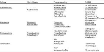
Preliminary characterization of the microbiome of the soft palate of swine has also been done with a larger number of animals, but using the less discriminatory method of T-RFLP analysis (Kernaghan et al. , Reference Kernaghan, Ojha, Slavic, Chen, Poljak and MacInnes2011; Table 3). Despite the use of labeled forward and reverse primers and a two restriction enzyme cut system, identification to the species level with this method is usually not possible. Moreover, the association of a particular pair of bands with a specific organism is not definitive. Nevertheless, T-RFLP analysis of 127 pigs from 29 farms gives a picture similar to that found by Lowe et al. (Reference Lowe, Marsh, Isaacs-Cosgrove, Kirkwood, Kiupel and Mulks2011, Reference Lowe, Marsh, Isaacs-Cosgrove, Kirkwood, Kiupel and Mulks2012). Pigs within a farm tended to be more similar to each other than to pigs in other farms. The identifications most frequently made with T-RFLP compared well with both the clone library and pyroseqencing data with Proteobacteria, Actinobacteria, and Firmicutes seen consistently in all pigs on all farms studied (Table 3); however, even at the phylum level, definitive identifications with this method require confirmation as T-RFLP data can, at best, be considered as providing presumptive identification (Schutte et al. , Reference Schutte, Abdo, Bent, Shyu, Williams, Pierson and Forney2008).
Comparison of phyla detected in the tonsil of the soft palate by culture (Table 1), clone library (Lowe et al. , Reference Lowe, Marsh, Isaacs-Cosgrove, Kirkwood, Kiupel and Mulks2011), 16S rRNA pyrosequencing (Lowe et al. , Reference Lowe, Marsh, Isaacs-Cosgrove, Kirkwood, Kiupel and Mulks2012), and T-RFLP analysis (Kernaghan et al. , Reference Kernaghan, Ojha, Slavic, Chen, Poljak and MacInnes2011)

Underlined phyla have also been reported in human tonsil (Kasenõmm et al. , Reference Kasenõmm, Kull and Mikelsaar2002; Aas et al. , Reference Aas, Paster, Stokes, Olsen and Dewhirst2005).
The microbiome in the tonsils of other hosts
Unlike other anatomical sites, there are few data available for comparison of the microbiota of the tonsil of the soft palate with those of various mammals (or other species) with one exception. The human oral microbiome has been very well characterized in recent years. An estimated 500–700 common species are thought to be common residents with more than 600 species or phylotypes deposited in the Human Oral Microbiome Database (HOMD) and ~280 species isolated in culture (Kasenõmm et al. , Reference Kasenõmm, Kull and Mikelsaar2002; Aas et al. , Reference Aas, Griffen, Dardis, Lee, Olsen, Dewhirst, Leys and Paster2008; Chen et al. , Reference Chen, Yu, Izard, Baranova, Lakshmanan and Dewhirst2010). There is, however, only one publication in which the microbiome of ‘tonsil of soft tissue surfaces’ is explicitly described (Aas et al. , Reference Aas, Paster, Stokes, Olsen and Dewhirst2005). Further, in this 16S clone library study of five healthy subjects, Aas et al. (Reference Aas, Paster, Stokes, Olsen and Dewhirst2005) found that within the oral cavity, there are typically 25–30 predominant species at a given site and some species are quite site- and individual-specific. In tonsillar swabs of the individuals in the Aas et al. (Reference Aas, Paster, Stokes, Olsen and Dewhirst2005) study, Gemella haemolysans, Gemella sanguinis, Granulicatella adiacens, Streptococcus mitis, and S. mitis bv. 2 were most frequently recovered (present in ≥15% of clones assayed); while Granulicatella elegans, Neisseria spp., Prevotella Clone BE073, Prevotella Clone HF050, Prevotella melaninogenica, Simonsiella muelleri, Streptococcus australis, Streptococcus intermedius, and Streptococcus parasanguinis were detected, but were represented by <15% of the clones assayed. It is perhaps noteworthy that 23 out of 25 families reported in human tonsil have also been detected in swine tonsil (Table 2). Although there are many quantitative and qualitative differences, especially at the genus and species levels, it is tempting to speculate that pigs might provide a useful model for the study of human tonsillar microbiota.
Other members of the microbiome
To date, there have been no published reports of Fungi, Protista, Archaea, or phage associated with the tonsil of the soft palate in swine, but studies in other species/other sites suggest that such organisms are likely present (Avila et al. , Reference Avila, Ojcius and Yilmaz2009; Pride et al. , Reference Pride, Salzman, Haynes, Rohwer, Davis-Long, White, Loomer, Armitage and Relman2011; Shan et al. , Reference Shan, Li, Simmonds, Wang, Moeser and Delwart2011). A number of viruses including species-specific pathogens such as porcine reproductive and respiratory syndrome (PRRS) virus and porcine circovirus type 2 (PCV-2) are well-known colonizers of the tonsil of the soft palate in swine. For example, in a recent study by O'Sullivan et al. (Reference O'Sullivan, Friendship, Blackwell, Pearl, McEwen, Carman, Slavic and Dewey2011) PRRSV and PCV-2 were detected in 22% and 11.9%, respectively, of the samples tested. Further, it has been demonstrated that various viruses found in the tonsil (e.g., PRRSV, SIV, and PCV-2) are able to interact with bacterial residents of the tonsil (e.g., M. hyopneumoniae and P. multocida) and cause polymicrobial respiratory disease complex (PRDC) in pigs (Opriessnig et al. , Reference Opriessnig, Gimenez-Lirola and Halbur2011). Although there is an emerging literature on PRDC, there are many unanswered questions about which microbe colonizes which site and in what order or how the pathogenic species, e.g., P. multocida interact with commensal microbes in the tonsil.
Manipulating the microbiome of the tonsil of the soft palate
With the increasing incidence of antibiotic resistance, the ban on prophylactic antibiotic use in some countries, and consumer resistance to ‘chemicals’ in food, there is a growing interest in the use of probiotics. Probiotics are defined as live organisms which when administered in adequate amounts, confer a health benefit on the host (FAO/WHO, 2002). Much of the work on probiotics has focused on their use in manipulating gut microbiota (Huyghebaert et al. , Reference Huyghebaert, Ducatelle and Van Immerseel2011; Kenny et al. , Reference Kenny, Smidt, Mengheri and Miller2011; Prakash et al. , Reference Prakash, Tomaro-Duchesneau, Saha and Cantor2011). They are thought (at least in the gut) to exert their effects by competing with other microbes for attachment sites and nutrients, producing antibacterial substances, helping to initiate repair of the epithelial damage, and modulating the host immune response (Wohlgemuth et al. , Reference Wohlgemuth, Loh and Blaut2010).
To date, no research has been done to examine the effects of probiotics on tonsil microbiota, but several studies have been undertaken to investigate the use of probiotics in the prevention or amelioration of respiratory tract disease (Vouloumanou et al. , Reference Vouloumanou, Makris, Karageorgopoulos and Falagas2009). Although mixed results were reported in some early studies, more recent work by Guglielmetti et al. (Reference Guglielmetti, Taverniti, Minuzzo, Arioli, Stuknyte, Karp and Mora2010) and Santagati et al. (Reference Santagati, Scillato, Patane, Aiello and Stefani2012) suggests that specific strains of Streptococcus spp. are safe and have potential in providing protection against respiratory pathogens. As the site of colonization of many primary, opportunistic, and zoonotic pathogens, it would be very interesting to investigate the effects of probiotics on the tonsil of the soft palate.
Composite view
Although the sample size was limited and the variables many, the studies of Lowe et al. (Reference Lowe, Marsh, Isaacs-Cosgrove, Kirkwood, Kiupel and Mulks2011, Reference Lowe, Marsh, Isaacs-Cosgrove, Kirkwood, Kiupel and Mulks2012) are the first comprehensive description of the bacterial species present in the tonsil of the soft palate of swine. Their work suggests that a wide range of species belonging to the family Pasteurellaceae are the predominant residents; however, the relative proportion of Actinobacillus, Haemophilus, and Pasteurella varies between (and even within) herds. As with all studies using 16S rRNA approaches, some errors arise due to differences in the efficiency of lysis and amplification, but in general there was good correlation between culture, clone library, and pyrosequencing data. Even at the phylum level, there are, however, differences in the predicted composition of the microbiome of the tonsil of the soft palate of swine based on the method used for analysis (Table 3). Proteobacteria and Firmicutes (seen by all methods) are almost certainly part of the core microbiome. In addition, Fusobacteria, Bacteroidetes, and Spirochaetes (seen by pyrosequencing and in clone libraries) and Actinobacteria and Tenericutes are also likely to be present. On the other hand, caution is needed in determining the significance of phyla seen only by pyrosequencing as the method and interpretation of these data are not without error (Quince et al. , Reference Quince, Lanzen, Curtis, Davenport, Hall, Head, Read and Sloan2009, Reference Quince, Lanzen, Davenport and Turnbaugh2011). Five of the phyla seen in pig tonsil by a variety of methods have been reported in association with human tonsil (Kasenõmm et al. , Reference Kasenõmm, Kull and Mikelsaar2002; Aas et al. , Reference Aas, Paster, Stokes, Olsen and Dewhirst2005). It should be noted, however, that very little work has been done on the microbiota of human tonsil and further work might reveal greater similarities.
Future studies
Despite its potential importance in health and disease, very little is known about the microbiome of the tonsil of the soft palate in swine (or equivalent tissue in other species). Studies of greater numbers of animals are needed so that the effects of different variables (e.g., age, antibiotic treatment, vaccination, diet, and health status) on the microbiome can be measured. Many basic questions need to be addressed such as how does the microbiome develop? Which organisms are early or late colonizers? What are the keystone species? What is the relationship between the microbiome of the piglet and that of the sow or its environment? What is the relation between the microbiome of the gastrointestinal tract and that of the tonsil? Are antibiotic or virulence genes being exchanged among members of the tonsil microbiome, and if so, which members? In addition to the bacteria and few viral species described to date, what other microbes are present? A better understanding of the role of phage and how they might affect the presence of bacterial species and transfer genetic information is needed (Pride et al. , Reference Pride, Salzman, Haynes, Rohwer, Davis-Long, White, Loomer, Armitage and Relman2011). It has been suggested that comparatively benign members of the microbiome can help in the control of more inimical microbes (Pennisi, Reference Pennisi2005), but much more needs to be learned about how inhabitants compete and the mechanism of that competition. Another large gap in our understanding of host–microbiome interactions is how microbes in the tonsil interact with the host immune system. More practically, we need to learn what factors (on both the host and microbe sides) are involved in the switch of organisms residing as benign commensals one minute and as harmful pathogens the next. Although there are obvious and demonstrated physiological roles of the microbiome in sites such as the gut, in the tonsil such activities (if any) need to be demonstrated.
As with the study of any microbiome, a variety of biological, molecular, and bioinformatic tools need to be applied for us to gain a better understanding of the microbes of the soft palate. It will be especially important to do metagenomic studies to get a greater insight into the metabolic potential of the microbial community and to gain some insight into how its members may interact with the host and one another. With such information it may be possible one day to manipulate the microbiome of the tonsil of the soft palate so as to promote health and prevent disease.
Acknowledgments
The authors thank the Natural Sciences and Engineering Research Council of Canada and the Ontario Ministry of Food and Rural Affairs Animal Health Strategic Investment program for their support; Rebecca Palmer for the composite drawing in Fig. 1; and Dr Brandon Lillie for sharing his expertise in anatomy and histology.