Introduction
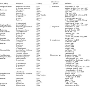
Adults of Clinostomum spp. are digenetic trematodes found in the oral cavity, pharynx or oesophagus of fish-eating birds, reptiles and occasionally mammals, including humans. Freshwater snails are first intermediate hosts, and many fishes and amphibians are second intermediate hosts (Kanev et al., Reference Kanev, Radev, Fried, Gibson, Jones and Bray2002). Metacercariae of Clinostomum spp. have been reported in amphibians, including urodeles and anurans, with almost all records limited to North America (table 1). In contrast, there are no records of Clinostomum among these hosts in Europe. Infections in newts are of particular interest because several species are protected at the national level and have been designated near-threatened or endangered by the IUCN Red List of Threatened Species (www.iucnredlist.org).
List of Clinostomum species detected in amphibians.

The Italian crested newt (Triturus carnifex) is the largest of the four species of newts in Italy; females can exceed 20 cm in length while males reach a maximum of 15 cm. In both sexes dorsal colouration varies from grey-brown to yellowish-brown to brown-greenish and blackish-brown, generally with dark roundish dots. The ventral surface is brightly coloured, yellow-orange with dark black or bluish spots which are variable in number, shape and arrangement (Vanni & Nistri, Reference Vanni and Nistri2006).
The smooth newt (Lissotriton vulgaris) reaches 7–9 cm in total length. Dorsal surfaces are usually brown or grey; during the aquatic phase the male shows some dark-green dots on the body and streaks of the same colour on the head. The ventral parts are whitish in both sexes with dark spots and with a more-or-less evident central orange stripe. The Tuscan populations belong to the subspecies L. vulgaris meridionalis (Vanni & Nistri, Reference Vanni and Nistri2006).
Both species show a terrestrial and an aquatic phase, the latter coinciding with the breeding season. During the breeding season, sexual dimorphism is evident in cloacal morphology and ornamentation in the male, including secondary sexual characteristics such as a dorsal crest and bright tail colouration. The aquatic phase of the smooth newt is limited, and (in Tuscany) usually begins in December and ends in April–May, while crested newt adults may remain in the water all year. In permanent water reservoirs, newts may undergo paedomorphism, i.e. achievement of sexual maturity without complete metamorphosis; this process is reversible and newts can complete the metamorphosis later (Denoël et al., Reference Denoël, Joly and Whiteman2005). Paedomorphosis is quite frequent in L. vulgaris but very rare in T. carnifex (Kalezic et al., Reference Kalezic, Cvetkovic, Djorovic and Dzukic1994).
Even in protected areas, newt populations face risks associated with climate change, anthropogenic impacts, loss of aquatic habitats (especially breeding sites) to agricultural intensification and agrochemical pollution, and the introduction of predatory fish and pathogenic agents, including parasites.
In this study, we report Clinostomum complanatum in the smooth (L. vulgaris) and Italian crested newt (T. carnifex), using morphological and molecular methods to identify the parasites. This is the first report of C. complanatum in European newts and, more generally, in European amphibians.
Materials and methods
Study area
The study site was an artificial pond, which is located in the Area Naturale Protetta di Interesse Locale ‘Podere la Querciola’ (Sesto Fiorentino, Tuscany, 43.824703N, 11.173299E). This is an area of approximately 55 ha containing five wetlands of great importance as staging grounds for migratory birds. It was designated a protected area in February 1998 by resolution no. 72 of the Town Council of Sesto Fiorentino, and is part of the Sites of Regional Importance (SIR) ‘Stagni della Piana Fiorentina e Pratese’ (SIC-ZPS IT51140011). The artificial pond was created in 2008 and was designed to safeguard local amphibian species from habitat fragmentation, destruction of breeding sites and to provide a refuge from alien species such as mosquito fish (Gambusia holbrooki) and red swamp crayfish (Procambarus clarkii). The pond is separated from the other ponds by a small protective wall which prevents the entrance of crayfish but allows access by amphibians.
Collection and examination of newts
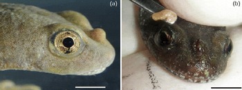
From October 2010 to March 2012, 18 Italian crested newts T. carnifex (4 metamorphic males, 4 metamorphic females, 2 paedomorphic females and 8 larvae) and 4 smooth newts L. vulgaris (1 paedomorphic male, 3 paedomorphic females) were collected with dipnets and transported alive, in a small plastic container (fauna box) with water, to a veterinary clinic. Cysts with yellow grub were evident (fig. 1a). Newts were classified on the basis of the developmental stage: metamorphic (sexually mature, with the gill slits closed); paedomorphic (sexually mature, gill slits open); and larval (immature, gill slits open). Sexual maturity and sexes were established by inspection of the cloaca and based on secondary sexual characteristics. Newts were placed in a bath of tricaine methanesulphonate (1 g/l) buffered with sodium bicarbonate at pH 7.0, which allowed smooth induction and did not irritate the skin and gills of the amphibians (Downes, Reference Downes1995). While anaesthetized, cysts were incised with a 21G sterile syringe needle and the parasites were removed using an ophthalmological Kimura spatula and a small forceps (fig. 1b). Wounds were left open to heal by second intention, and a small amount of antibiotic eye ointment (Oftacilox® 0.3%, Alcon Italia) was applied to prevent infection (Wright & Whitaker, Reference Wright, Whitaker, Wright and Whitaker2001). Finally, newts were rinsed with clean, fresh water until they recovered and were released into the wild after 1 day of supervision. Parasites were excysted, identified as digenean metacercariae and fixed in 90% ethanol. Mean intensity was calculated according to Bush et al. (Reference Bush, Lafferty, Lotz and Shostak1997).
(colour online) Triturus carnifex with an evident cyst in the head: (a) metamorphic female; (b) extraction of the parasite with Kimura's spatula. Scale bars = 6 mm.

Morphological and molecular analysis
Morphological studies were carried out on 11 metacercariae stained with Mayer's acid carmine (Pritchard & Kruse, Reference Pritchard and Kruse1982). Line drawings were made with the aid of a drawing tube; measurements were taken following Matthews & Cribb (Reference Matthews and Cribb1998). Before staining, the posterior part was cut and processed for molecular analysis after measurement of the total length and maximum width. Total DNA was extracted using a PureLink Genomic DNA Kit (Invitrogen, Carlsbad, California, USA) following the manufacturer's protocol. Internal transcribed spacer (ITS) rRNA was amplified as reported by Gustinelli et al. (Reference Gustinelli, Caffara, Florio, Otachi, Wathuta and Fioravanti2010), and a fragment of cytochrome c oxidase I (COI) mtDNA was amplified using the protocol and degenerate primers of Moszczynska et al. (Reference Moszczynska, Locke, McLaughlin, Marcogliese and Crease2009). Polymerase chain reaction (PCR) products were sequenced with an ABI 3730 DNA Analyzer at StarSEQ GmbH (Mainz, Germany).
Results
The 18 Italian crested newts had 30 cysts (mean intensity of infection 1.67) distributed in different parts of the body, such as the head (14), snout (6), gular region (4), mouth (5) and tail (1). In the four smooth newts, a total of five cysts (mean intensity of infection 1.25) were recovered, all in the head. The distribution of infections among developmental stages of the newts is reported in table 2. No pathological changes were observed in the newts examined, other than parasitic nodules.
Distribution of metacercariae of Clinostomum complanatum in developmental stages of each newt species.

F, female; M, male.
Morphology
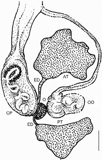
The main morphological characters of the metacercariae were (min.–max. (mean ± SD) μm): body stout, oval, elongated with flattened posterior end, 2637–4181 (3744 ± 401.7) long, 1103–1522 (1272 ± 138.41) wide, covered by thick cuticular spines (13.3–15.9 × 2.7–3.4). Oral collar not evident. Oral sucker, 154–263 (221 ± 34.01) long × 231–335 (294 ± 25.23) wide, smaller than ventral sucker, 461–796 (685 ± 90.80) long × 482–776 (677 ± 76.95) wide. Intestinal cecae lateral to ventral sucker and genital complex. Testes slightly triangular, facing each other with a smooth concave face. Anterior testis, 168–317 (221 ± 40.23) long × 232–309 (268 ± 23.10) wide, bigger and lobed, placed in the posterior end of the middle part of the body. Posterior testis smooth, 140–395 (229 ± 78.10) long × 212–358 (287 ± 47.50) wide, set in the anterior part of the posterior end of the body. Efferent ducts originating from lateral right margin of testes and ending in cirrus pouch (fig. 2). Cirrus pouch, 221–293 (249 ± 24.81) long × 105–142 (121 ± 11.11) wide, extending from intertesticular space to middle right margin of the anterior testis, opening into genital atrium. Ovary, 57–95 (82 ± 11.52) long × 77–114 (98 ± 11.90) wide, fairly oval, set in right portion of the intertesticular space. Uterine sac tubular, with metraterm approaching the right margin of the anterior testis.
Clinostomum complanatum genital complex. AT, anterior testis; PT, posterior testis; OO, ootype; O, ovary; ED, efferent ducts; CP, cirrus pouch. Scale bar = 100 μ_rm;m.

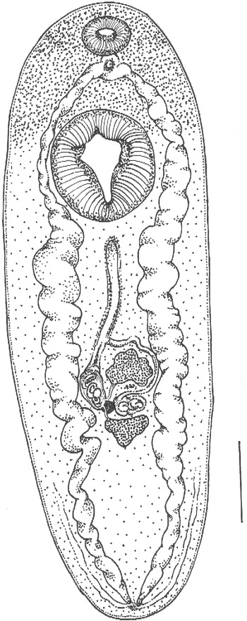
These morphological observations indicated that the metacercariae collected were of the species C. complanatum (Digenea: Clinostomidae) (fig. 3).
Clinostomum complanatum metacercaria. Scale bar = 500 μ_rm;m.

Molecular analysis
In total, ten sequences of the COI mtDNA gene (650 bp) and five from the ITS rDNA (1030 bp) were obtained. The BLAST search, for both molecular markers, gave 99.9–100% identity with C. complanatum and 83% with C. marginatum, the only Clinostomum species described in amphibians whose sequences were available in GenBank.
Discussion
Metacercariae of Clinostomum spp. have frequently been observed in North American amphibians, but records are confusing, with authors variously reporting C. marginatum, C. attenuatum, C. complanatum and unidentified Clinostomum species. McAllister (Reference McAllister1990) and McAllister et al. (Reference McAllister, Bursey, Niemiller and Miller2007) described C. complanatum in the Texas salamander (Eurycea neotenes) from Texas and in the cave salamander (E. lucifuga) from north central Tennessee. These authors provided a brief morphological description and considered C. marginatum a junior synonym of C. complanatum, following Ukoli (Reference Ukoli1966), Dowsett & Lubinsky (Reference Dowsett and Lubinsky1980) and Feizullaev & Mirzoeva (Reference Feizullaev and Mirzoeva1983), but in contradiction to Dzikowski et al. (Reference Dzikowski, Levy, Poore, Flowers and Paperna2004). Dzikowski et al. (Reference Dzikowski, Levy, Poore, Flowers and Paperna2004) showed that C. complanatum and C. marginatum were distinct species on the basis of the 18S rDNA sequences, a result later supported by interspecific differences in morphology (mainly in the genital complex) and in sequences of COI and ITS rDNA (Caffara et al., Reference Caffara, Locke, Gustinelli, Marcogliese and Fioravanti2011). Etges (Reference Etges1991) reported C. attenuatum in the eye of Bufo marinus and presented a picture of the parasite, but no morphometric data. Miller et al. (Reference Miller, Bursey, Gray and Smith2004) reported metacercariae of C. attenuatum in three species of amphibians, basing their identification on the position of the primordial genitalia, which were in the posterior end of the body, even if this arrangement is typical of the adult stage only. McAllister et al. (Reference McAllister, Bursey, Crawford, Kuhns, Shaffer and Trauth2010) reported C. marginatum and C. attenuatum in an ambystomatid salamander, but did not provide morphological data to support their identifications. Bonett et al. (Reference Bonett, Steffen, Trujano-Alvarez, Martin, Bursey and McAllister2011) described C. marginatum in two species of the plethodontid salamander using morphological and molecular methods. In addition to these reports, many authors have reported unidentified metacercariae of Clinostomum during surveys of the helminths of amphibians (Goldberg et al., Reference Goldberg, Bursey and Cheam1998, Reference Goldberg, Bursey and Gergus2001; Goldberg & Bursey, Reference Goldberg and Bursey2001; Muzzall et al., Reference Muzzall, Gilliland, Summer and Mehne2001; Yoder et al., Reference Yoder, Coggins and Reinbold2001; Kuperman et al., Reference Kuperman, Matey, Fisher, Ervin, Warburton, Bakhireva and Lehman2004; Léon-Règagnon et al., Reference León-Règagnon, Martínez-Salazar, Lazcano-Villareal and Rosas-Valdez2005; King et al., Reference King, Gendron, McLaughlin, Giroux, Brousseau, Cyr, Ruby, Fournier and Marcogliese2008; Lemke et al., Reference Lemke, Dronen, Fox and Namhiar2008; Cabrera-Guzmán et al., Reference Cabrera-Guzmán, Garrido-Olvera and León-Règagnon2010; Perpiñán et al., Reference Perpiñán, Garner, Trupkiewicz, Malarchik, Armstrong, Lucio-Forster and Bowman2010; Schotthoefer et al., Reference Schotthoefer, Rohr, Cole, Koehler, Johnson, Johnson and Beasley2011). The morphological features observed in all the specimens from the newts examined during the present survey were consistent with C. complanatum metacercariae.
Although the parasite measurements were, on average, smaller than those of metacercariae from fish, the arrangement of the genital complex and of the efferent ducts, clearly visible in some specimens, was unequivocally consistent with C. complanatum. Maccagno (Reference Maccagno1934) emphasized the importance of the efferent ducts for differentiating C. complanatum and C. marginatum, but subsequent authors have ignored this.
In some specimens cleared with Amman's lactophenol, the presence of thick cuticular spines (13.3–15.9 μm × 2.7–3.4 μm) on the body surface was observed. These spines, described by Cort (Reference Cort1913) as typical of C. attenuatum and useful in differentiating it from C. marginatum, were never visible in the stained specimens. We speculate that during the staining procedure, most of the cuticular spines are lost and are thus no longer detected in the permanent slide.
It should be pointed out that the taxonomy of the genus Clinostomum is still confusing due to the presence of some morphological features which may show high intraspecific variability, so that the morphology per se is sometimes ineffective in reaching correct identification at the species level. For this reason, the DNA-based approach combined with traditional methods seems to be a better method of accomplishing species identification, within both Clinostomum and digeneans in general (Nolan & Cribb, Reference Nolan and Cribb2005). The molecular markers used in this study, COI and ITS rDNA, are commonly used to infer species boundaries among flatworms (Morgan & Blair, Reference Morgan and Blair1995, Reference Morgan and Blair1998; Léon-Règagnon et al., Reference León-Règagnon, Brooks and Pérez-Ponce de León1999; Tkach et al., Reference Tkach, Pawlowski and Sharpilo2000; Kostadinova et al., Reference Kostadinova, Herniou, Barrett and Littlewood2003; Scholz et al., Reference Scholz, Skeríková, Shimazu and Grygier2004; Vilas et al., Reference Vilas, Criscione and Blouin2005) and they allowed us to confirm morphological identifications.
Other than the parasitic nodules, no pathological changes attributable to C. complanatum metacercariae were observed in the 22 newts examined. In a study on tiger salamander (Ambystoma tigrinum), Perpiñán et al. (Reference Perpiñán, Garner, Trupkiewicz, Malarchik, Armstrong, Lucio-Forster and Bowman2010), described mild infestations (1–5 nodules/subject) of Clinostomum sp. metacercariae without evident clinical signs, and one subject, having multiple nodules in the skin over its entire body, which was affected by scoliosis.
A number of European newt species are designated near-threatened or endangered by the IUCN Red List of Threatened Species (http://www.iucnredlist.org). The parasitosis reported here, although not in species of concern, may represent a potential threat for the conservation of more sensitive newt species. Parasites are among the causes of population declines, local extirpations and, in a few species, possible extinction of amphibians (Garner et al., Reference Garner, Fisher, Bosch and Bielby2012). In this regard, two hypotheses have been proposed: (1) in an increasingly globalized world, parasite introductions may cause naïve hosts to decline rapidly; and (2) environmental changes could alter previously balanced host/parasite interactions (Garner et al., Reference Garner, Fisher, Bosch and Bielby2012). Moreover, anthropogenic landscape alterations could have a direct or indirect effect on the transmission and abundance of certain amphibian parasites (McKenzie, Reference McKenzie2012).
This study represents the first description of C. complanatum metacercariae in T. carnifex and L. vulgaris and, more generally, in amphibians of the European area. Although no pathological changes and clinical signs were observed, except for parasitic nodules, at higher infection intensities C. complanatum may represent a health risk for both individuals and populations, e.g. by making infected animals more prone to predation by aquatic birds. This may lead to a population decline and, at the same time, to widespread parasitosis. In this context, accurate species identification is of potentially high importance.
Acknowledgements
This research was supported by the Italian Ministry of Education, University and Research (MIUR-PRIN08). We thank Giacomo Radi for providing images, Daniele Nembi (Legambiente) and the Municipal Administration of Sesto Fiorentino for creation of the artificial pond, and Dr Enrico Buriola (Department of Animal Biology, University of Turin) for his early input in this study and Dr Sean Locke (Environment Canada, St. Lawrence Centre) for English revision and valuable suggestions.