Introduction
With any scientific study in which contamination is a concern, researchers take preventative steps to mitigate the perceived risk. Standard protocols for sampling insect populations often involve the use of attractants to draw specimens into pitfall, light, pheromone, and carbon dioxide traps. With these methods, trapped insects are killed en masse, collectively preserved in ethanol, and returned to the laboratory. If specimens are to be analysed at the molecular level, individual insects are separated to reduce the potential for cross-contamination.
For our study, we focussed on determining the contamination risk of combined insect storage when using molecular techniques to assess the presence of an endosymbiotic bacterium, Wolbachia (Anaplasmataceae). In one study, researchers found this risk to be negligible (Duploy et al. Reference Duploy, Vermenot, Davies, Roderick, Hurst and Charlat2009). However, as we recently incorporated surveys of Wolbachia in local insect populations into our biology curriculum (National Science Foundation Division of Undergraduate Education; NSF DUE-0856593), we wanted to reassess the risk of contamination using the methodology and reagents employed by our students and that differ from the design of this earlier study. Wolbachia is a gram-negative bacterium that was first observed by Hertig and Wolbach (Reference Hertig and Wolbach1924) within the stained reproductive organs of Culex pipiens Linnaeus mosquitoes (Diptera: Culicidae). Later, formally described as Wolbachia pipientis (Hertig Reference Hertig1936), the study of this obligate endosymbiotic microbe did not gain momentum until Yen and Barr (1971) determined that it inhibited egg development in infected host mosquitoes. Since then, Wolbachia has been found to manipulate the survival and fitness of Diptera and other insect orders with a successful strategy to enhance its own horizontal transmission (Werren Reference Werren1997). Recent advances in molecular techniques are allowing scientists to identify the ubiquity of Wolbachia in arthropods (reviewed by Hilgenboecker et al. Reference Hilgenboecker, Hammerstein, Schlattmann, Telschow and Werren2008). Estimates show that more than 50% of aquatic insects (Sazama et al. Reference Sazama, Bosch, Shouldis, Ouellette and Wesner2017) and 40–66% of terrestrial species (Hilgenboecker et al. Reference Hilgenboecker, Hammerstein, Schlattmann, Telschow and Werren2008; Zug and Hammerstein Reference Zug and Hammerstein2012; Weinert et al. Reference Weinert, Araujo-Jnr, Ahmed and Welch2015) harbour the endoparasite. Interest in Wolbachia and other endosymbionts has expanded as their use to directly control insect pest populations has met with success (Mains et al. Reference Mains, Kelly, Dobson, Petrie and Dobson2019). This is especially intriguing because Wolbachia has also been shown to inhibit the transmission of insect vector–borne pathogens responsible for human diseases such as dengue (Frentiu et al. Reference Frentiu, Zakir, Walker, Popovici, Pyke and van den Hurk2014) and Zika (Dutra et al. Reference Dutra, Rocha, Dias, Mansur, Caragata and Moreira2016).
Although Wolbachia’s interactions with its insect host are well documented, other, less-understood endosymbionts are now garnering interest. Expanded surveys of arthropods show that the endoparasite Cardinium (Flexibacteraceae) has a wider host range than was previously understood (Nakamura et al. Reference Nakamura, Kawai, Yukuhiro, Ito, Gotoh and Kisimoto2009; Martin et al. Reference Martin, Puniamoorthy, Gubler, Wimmer, Germann and Bernasconi2013). Not surprisingly, screens of single insect specimens have shown that they can be co-infected with multiple endosymbionts (Skaljac et al. Reference Skaljac, Zanic, Ban, Kontsedalov and Ghanim2010; Mee et al. Reference Mee, Weeks, Walker, Hoffmann and Duchemin2015). However, the potential interspecific interactions occurring between these bacteria and the effects they may independently or collectively have on their insect host are unclear (White et al. Reference White, Kelly, Perlman and Hunter2011). Given the ubiquity of Wolbachia and other bacterial endosymbionts, and their potential to be used in biological control, we sought to determine if the storage of intact insect specimens can lead to cross-specimen Wolbachia DNA contamination of samples stored collectively in ethanol. In addition, we wanted to determine if the duration of storage correlates with contamination risk. Any contamination risks might be applicable to studies involving other endosymbionts.
Materials and methods
We obtained two sets of laboratory-reared fruit flies Drosophila melanogaster Meigen (Diptera: Drosophilidae; yw strain) from the Bordenstein Lab (Vanderbilt University, Nashville, Tennessee, United States of America), the original founders of the NSF-funded Wolbachia Project Discover the Microbes Within (Bordenstein et al. Reference Bordenstein, Brothers, Wolfe, Bahr, Minckley and Clark2010). The positive control group (Wolbachia + flies) was reared from flies naturally infected with wMel Wolbachia (Wu et al. Reference Wu, Sun, Vamathevan, Riegler, Deboy and Brownlie2004), whereas the negative control group (Wolbachia – flies) was reared from the same wMel-infected yw strain but treated with three generations of the antibiotic tetracycline. These control fly lines were developed in 2011 and have been maintained separately ever since. The 100% presence or absence of Wolbachia is confirmed every one to two years by polymerase chain reaction (PCR) screening (Bordenstein, personal communication). Infected (+) and uninfected (−) flies were shipped and stored in separate tubes of ethanol at –20 °C until use. For each co-storage study, intact flies were removed from each tube and air dried to remove residual ethanol. Featherweight forceps were used to transfer individual specimens into 1.5-mL microcentrifuge tubes containing 0.5 mL of 75% ethanol. Between specimens, forceps were rinsed in ethanol and dried. To determine if the quantity of flies co-stored in a single tube could influence cross-contamination, two ratio combinations (1:1 and 1:5) were used. For the 1:1 ratio, one infected (+) fly and one uninfected (−) fly were stored together into a single tube. For the 1:5 ratio, a single infected (+) fly was added to a tube containing five uninfected (−) specimens. All tubes were stored at –20 °C for three hours, 24 hours, or one week before DNA extraction to test if increased duration of co-storage increases the risk of cross-contamination. For all experimental conditions, five replicates of each trial were conducted, for a total of 120 flies tested.
Total genomic DNA was extracted individually from all flies using the QIAGEN DNeasy® blood and tissue kit (Germantown, Maryland, United States of America) according to the manufacturer’s instructions. Final DNAs were eluted with 100 µL elution buffer and stored at −20 °C until PCRs were performed. All PCR primers were custom ordered from Fisher Scientific (Pittsburgh, Pennsylvania, United States of America). All DNA samples were screened for the presence of Wolbachia 16S rDNA using the W-Spec-F (5´-CATACCTATTCGAAGGGATAG) and W-Spec-R (5´-AGCTTCGAGTGAAACCAATTC) primers originally designed by the Bordenstein lab (personal communication). All genomic DNAs were screened for the presence of the insect CO1 mitochondrial gene (CO1-F 5´-GGTCAACAAATCATAAAGATATTGG and CO1-R 5´-TAAACTTCAGGGTGACCAAAAAATCA) as a positive control and DNA quality check for each sample. Any samples without an insect CO1 signal were removed from further analyses. Wolbachia DNA was amplified using 16s rRNA primers. Polymerase chain reactions were performed using QIAGEN’s HotStarTaq Plus master mix according to the manufacturer’s recommendation. Briefly, each PCR reaction had a final volume of 25 µL and was composed of 1 × HotStarTaq Plus Master mix (1 × CoralLoad PCR Buffer, which contains 1.5 mM magnesium chloride2), 200 µM of each deoxynucleoside triphosphate (dNTP), 1 unit HotStarTaq Plus DNA polymerase, 0.5 µM each forward and reverse primer (four primers total for insect CO1 and Wolbachia 16S rRNA), and 2 µL of template DNA (or sterile H2O for no template controls). Reactions were run in a Bio-Rad Laboratories MyCycler® (Hercules, California, United States of America) using the following conditions: denaturation at 95 °C for 10 minutes, followed by 30 cycles of amplification (94 °C for 30 seconds, 50 °C for 45 seconds, 72 °C for 1 minute) and a final extension of 7 minutes at 72 °C. All PCR-amplified samples (10 µL each) were subjected to electrophoresis through a 1% agarose (GOLDBIO, St. Louis, Missouri, United States of America) tris-acetate ethylenediamine tetraacetic acid (TAE) gel containing 0.5 µg/mL ethidium bromide (Thermo Scientific, Rockford, Illinois, United States of America). The O’GeneRuler 100-bp Plus DNA ladder (Thermo Scientific) was run concurrently to provide a size estimation of any amplified products. Bands were visualised, and gel images were captured using a Bio-Rad ChemiDoc® Imager. Because we were determining if storage conditions affect contamination risk, we treated tubes rather than individual flies within the containers as units of comparison. Data were subjected to statistical analyses using the Fisher’s exact test, where P values < 0.05 were considered significant.
Results and discussion
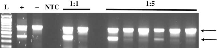
In this study, co-storage with infected (+) flies resulted in cross-contamination of uninfected (−) flies with Wolbachia DNA. The results from a representative gel are shown in Figure 1, which illustrates cross-contamination within a 1:5 co-storage specimen set. In total, cross-contamination was observed in eight of 30 tubes containing co-stored specimens, regardless of the duration of co-storage (Table 1). Contamination with Wolbachia DNA was detected in at least one replicate tube for all time frames for the 1:5 ratio and in one replicate tube at the 1:1 ratio after one week of co-storage. In each tube showing contamination, a total of two flies clearly tested positive for Wolbachia. These data indicate that the infected (+) specimen in the co-stored samples contaminated an uninfected (−) insect within the tube during the storage period. It is always possible that more than one specimen was contaminated during co-storage and that the levels of contamination were below the level of detection by electrophoresis. We did not attempt to quantify the risk, as we sought only to establish that contamination was possible.
Representative electrophoresis results demonstrating evidence of cross-contamination. Insect CO1 (708 bp; indicated by top arrow) is detected in all samples containing insect DNA but not in the no template control (NTC). The 438-bp Wolbachia 16S rRNA band (indicated by bottom arrow) is observed in the Wolbachia-infected positive control (+) but absent from the uninfected (–) and no-template controls. In the 1:1 co-storage treatment tube, no evidence of cross-contamination occurred, because the Wolbachia 16S rRNA band is seen in only one sample, as expected. However, in the 1:5 co-storage treatment tube, the first and fourth samples of the group clearly tested positive for Wolbachia 16S rRNA, indicating that cross-contamination occurred under the storage conditions. L: DNA ladder.

The number of tubes in which Wolbachia contamination was detected in flies stored together in a single container of ethanol. The ratio of infected (+) flies to uninfected (−) flies ranged from 1:1 (total of two flies per tube) to a more crowded 1:5 (total of six flies per tube). Samples were stored at −20 °C for three hours, 24 hours, and one week, with five replicates for each trial, for a total of 30 tubes containing co-stored flies. A total of 120 co-stored flies were tested for the presence of Wolbachia.

Although Wolbachia’s presence in the reproductive tissues of insects is well documented, Wolbachia has also been found in somatic tissues, with its distribution varying between different Wolbachia–host associations (Dobson et al. Reference Dobson, Bourtzis, Braig, Jones, Zhou, Rousset and O’Neill1999). Wolbachia possibly could contaminate the surrounding preservatives as haemolymph, digestive, and reproductive tract contents leak from dead insects. In our study, placing a whole, intact infected (+) insect specimen in the same tube for as little as three hours can result in transfer of Wolbachia DNA between co-stored samples. In addition, when comparing the total number of contaminated tubes with the time-duration trials, we found that the density of specimens stored together in a single container significantly increased contamination risk (1/15 for the 1:1 and 7/15 for the 1:5; Fisher’s exact test 2 × 2 contingency table, P = 0.035; Table 1). Although there was a trend for increased cross-contamination of flies stored in tubes for longer periods (2/10 for three hours, 1/10 for 24 hours, and 5/10 for one week), these results were not significant for the number of tubes tested (Fisher’s exact test 2 × 3 contingency table, P = 0.094; Table 1).
Our study appears to be the first to document the risk of cross-contamination of endosymbiotic bacterial DNA in whole-insect specimen storage. In contrast, Duploy et al. (Reference Duploy, Vermenot, Davies, Roderick, Hurst and Charlat2009) report an absence of cross-contamination between insects that were intentionally dissected to mimic damage that might occur to specimens during field collection and preservation. Additionally, infected cell culture material was used in that study to simulate complete maceration of tissues liberated in the mass storage preservative. Neither of their experiments resulted in detectable DNA transfer from infected (+) to uninfected (−) specimens. A close comparison of Duploy’s study with ours reveals several discrepancies in experimental design that could explain the conflicting results. Both studies used different species of Drosophila (Duploy: D. simulans and D. willistoni (both Diptera: Drosophilidae) versus our D. melanogaster) from different laboratory-reared stocks. The D. simulans stocks were infected with Wolbachia wRi (Giordano et al. Reference Giordano, O’Neill and Robertson1995), whereas the D. willistoni stocks were infected with wWil (Miller and Riegler Reference Miller and Riegler2006). The D. melanogaster stocks from the Bordenstein laboratory were infected with the wMel strain (Wu et al. Reference Wu, Sun, Vamathevan, Riegler, Deboy and Brownlie2004). Variations in infection loads, bacterial strains, and fly species could result in variable risks of contamination. Presumably, flies with lower infection levels would be less likely to transfer Wolbachia DNA between samples. Additionally, Duploy’s study tested a total of 22 flies in 5 mL of ethanol (1 fly/227 µL), whereas our flies were combined with up to six flies in 0.5 mL ethanol (1 fly/83 µL), resulting in a higher fly density per co-storage experiment. Although differences in the co-storage variables could explain the divergent results, the conflicting data could also be the result of detection thresholds. One of the more striking differences between studies is the methods of DNA extraction used: Chelex versus QIAGEN extraction. The Chelex method (Bio-Rad Laboratories) is quicker and facilitates faster processing of multiple samples; however, the Chelex resin can directly inhibit PCR by binding the Mg2+ ions required for Taq polymerase function (Willard et al. Reference Willard, Lee and Holland1998). In a later study, Phillips et al. (Reference Phillips, McCallum and Welch2012) demonstrated that QIAGEN kits more efficiently removed inhibitors, thereby yielding higher-quality DNA products, especially when DNA is more concentrated. If inhibitors were present, they would impact amplification of low-abundance templates (such as those from Wolbachia) moreso than those templates in high abundance (insect CO1). It is reasonable to speculate that PCR amplification from Chelex-extracted DNA might not yield Wolbachia products, whereas it might be apparent in amplified products from QIAGEN-extracted DNA. Additionally, the PCR primers used for amplification of CO1 and Wolbachia DNA were unique in both studies, which represents another variable in which sensitivity of detection could vary based on primer specificities.
In conclusion, our study confirms that when researchers are collecting insects for molecular studies, efforts should be made to isolate specimens in single storage tubes before preserving them. We selected the duration of storage (three hours, 24 hours, and one week) to mimic what would realistically occur when sampling populations of insects in the field and returning them to the lab for DNA extraction. We found that cross-contamination can occur in as little as three hours of co-storage in ethanol. Insect-trapping methods should also be considered when obtaining samples used in molecular studies. Traditional sampling methods, such as pitfall, Malaise, funnel, pan, barrier, and Berlese traps that lure and then preserve insects in a single container, might also contribute to cross-contamination. To minimise this risk, alternative collection and storage methods may be necessary if single insect specimens are to be accurately screened for endosymbiont presence. If insects have been collected in bulk or co-stored, rinsing specimens in ethanol before extracting DNA may minimise cross-contamination. Because we observed a trend that contamination may increase with storage duration, co-storage of samples should be restricted to short periods. Finally, using collection and storage containers filled with ample volumes of preservative may reduce cross-contamination of more densely stored specimens.
Acknowledgements
The authors thank the Department of Biology and the College of Science and Mathematics at Georgia Southern University for project funding. Drosophila controls were kindly donated by the Bordenstein Lab and Wolbachia Project at Vanderbilt University.