Introduction
Minasgeraisite-(Y), end-member formula Y2CaBe2Si2O10, belongs to the gadolinite subgroup (Be dominant at the Z site) of the gadolinite–datolite group that has the general formula (Foord et al., Reference Foord, Gaines, Crock, Simmons and Barbosa1986, Bačik et al., Reference Bačik, Fridrichová, Uher, Pršek and Ondrejka2014): W 2XZ 2T 2O 8V 2 where W = Ca, REE (Y + lanthanides), Bi; X = Fe2+, □ (vacancy), Mg, Mn, Zn, Cu, Al, Fe3+; Z = B, Be, Li; T = Si, B, Be, S, P; V = O(5) = O, OH, F.
Minasgeraisite-(Y) is the only member of this group with Ca assigned to the octahedrally coordinated X site, which has been of some concern as Ca is generally assigned to the larger [8]-coordinated W site in all other members of the group (Demartin et al., Reference Demartin, Minaglia and Gramaccioli2001, Bačik et al., Reference Bačik, Fridrichová, Uher, Pršek and Ondrejka2014). All valid minerals in the gadolinite–datolite group have been assigned the monoclinic space group P21/a (standard setting P21/c); as the latest review by Bačik et al. (Reference Bačik, Fridrichová, Uher, Pršek and Ondrejka2014) uses the P21/a setting, and the original description of minasgeraisite-(Y) (Foord et al., Reference Foord, Gaines, Crock, Simmons and Barbosa1986) also uses the P21/a setting, we retain this cell setting in the present work. The minerals of the gadolinite–datolite group are well known for their chemical inhomogeneity, and minasgeraisite-(Y) is no exception (Foord et al., Reference Foord, Gaines, Crock, Simmons and Barbosa1986). The lack of a crystal structure for minasgeraisite-(Y) was brought to our attention by (the late) Luis Menezes, and Luis gave us a sample of what he considered to be minasgeraisite-(Y) from the type locality to characterize the crystal structure. We were surprised to find that the crystals given to us are triclinic rather than monoclinic, and further work showed that some other members of the gadolinite–datolite group are also triclinic. Obviously, this difference in symmetry affects the nomenclature and classification of the minerals of the gadolinite–datolite group, but additional data is required on the symmetry of the constituent minerals before this issue can be addressed. Thus we choose not to assign a formal name to the material we examine here, but need a suitable soubriquet to allow description of the structure in this paper: we will write the name as ‘minasgeraisite-(Y)’, indicating that we are not implying that the material is minasgeraisite-(Y).
Sample
‘Minasgeraisite-(Y)’ forms minute sheaf-like crystals typically < 10 µm in maximum dimension and occurring as rosettes, 0.2 to 1.0 mm in diameter, coating and intergrown with milarite, albite, quartz and muscovite in a late-stage complex granitic pegmatite at Jaguaraçu, Minas Gerais, Brazil (Foord et al., Reference Foord, Gaines, Crock, Simmons and Barbosa1986).
Chemical composition
It would be desirable to have a chemical analysis of the crystal used for collection of the X-ray intensity data in order to constrain the site-assignments, but ‘minasgeraisite-(Y)’ forms minute sheaf-like crystals typically < 10 µm, and this was not feasible. The crystal used here to collect the X-ray intensity data is ~4 µm thick and it was not possible to mount and polish this crystal. Thus our evaluation of the ‘minasgeraisite-(Y)’ structure uses the chemical composition determined by Foord et al. (Reference Foord, Gaines, Crock, Simmons and Barbosa1986) on material from the same locality.
X-ray data collection
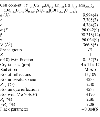
A crystal of ‘minasgeraisite-(Y)’ was attached to a tapered glass fibre and mounted on a Bruker D8 three-circle diffractometer equipped with a rotating-anode generator (MoKα), multilayer optics and an APEX-II detector. A total of 13,109 reflections (4288 in the Ewald sphere) was collected to 60°2θ using 24 s per 0.2° frame with a crystal-to-detector distance of 5 cm. Empirical absorption corrections (SADABS; Sheldrick, Reference Sheldrick2008) were applied, identical reflections (collected at multiple positions along the diffraction vector) were corrected for Lorentz, polarization and background effects, averaged and reduced to structure factors. The unit-cell dimensions were obtained by least-squares refinement of the positions of 4081 reflections with I > 10σI and are given in Table 1, together with other information pertaining to data collection and structure refinement.
Miscellaneous crystallographic data for ‘minasgeraisite-(Y)’.

R 1 = Σ(|Fo| – |Fc|)/Σ|Fo|; wR 2 = [Σw(F 2o – F2c)2/Σw(F 2o)2]½, w = 1/[σ2(F 2o) + 0.0494 P)2 + 0.56 P] where P = (max(F 2o,0) + 2F 2c)/3
Triclinic vs. monoclinic character of the single-crystal X-ray data
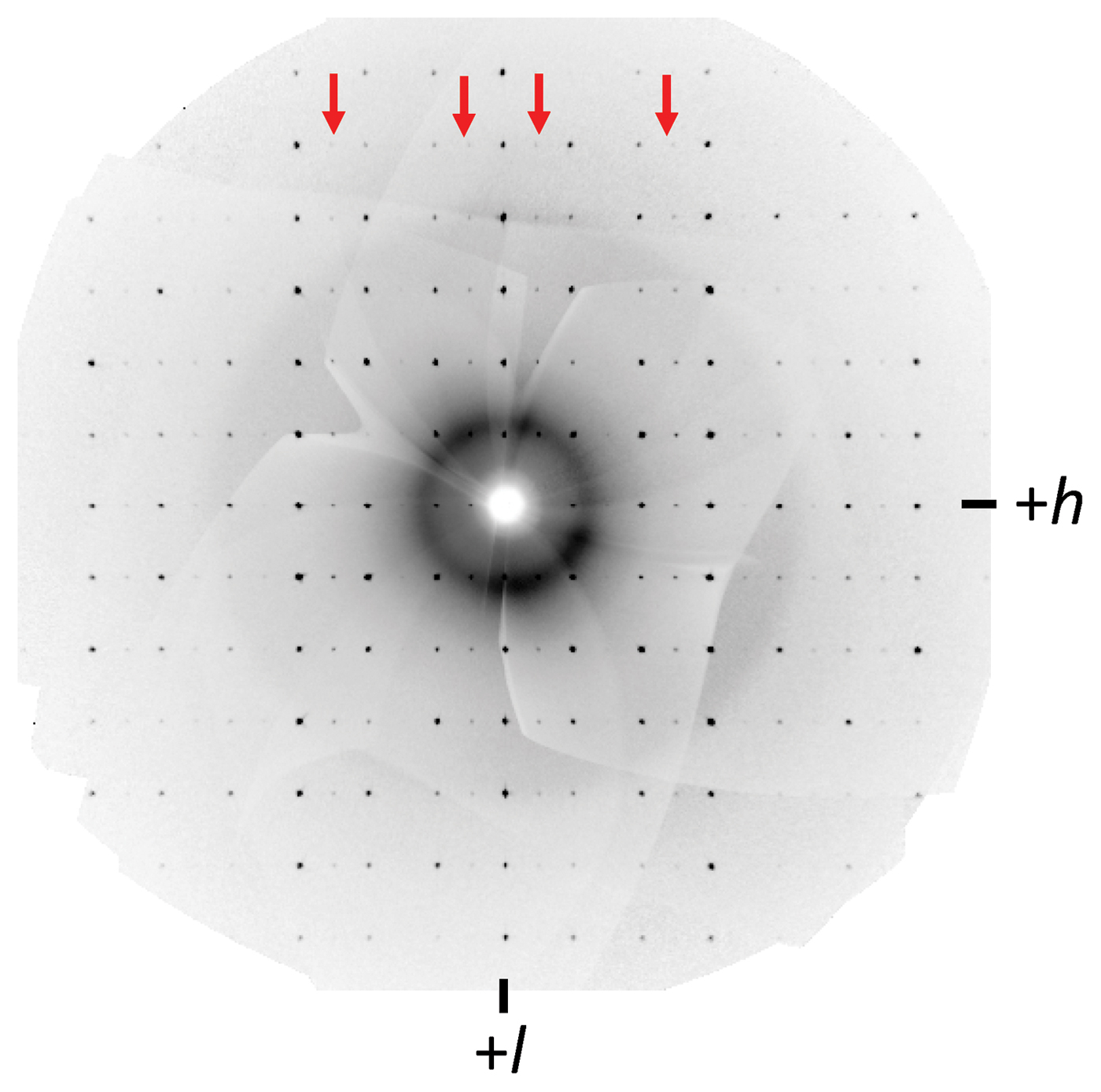
As noted above, all structure work on the minerals of the gadolinite–datolite group has been done in the space group P21/a. However, in our data for ‘minasgeraisite-(Y)’, there are many reflections with significant intensity (|F o| > 40–60 σ|F|) that violate both the presence of a 21-screw axis (0k0; k = 2n) and an a-glide plane (h00, h0l; h = 2n). On the (h0l) precession slice constructed from the raw data frames, there are prominent rows of observed diffraction spots with h ≠ 2n (Fig. 1) that violate the presence of an a-glide plane.
The (h0l) precession slice generated from the ‘minasgeraisite-(Y)’ single crystal X-ray data. The red arrows highlight the observed reflections (h00, h0l) with h ≠ 2n.

The distinction between triclinic and monoclinic symmetry can be made by careful assessment of the X-ray data via: (1) the refined unit-cell parameters; and (2) the Laue-merging results.
Careful observation on our instrument over time has shown that, when the following conditions hold (as for the data set for ‘minasgeraisite-(Y)’): (1) the instrument is well aligned and the crystal is accurately centred within the X-ray beam path; (2) the crystal is of high quality with relatively low mosaicity; and (3) reflections are systematically sampled from a full sphere of diffraction data, then the resulting unconstrained refined unit-cell parameters provide accurate cell metrics to within ≤0.1° on the angles (α, β, γ). Thus if α or γ fall outside the range 89.9–90.1° (for b as the unique monoclinic axis), then the possibility of lower triclinic symmetry needs to be further investigated. For ‘minasgeraisite-(Y)’, our unconstrained α and γ angles deviate < 0.05° from 90, and therefore our observed unit-cell metrics are compatible with monoclinic (and triclinic) symmetry (Table 1).
Next, we investigated the merging of potentially equivalent reflections for the space groups P21/a and P  $\bar 1$. The full Ewald sphere of diffraction data contains 4288 reflections. For P21/a symmetry, there are 1075 unique reflections with R merge(σ) = 9.68 (3.00)%; for P
 $\bar 1$ symmetry there are 2147 unique reflections with R merge(σ) = 5.68 (2.80)%. In itself, this difference in data merging supports P
 $\bar 1$ as the correct space group. However, we have found on our instrument that the relative significance of these R merge results should always be weighed against the general quality of the data. The best indicator of the general quality of the diffraction data for a given experiment is expressed by a term (referred to here as R psi) that is generally not reported for structure refinements; it is the internal agreement index from the merging of identical reflections (i.e. averaged individual reflections acquired from multiple positions on the diffraction vector) following absorption correction. There are many factors related to the diffractometer, the sample and the data-collection strategy that can influence R psi, but R psi will always provide a suitable baseline agreement-index independent of any symmetry argument. Additionally, X-ray data collected with modern area detectors are generally characterized by significant redundancy, allowing evaluation of R psi, the agreement between the intensities of identical reflections measured several times at different positions around their diffraction vectors. The value of R psi is a measure of the intrinsic reproducibility of ones measurements. If symmetry is present in the data, symmetry averaging should give a value of R merge ≈ R psi. If R merge is significantly greater than R psi, the true symmetry of the data may be lower than the assigned space-group symmetry. For ‘minasgeraisite-(Y)’, R psi(σ) is 2.40 (2.63)% whereas R merge(σ) is 5.68 (2.80)% for
 $\bar 1$ symmetry. As centrosymmetrically related reflections show a much lower measure of agreement, it follows that reflection averaging in P
 $\bar 1$ does not conform to the symmetry of the crystal. It should be noted that for the ‘minasgeraisite-(Y)’ data, the magnitude of R psi (2.40%) is in accord with typical R psi values from similar high-quality diffraction-data collected on our instrument that is well observed (i.e. has a mean I/σ for all data-resolution shells ≥ ~15). Moreover, a non-centrosymmetric space group is supported by the E-statistics, as |E 2–1| = 0.773 for the ‘minasgeraisite-(Y)’ data. Thus we conclude that the space group of ‘minasgeraisite-(Y)’ is P1.
‘Minasgeraisite-(Y)’ unit-cell parameters
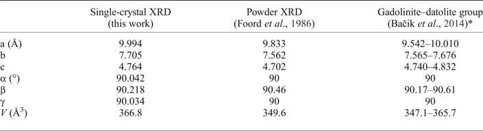
Table 2 lists our unit-cell parameters for ‘minasgeraisite-(Y)’ refined from single-crystal X-ray diffraction (XRD) data alongside those reported by Foord et al. (Reference Foord, Gaines, Crock, Simmons and Barbosa1986) refined from powder XRD data. Our cell-dimensions are significantly larger than those of Foord et al. (Reference Foord, Gaines, Crock, Simmons and Barbosa1986). In particular, our b dimension is larger than that reported for any mineral of the gadolinite–datolite group (Bačik et al., Reference Bačik, Fridrichová, Uher, Pršek and Ondrejka2014), whereas the b and c dimensions reported by Foord et al. (Reference Foord, Gaines, Crock, Simmons and Barbosa1986) for minasgeraisite-(Y) are the smallest for any mineral of the gadolinite–datolite group. This discrepancy in reported ‘minasgeraisite-(Y)’ unit-cell parameters is difficult to understand, as our chemistry inferred from structure is in general agreement with the experimental chemistry of Foord et al. (Reference Foord, Gaines, Crock, Simmons and Barbosa1986); the significant presence of the large Bi3+ cation as a W-site substituent would be expected to increase the unit-cell parameters relative to the analogous non-Bi-bearing minerals.
‘Minasgeraisite-(Y)’ and related unit-cell parameters.

* excluding ‘minasgeraisite-(Y)’
P1 structure model
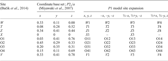
An initial structure was first solved in P1, and then the coordinate set was adapted to conform to the P21/a hingganite-(Y) model of Miyawaki et al. (Reference Miyawaki, Matsubara, Yokoyama and Okamoto2007), and the site labelling expanded to conform with the most recent review of the minerals of the gadolinite–datolite group (Bačik et al., Reference Bačik, Fridrichová, Uher, Pršek and Ondrejka2014). All general sites in P21/a split into four non-equivalent sites in P1, and the X site at the origin splits into two non-equivalent sites. Note that we have physically restrained the primary X site (X1) cation to the origin to enable direct comparison with the P21/a structure model. The P21/a → P1 model expansion with accompanying site label / symmetry-operator relations is given in Table 3.
‘Minasgeraisite-(Y)’ P1 structure model.

Structure refinement
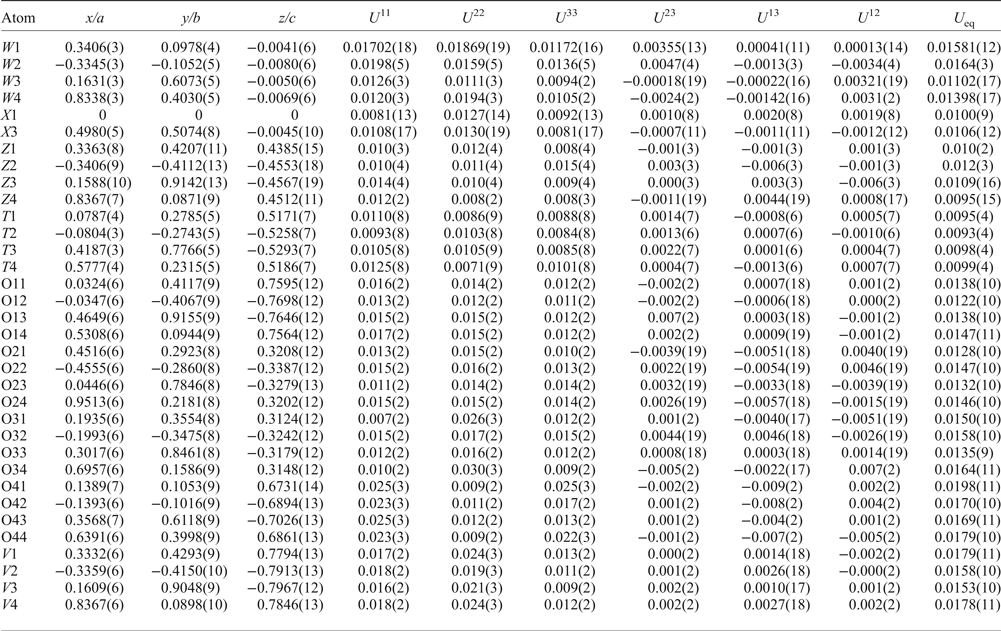
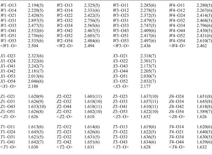
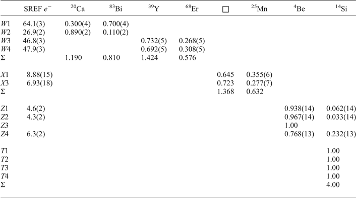
The initial refinement model used single scattering factors and refined unconstrained the occupancies for all cation sites. Each site was assessed in turn, and refining occupancies were either retained with multiple scattering species, or fixed for a single scatterer. Our fully anisotropic structure-model refined to R 1 = 3.41%, with the great majority of the worst-fit reflections classed as |F o| > |F c|. In turn, we tried all simple twin-laws, of which, only reflection across (010) gave a significantly improved R value. The final twin refinement for a complete anisotropic model refined to R 1 = 2.86% with a twin fraction of 0.157(3). The Flack parameter refined to –0.004(6), confirming the correct absolute configuration of the refinement model. Refined atom coordinates and anisotropic-displacement parameters are listed in Table 4, selected interatomic distances are given in Table 5 and refined site-scattering values are given in Table 6.
Atom coordinates and displacement parameters for ‘minasgeraisite-(Y)’.

Selected interatomic distances for ‘minasgeraisite-(Y)’.

Final site-scattering values (epfu) and assigned site-populations (apfu) for ‘minasgeraisite-(Y)’.

SREF – Single-crystal structure refinement.
Crystal structure: structural connectivity
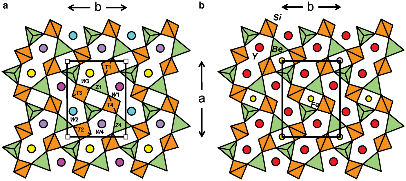
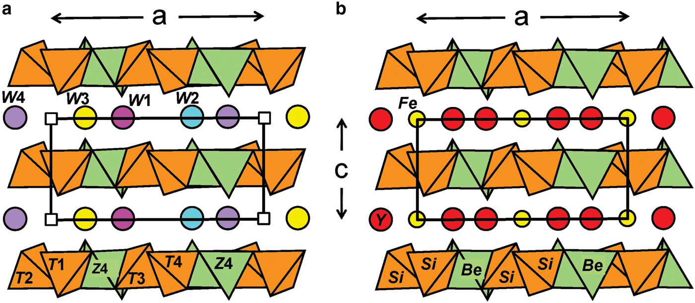
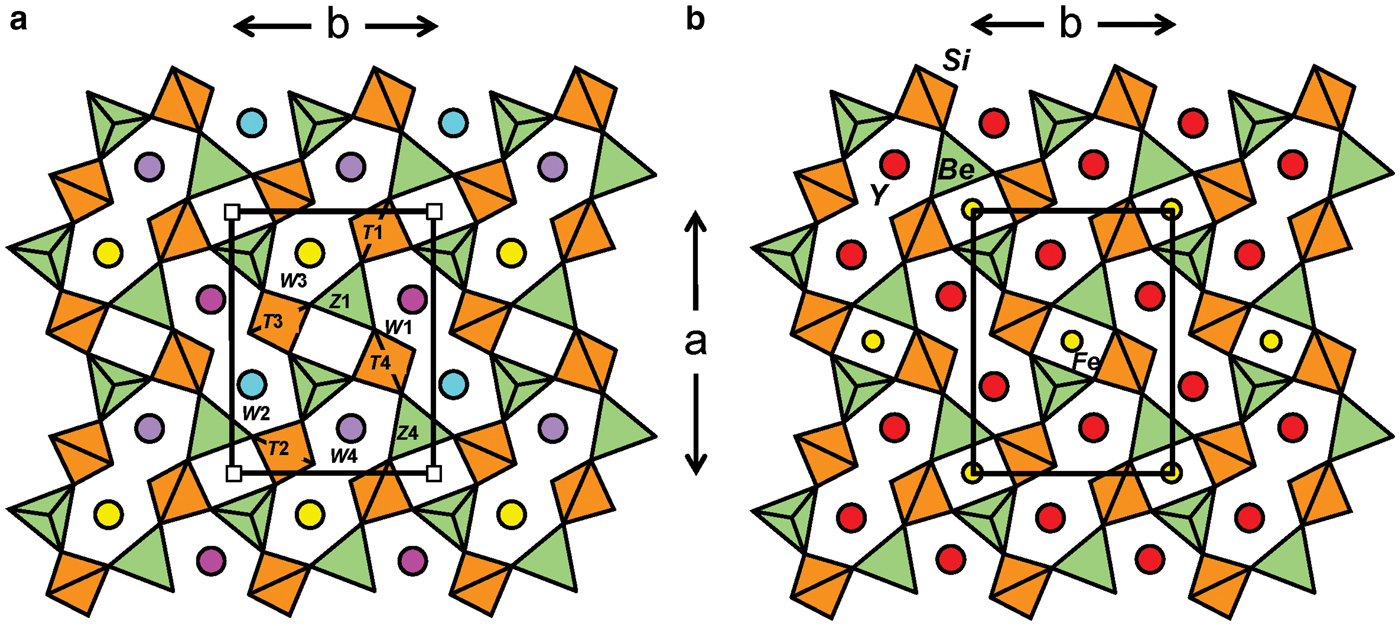
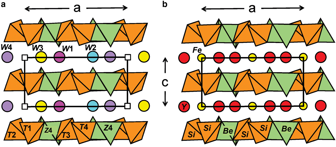
The structure of ‘minasgeraisite-(Y)’ has a bond topology identical to that of gadolinite and datolite (Ito and Mori, Reference Ito and Mori1953; Foit et al., Reference Foit, Phillips and Gibbs1973; Miyawaki et al., Reference Miyawaki, Nakai and Nagashima1984; Demartin et al., Reference Demartin, Pilati, Diella, Gentile and Gramaccioli1993; Rinaldi et al., Reference Rinaldi, Gatta and Angel2010). The structures of the datolite-group minerals are based on tetrahedra at the vertices of the 4.82 net. There are both four-membered and eight-membered rings of tetrahedra (Fig. 2) and Si4+ and Be2+ or B3+ alternate throughout the sheet. In ‘minasgeraisite-(Y)’ (Fig. 2a) and gadolinite-(Y) (Fig. 2b), the sheets are very similar. The lower symmetry in ‘minasgeraisite-(Y)’ allows order over tetrahedrally coordinated sites that are symmetrically equivalent in gadolinite-(Y). The interstitial cations occur between adjacent sheets (Fig. 3). In gadolinite-(Y), Fe2+ occurs at the Fe site at the origin of the unit cell (Figs 2b, 3b). Comparison with the structure of ‘minasgeraisite-(Y)’ (Fig. 2a, 3a) shows that the equivalent sites, labelled X1 and X3 in ‘minasgeraisite-(Y)’, are dominated by vacancies (shown by the white squares in Figs 2a and 3a). There is one W site in gadolinite-(Y) (Figs 2b, 3b); this splits into four non-equivalent sites in ‘minasgeraisite-(Y)’: W1: pink; W2: blue; W3: yellow; W4: lilac in Figs 2a and 3a.
Comparison of the structures of (a) ‘minasgeraisite-(Y)’ and (b) gadolinite projected onto the ab plane. ‘minasgeraisite-(Y)’ – orange: T tetrahedra occupied by Si4+; green: Z tetrahedra occupied by Be2+ and minor Si4+; fuchsia: W1 site occupied dominantly by Bi3+; blue: W2 site occupied dominantly by Ca2+; yellow and lilac: W3 and W4 sites occupied dominantly by Y3+; and white squares: X sites occupied dominantly by □ (vacancies). Gadolinite – orange: Si tetrahedra; green: Be tetrahedra; red circles: Y3+; and yellow circles: Fe2+.
Comparison of the structures of (a) ‘minasgeraisite-(Y)’ and (b) gadolinite projected onto the ac plane; legend as in Fig. 2.
Crystal structure: cation assignment and order
The Z sites
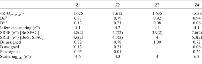
The four Z sites were assigned the Be scattering factor, and the site occupancies were allowed to refine unconstrained, giving 4.8(2), 4.7(2), 3.9(2) and 7.6(2) eps (electrons per site) for the Z1, Z2, Z3 and Z4 sites, respectively. This suggests full occupancy of the Z3 site by Be, and excess scattering (i.e. > 4 eps) at the other Z sites due to one or more heavier elements, typically B (Demartin et al., Reference Demartin, Minaglia and Gramaccioli2001); moreover, some Si must be present at Z4. Demartin et al. (Reference Demartin, Minaglia and Gramaccioli2001) derived the following linear relation for the gadolinite-group minerals:
 $$^Z {\rm B} = 1\,-\,\left[ {\left( { \lt\! Z-{\rm O}\!\! \gt _{{\rm O}2,3,4} -{\rm} 1.475} \right)/0.173} \right]$$
where <Z–O>O2,3,4 is derived from the bond lengths involving the three non-(OH) anions O2, O3 and O4, and ZB is the content of tetrahedrally coordinated B. Applying this relation to ‘minasgeraisite-(Y)’ gives the following compositions / inferred scattering for the Z sites: Z1(Be0.87B0.13) / 4.1 e −, Z2(Be0.79B0.21) / 4.2 e −, Z3(Be0.92B0.08) / 4.1 e − and Z4(Be0.94B0.06) / 4.1 e −. The inductively coupled plasma (ICP) analysis of the bulk minasgeraisite-(Y) sample (Foord et al., Reference Foord, Gaines, Crock, Simmons and Barbosa1986) reports 1.5 wt.% B2O3, so significant B is anticipated in our X-ray crystal. In Table 7, we give the bond-length, site-scattering and site-assignment details for the Z sites. The inferred site-scattering at Z1, Z2 and Z4 from application of the <Z–O> – Be/(B + Be) relation gives values too low in comparison with the actual refined site-scattering values; this indicates that a third heavier (i.e. > 5 e −) scattering species must be present, and Si is the presumed constituent. As Si has an effective ionic radius (0.26 Å) that is nearly the same as Be (0.27 Å), Si will proxy for Be in terms of size, but Si (14 e −) is a considerably stronger scattering species than Be (4 e −); moreover, Al is not significant and can be excluded as a Z-site substituent (Foord et al., Reference Foord, Gaines, Crock, Simmons and Barbosa1986). Prior to the assignment of Si to the Z1, Z2 and Z4 sites, the refinement model was updated with the inclusion of coupled Be–Si occupancy, as a slightly more accurate total scattering will result; the Z3 site-occupancy was fixed at Be = 1.00. Slightly lower total refined site-scattering results at the Z1, Z2 and Z4 sites with inclusion of the variable Si scattering component, and final Be, B and Si site assignments are given (same B content as earlier) with adjusted Be content resulting from the inclusion of Si (Table 7). The combined Z-site composition for our X-ray crystal is (Be3.32B0.40Si0.28)4.00. A significant P2O5 content (1.21 wt.%) is given for the bulk minasgeraisite-(Y) chemical analysis, but no P2O5 content was given for the electron microprobe (EMP) analysis (Foord et al., Reference Foord, Gaines, Crock, Simmons and Barbosa1986). Our Z4 site shows the greatest excess scattering relative to Be, but the <Z4–O > distance supports minor Si → Be and not P → Be (i.e. [4]P5+ has a radius of 0.17 Å and its presence would reduce <Z4–O>); we therefore regard the reported P2O5 content from the chemical analysis as questionable.
Z-site development in ‘minasgeraisite-(Y)’.

[1]: ZB = 1 – [(<Z–O>O2,3,4–1.475)/0.173], ZBe = (1–ZB) ; Demartin et al. (Reference Demartin, Minaglia and Gramaccioli2001).
SREF – Single-crystal structure refinement.
The T sites
The <T–O> distances range from 1.628–1.632 Å for our X-ray crystal and fall within the range 1.621–1.633 Å reported for various gadolinite-group minerals by Demartin et al. (Reference Demartin, Minaglia and Gramaccioli2001). Moreover, there was no significant departure from full occupancy in the refinement. Thus the combined T-site composition for our X-ray crystal is Si4.00 atoms per formula unit (apfu).
The (Z + T)-site composition
Combining our T-site assignments gives (Si4.28Be3.32B0.40)8.00, which is in close agreement with the average of the bulk ICP and EMP analyses of Foord et al. (Reference Foord, Gaines, Crock, Simmons and Barbosa1986) (20 oxygen basis) which gives (Si4.36Be3.21B0.44)8.01.
The X sites
The X-site composition given for the minasgeraisite-(Y) empirical formula (20 oxygen basis) is as follows: (Ca0.90Mn0.40□0.38Mg0.16Fe0.10Zn0.04Cu0.02)2.00 (Foord et al., Reference Foord, Gaines, Crock, Simmons and Barbosa1986). For our P1 structure model, we would anticipate that the X1 and X3 sites both contain a subordinate vacancy and have a combined site-scattering of ≈ 34 electrons pfu (Z = 1). As Ca was expected to be the dominant cation species, we first refined the site scattering at both X sites using the Ca scattering factor, which gave 9.56(18) eps for the X1 site and 7.60(18) eps for the X3 site. Our combined site-scattering at the X sites (17.2 epfu) falls well short of the anticipated value (34 epfu). Note that the low observed site-scattering at both X sites precludes the possibility of either X site being Ca-dominant in the ‘minasgeraisite-(Y)’ crystal refined here. The observed <X1–O> and <X3–O> distances of 2.188 and 2.177 Å, respectively, fall within the range 2.174–2.200 Å reported for other gadolinite-group minerals with variable Fe-occupancy at the X site (Demartin et al., Reference Demartin, Minaglia and Gramaccioli2001). If we exclude all Ca from the X site, the remaining X-site constituents from the empirical formula of Foord et al. (Reference Foord, Gaines, Crock, Simmons and Barbosa1986) sum to 16.3 epfu, which is in much better agreement with our observed total site-scattering value of 17.2 epfu. If we replace the Ca scattering factor with the Mn scattering factor at both X sites (as in our final model), the combined site-scattering over the two X sites is 15.8 epfu. Thus the allocation of excess Ca (i.e. after filling the W site) to the X site seems unjustified, and the high Ca content in the chemical analysis is viewed as questionable. The assigned Mn scattering factor results in a combined X-site composition of (□1.37Mn0.63)2 for our X-ray crystal, where both individual X sites have dominant vacancy.
The W sites
There are four [8]-coordinated W sites that are expected to contain significant 39Y, 57–71Ln, 20Ca and 83Bi which may be regarded as four distinct scattering species (the Ln content is dominated by the heavy REE and has a mean electron count of 68Er). The effective ionic radii [CN = 8] for the four scattering species are: Y3+ = 1.019, Er3+ = 1.004, Ca2+ = 1.12 and Bi3+ = 1.17 Å (Shannon, Reference Shannon1976). There are two larger cations (Bi3+ and Ca2+) and two smaller cations (Y3+ and Er3+). However, it was noted by Demartin et al. (Reference Demartin, Minaglia and Gramaccioli2001) that the observed range of < W–O > of 2.420–2.438 Å for various gadolinite-group minerals does not show a reasonable bond-length variation with respect to Ca content. If our four constituent scattering species (20Ca, 39Y, 68Er and 83Bi) were to show any W-site ordering, we would therefore expect this to be best indicated by site-scattering differences at the individual W sites.
As Y3+ and Ln 3+ have the same charge and similar size, we assumed initially that they will be completely disordered. The two chemical compositions of Foord et al. (Reference Foord, Gaines, Crock, Simmons and Barbosa1986) give (REE)2O3 (REE = Y + Ln) totals of 32.2 and 23.1 wt.%, respectively, with a constant Ln/(Ln + Y) ratio of ~0.37. Our initial refinement model used the Y scattering factor at each W site with freely refining site-occupancies to test for any significant difference in site scattering among the W sites. It was immediately apparent that there was a single ‘heavy’ site (W1), one ‘light’ site (W2) and two similar ‘intermediate’ sites (W3 and W4). The initial refined site-scattering values (epfu) and observed mean bond-lengths (Å) were as follows: W1: 71.3(4), 2.499; W2: 29.3(2), 2.488; W3: 49.6(3), 2.440; and W4: 50.9(3), 2.465. From the available chemistry and these site-scattering values, it is apparent that Bi must be ordered at W1 and Ca must be ordered at W2. The Ln/(Ln + Y) ratio of 0.37 using the scattering values for 68Er and 39Y (see above) gives 49.7 epfu, which is in close agreement with the refined values of 49.6 and 50.9 epfu at W3 and W4, suggesting that these sites are occupied dominantly by the REEs. Additionally, the larger <W1–O> and <W2–O> distances are in accord with the larger radii of Bi3+ and Ca2+. The refinement model was then modified to include the following site-scattering assignments: W1 = Bi and Ca; W2 = Bi and Ca; and W3, W4 = Y and Er. The final site-scattering values and site assignments for the W sites are given in Table 6. The W1 site is Bi3+ dominant, the W2 site is Ca2+ dominant, and the W3 and W4 sites are Y3+ dominant. The combined W-site composition from structure refinement is (Y1.42Ca1.19Bi0.81Er0.58)4.00.
The structural formula of ‘minasgeraisite-(Y)’
We have expanded the structural formula of ‘minasgeraisite-(Y)’ relative to the general formula of the minerals of the gadolinite–datolite group, W 2XZ 2T 2O8V 2, to 20 anions per pfu (Z = 1): (Y1.42Ca1.19Bi0.81Er0.58)Σ 4(□1.37Mn0.63)Σ 2(Be3.32B0.40Si0.28)Σ 4Si4O16[(OH)2.74O1.26]Σ 4
The lanthanides are represented by Er; the X-site occupancy has been simplified to the inferred dominant cation scattering species, Mn2+, and the (OH) content at V is calculated from the vacancy content at X using the assumption that 2(OH) groups correspond to each ‘vacancy’ at the X site (Demartin et al., Reference Demartin, Minaglia and Gramaccioli2001). Although the development of this formula has used several simplifications, the formula is close to electroneutral [0.23– charge]. Some possible reasons for the slight deficiency in positive charge are: (1) overestimation of Ca at the W sites; and (2) some fraction of the X-site cations may be trivalent (i.e. Mn3+, Fe3+). In the context of the minerals of the gadolinite-datolite group, if the cation order caused by lower (P1) symmetry were temporarily ignored and the formula collapsed to the usual 10 anions, the mineral could be referred to as (Ca2+,Bi3+,Mn2+)-bearing hingganite-(Y), with W = Y, X = □, Z = Be, T = Si and O8(OH)2 anions (c.f. Demartin et al., Reference Demartin, Minaglia and Gramaccioli2001). However, the correct P1 structure of ‘minasgeraisite-(Y)’ represents a triclinic subgroup within the gadolinite–datolite group. The following simplified formula for ‘minasgeraisite-(Y)’ highlights the W-site ordering: BiCa(Y,Ln)2(□,Mn)2(Be,B,Si)4Si4O16[(OH),O]4.
In ‘minasgeraisite-(Y)’, ~0.67 of the Be at the Z sites is replaced by ~0.33 B + 0.33 Si, which is compensated by ~1 Ca replacing trivalent cations at the W sites. These substitutions have two important implications: (1) the incorporation of significant Si (in addition to B) at the Z sites represents a new substitution mechanism, and will perturb the simple 1:1 (Ca:B) ratio inferred by Demartin et al. (Reference Demartin, Minaglia and Gramaccioli2001); (2) the assumption of 4 Si pfu (20 anion basis) in relation to T sites filled with Si and no Si at the Z sites, leading to a normalization scheme of 4 Si pfu, is not valid for ‘minasgeraisite-(Y)’ (c.f. Demartin et al., Reference Demartin, Minaglia and Gramaccioli2001).
The indication is that all gadolinite subgroup members with significant W-site cations of different size and/or charge in relation to REE(Y, Ln), needs to be examined carefully by structure refinement for the possibility of W-site cation ordering in a symmetry lower than P21/a (e.g. gadolinites or hingganites with significant Ca).
Bi–Ca ordering at the W site
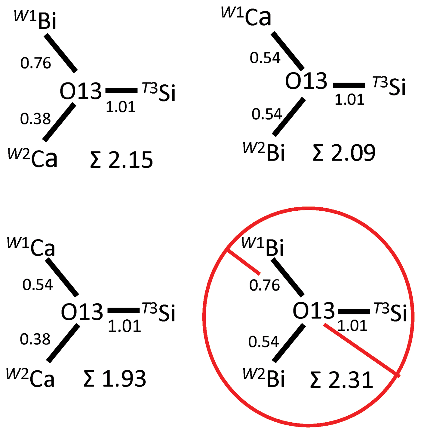
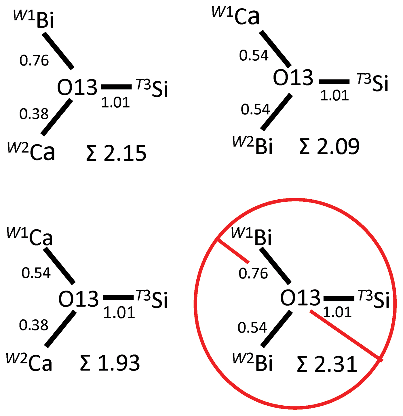
The W1 site gave the largest refined site-scattering value (64.1 e −) and largest < W–O > distance (2.504 Å), consistent with occupancy by Bi0.700Ca0.300 (Tables 5, 6). All four W sites have quite asymmetric bond-length patterns: all contain two short distances to O1* anions (O1* = O11, O12, O13 or O14, see Table 5) and a third next-shortest bond to one O2* anion. These three anions all lie to one side of the coordination (CN = [8]) polyhedron. Of the four W sites, the W1 cation shows the greatest displacement off-centre, consistent with the lone-pair stereoactive behaviour generally observed for Bi3+. The O1* anions that bond to both W1 and W2 are O13 and O14, and local ordering of Bi–Ca between W1 and W2 was examined in terms of local bond-valence incident at the O13 and O14 anions. The O13 and O14 anions are [3]-coordinate O2– anions with a third bond to T3 and T4, respectively. The T sites are occupied solely by Si and the T3–O13 and T4–O14 distances of 1.619 and 1.620 Å, respectively, have a bond valence of 1.01 valence units. Thus the remaining bond-valence contributions to the O13 and O14 anions from neighbouring W1 and W2 cations must adjust to satisfy the bond-valence requirements at these anions. The four possible patterns of local order of Bi and Ca over the W1 and W2 sites are shown in Fig. 4, along with the associated bond-valence values incident at O13; a comparable situation (not shown) exists for the O14 anion. It is apparent from the incident bond-valence sums at O13 that the local arrangements W1Bi–W2Ca, W1Ca–W2Bi and W1Ca–W2Ca are favoured, whereas arrangement W1Bi–W2Bi is unlikely to occur. These three favoured local arrangements are in accord with the refined site populations W1 (Bi0.700Ca0.300) and W2 (Ca0.890Bi0.110), where the combined Bi content is < 1, and local W1Bi–W2Bi associations are avoided.
Local cation arrangements coordinating the O13 anion; bond valences (Brown and Altermatt, Reference Brown and Altermatt1985) shown in valence units.
What to do about minasgeraisite-(Y)
The discovery of triclinic symmetry in ‘minasgeraisite-(Y)’ raises questions as to (1) the possible symmetry of other related minerals, and (2) the classification of the minerals of the gadolinite–datolite group. These issues would be better addressed with additional experimental work on these minerals, and rather than submit a proposal to the International Mineralogical Association (IMA) to modify minasgeraisite-(Y), we decided to publish our results; hopefully this will stimulate further study such that the questions raised above can be addressed from a general perspective. Minasgeraisite-(Y) is currently an IMA-approved mineral of the gadolinite–datolite group, and it is defined with Ca dominant at the X site. This species distinction was approved by the Commission on New Minerals, Nomenclature and Classification of the IMA without an accompanying crystal-structure refinement establishing the dominance of Ca at the X site (Foord et al., Reference Foord, Gaines, Crock, Simmons and Barbosa1986). Our structure refinement of ‘minasgeraisite-(Y)’ shows that Ca does not occur at the X site(s), and that major cation order (involving Ca) occurs at a W site in lower triclinic (P1) symmetry. The assigned cations, summed over the P1 structure, suggest that ‘minasgeraisite-(Y)’ can be averaged to a (Bi–Mn)-bearing hingganite-(Y) with a significant datolite (i.e. Ca and B) component. Our P1 structure maintains the same bond topology as the other minerals of the gadolinite–datolite group, and accordingly ‘minasgeraisite-(Y)’ might be regarded as a triclinic polymorph of hingganite-(Y). According to Nickel and Grice (Reference Nickel and Grice1998), “the formal definition of polymorphism is restricted to substances with identical chemical compositions, this strict limitation is broadened somewhat to include relatively minor chemical variations when the topology is retained”. In this regard, ‘minasgeraisite-(Y)’ represents a rather extreme case with respect to “polymorphic chemical similarity” in that the chemical difference (i.e. the Bi content) from hingganite-(Y) is quite distinctive. In addition, the extent of cation order is very significant; one W site is clearly Bi dominant with no REEs, one W site is clearly Ca dominant with no REEs, and two W sites are Y dominant with only REEs. Thus, there is a 100% cation order at the W sites with respect to monoclinic minerals of the gadolinite–datolite group involving REEs and Ca (i.e. half of the W sites have no REEs). Do highly ordered lower-symmetry derivative species containing new site-dominant chemical constituents (e.g. Bi) justify distinct species status, or does the uncalibrated ‘common topology argument’ prevail and relegate them to adjectivally-modified polymorphic status? The question also arises as to whether what we have found for ‘minasgeraisite-(Y)’ is an isolated case or a common occurrence; any decision about how to deal with the name obviously involves how common is this phenomenon in these minerals.
Conclusions
In ‘minasgeraisite-(Y)’: (1) The correct unit-cell, based on single-crystal X-ray data, is as follows: a = 9.994, b = 7.705 c = 4.764 (Å), α = 90.042, β = 90.218, γ = 90.034 (°) and V = 366.8 (Å3), which is significantly larger than the unit-cell (refined from powder XRD data) previously reported for minasgeraisite-(Y). (2) The correct symmetry is triclinic P1. (3) The dominant constituent of the X sites is a vacancy, and Ca does not occur at the X sites. (4) There is significant ordering of Bi–Ca–REE over the W sites, with Bi dominant at W1, Ca dominant at W2 and Y dominant at W3 and W4. (5) There is significant B and Si at the Z sites.
Acknowledgements
We thank Ritsuro Miyawaki for discussions concerning ‘minasgeraisite-(Y)’ and the nomenclature and classification of the minerals of the gadolinite–datolite group, and for reviewing this manuscript. We thank the Principal Editor Peter Williams for his sagacity in dealing with this rather unusual ‘mineral’. This work was supported by a Discovery grant from the Natural Sciences and Engineering Research Council of Canada and by Innovation Grants from the Canada Foundation for Innovation to FCH.
Supplementary material
To view supplementary material for this article, please visit https://doi.org/10.1180/minmag.2017.081.043