INTRODUCTION
Microsporidia are obligate intracellular parasites of animals and protists. They are particularly common parasites of amphipod crustaceans (Terry et al. Reference Terry, Smith, Sharpe, Rigaud, Littlewood, Ironside, Rollinson, Bouchon, MacNeil, Dick and Dunn2004), in which they are transmitted both horizontally and vertically (MacNeil et al. Reference MacNeil, Dick, Hatcher, Fielding, Hume and Dunn2003; Haine et al. Reference Haine, Brondani, Hume, Perrot-Minnot, Gaillard and Rigaud2004) and in which they cause pathologies ranging from extensive muscle degeneration (Slothouber-Galbreath et al. Reference Slothouber-Galbreath, Smith, Terry, Becnel and Dunn2004) to sex-ratio distortion (Haine et al. Reference Haine, Motreuil and Rigaud2007; Mautner et al. Reference Mautner, Cook, Forbes, McCurdy and Dunn2007). These parasites were discovered in amphipod hosts for the first time in Germany (Pfeiffer, Reference Pfeiffer and Pffeifer1895) and more than 20 species were described till the mid-20th century (Spraque, Reference Spraque1977). However, their taxonomic and phylogenetic status has recently been reappraised following a series of studies using ultrastructural and molecular techniques (Terry et al. Reference Terry, Smith, Bouchon, Rigaud, Duncanson, Sharpe and Dunn1999, Reference Terry, MacNeil, Dick, Smith and Dunn2003, Reference Terry, Smith, Sharpe, Rigaud, Littlewood, Ironside, Rollinson, Bouchon, MacNeil, Dick and Dunn2004; Slothouber-Galbreath et al. Reference Slothouber-Galbreath, Smith, Terry, Becnel and Dunn2004; Franzen et al. Reference Franzen, Nassonova, Scholmerich and Issi2006).
The Ponto-Caspian amphipod Dikerogammarus villosus has recently colonized most large rivers of Western and Central Europe (Bij de Vaate et al. Reference Bij de Vaate, Jazdzewski, Ketelaars, Gollasch and Van der Velde2002; Bollache et al. Reference Bollache, Devin, Wattier, Chovet, Beisel, Moreteau and Rigaud2004; Grabowski et al. Reference Grabowski, Jazdzewski and Konopacka2007; Wattier et al. Reference Wattier, Haine, Beguet, Martin, Bollache, Musko, Platvoet and Rigaud2007). This amphipod can be considered an invasive species since the establishment of viable populations rapidly followed spread by man. Moreover, D. villosus is a predatory species which tends to outnumber native macrobenthic crustaceans in colonized areas (Dick and Platvoet, Reference Dick and Platvoet2000; Bollache et al. Reference Bollache, Devin, Wattier, Chovet, Beisel, Moreteau and Rigaud2004). Two microsporidia, Nosema dikerogammariOvcharenko and Kurandina, Reference Ovcharenko and Kurandina1987 and Microsporidium sp. D (Wattier et al. Reference Wattier, Haine, Beguet, Martin, Bollache, Musko, Platvoet and Rigaud2007) were described in both native and invasive populations of D. villosus, often at high prevalence.
Nosema dikerogammari was first described based on light microscopy, infecting abdominal musculature of D. villosus in the Dnieper Estuary, within its native range (Ovcharenko and Kurandina, Reference Ovcharenko and Kurandina1987). It was placed within the genus Nosema due to its diplokaryotic development and disporoblastic sporogony. Ultrastructural data obtained from D. villosus collected in the Kiev Reservoir on the River Dnieper, later confirmed typical characteristics of Nosema (Ovcharenko and Vita, Reference Ovcharenko and Vita1996). In addition, N. dikerogammari was detected in the Danube Estuarine Lakes, within the native range of D. villosus, and in eastern Poland (Bug River), a part of the invaded range of D. villosus (Ovcharenko et al. Reference Ovcharenko, Codreanu-Bălcescu, Grabowski, Konopacka, Wita and Czaplińska2009). In parallel, Wattier et al. (Reference Wattier, Haine, Beguet, Martin, Bollache, Musko, Platvoet and Rigaud2007) surveyed the main water bodies of Western Europe (invaded range) and the lower course of the Danube (southern native range) using a PCR-based molecular screening. This study indicated that D. villosus was infected by 4 microsporidian species. Three of them were close to parasites already described in other amphipods species: Nosema granulosis, Dictyocoela mulleri and D. sp. roeselum. They were found in few host populations, at very low prevalence and always outside the native range of the host. The fourth parasite, found in most D. villosus populations, was a new species of microsporidian which was provisionally named Microsporidium sp. D. Phylogenetic analysis based on a 476 bp region of SSU rDNA indicated that this species was closely related to an undescribed Microsporidium sp. JES2002G infecting a gammarid from Western Europe (Terry et al. Reference Terry, Smith, Sharpe, Rigaud, Littlewood, Ironside, Rollinson, Bouchon, MacNeil, Dick and Dunn2004) with a closer affinity to the genera Dictyocoela and Pleistophora than to Nosema to which it was definitely distantly related (Wattier et al. Reference Wattier, Haine, Beguet, Martin, Bollache, Musko, Platvoet and Rigaud2007). Microsporidium sp. D was found at high prevalence and showed the very same symptoms as N. dikerogammari parasites (Wattier et al. Reference Wattier, Haine, Beguet, Martin, Bollache, Musko, Platvoet and Rigaud2007).
These studies raise the question of whether N. dikerogammari and Microsporidium sp. D were 2 different species or merely a single microsporidian species described using 2 different techniques (light microscopy and DNA sequencing). This question is important because Dikerogammarus villosus is one of the most invasive freshwater species in Europe (Devin et al. Reference Devin, Piscart, Beisel and Moreteau2003; Van Riel et al. Reference Van Riel, Van der Velde, Rajagopal, Marguiller, Dehairs and Bij de Vaate2006) and has carried its microsporidian parasites during its rapid expansion into the main water bodies of Europe (Wattier et al. Reference Wattier, Haine, Beguet, Martin, Bollache, Musko, Platvoet and Rigaud2007; Ovcharenko et al. Reference Ovcharenko, Codreanu-Bălcescu, Grabowski, Konopacka, Wita and Czaplińska2009). Surveillance and early detection of parasites after ecological changes is important to detect potential emerging disease (Woolhouse, Reference Woolhouse2002). For this reason, it is crucial to ascertain the number and diversity of microsporidian species introduced into European watersheds through the invasion of D. villosus.
In this paper, we combine molecular phylogenetic and ultrastructural analysis to demonstrate that Nosema dikerogammari and Microsporidium sp. D are a single species, and cannot be related to the few Nosema granulosis parasites previously described in a few Western Europe populations. We further provide information on the parasite's tissue specificity and routes of transmission. On this basis, we propose to redescribe N. dikerogammari as Cucumispora dikerogammari, a member of the novel genus Cucumispora gen. nov.
MATERIALS AND METHODS
Sampling sites
Dikerogammarus villosus individuals were sampled from 3 sites: St Jean de Losne on the Saone River, France (N47°5′23·45″; E05°14′31·63″), Uraz on the Oder River, Western Poland (N51°14′54·79″; E16°50′47·61″) and Zegrze on the Zegrzynski Reservoir on the Bug River, Eastern Poland (N52°27′30·79″; E21°01′04·52″). All the sites were within the introduced range.
Light and electron microscopy
Light and electron microscopy were performed on individuals from the 2 Polish sites. The presence of spores in infected individuals was identified by dissection under the binocular microscope. Muscles of infected individuals were then stored in 100% ethanol for molecular analysis. Live spores and Giemsa-stained slides were examined under an Olympus BX50F4 light microscope. The software Analysis Pro 2.11 (Soft Imaging System GmbH, Germany) was used for measurements. For transmission electron microscopy, the infected muscle tissues were fixed in a 2·5% (v/v) glutaraldehyde in a 0·2 m sodium cacodylate buffer (pH 7·2) for 1–3 days. After washing and post-fixation in 2·0% (w/v) osmium tetroxide in cacodylate buffer for 1 h at 4°C, the pieces were dehydrated and embedded in Epon-Araldite solution using a standard procedure (Vávra and Maddox, Reference Vávra, Maddox, Bulla and Cheng1976). Blocks of embedded tissues were sectioned with an LKB III ultra-microtome. Semi-thin sections were stained with methylene blue. Ultrathin sections were then mounted on copper grids, double stained with uranyl acetate and lead citrate and examined with a JEM 100B electron microscope operated at 80 kV.
Molecular methods
DNA extraction
DNA was extracted from Polish and French infected D. villosus, including Polish individuals in which Nosema dikerogammari had beforehand been firmly identified by light and electron microscopy. Microsporidian DNA of French in dividuals was co-extracted with DNA from host muscle tissues. Around 4 mm3 of the host muscle were homogenized in a 1·5 ml tube containing 200 μl of Queen's lysis buffer (Seutin et al. Reference Seutin, White and Boag1991) using a plastic pestle and incubated overnight at 55°C with 5 μl of proteinase K (20 mg/ml). DNA was then extracted based on a standard phenol-chloroform method as described by Hillis et al. (Reference Hillis, Moritz and Mable1996). Air-dried DNA pellets were re-suspended in 100 μl of TE buffer, pH 8·00. Microsporidian DNA of Polish individuals was co-extracted from host muscle tissue of the remaining individuals, in which N. dikerogammeri had been identified, using a Qiagen DNeasy Blood and Tissue kit. Tissue was lysed in 180 μl of ATL buffer and 20 μl of proteinase K for 3 h at 55°C. DNA was then extracted as per the manufacturer's protocol.
Sequencing
DNA sequences of the complete microsporidian small subunit (SSU) rDNA, the internal transcriber spacer (ITS) and part of the large subunit rDNA (LSU) were obtained for 1 individual from each of the 3 sampled sites (St Jean de Losne, Uraz and Zegrze). In order to obtain overlapping fragments, the set of primers described in Table 1 was used. The product was amplified under the following PCR conditions: an initial denaturing step at 95°C for 5 min was followed by 35 cycles of 95°C for 1 min, 50°C for 1 min and 72°C for 1 min. These cycles were followed by a final extension at 72°C for 5 min. Direct sequencing was performed using BigDye technology either by Macrogen Inc., Korea or IBERS, Aberystwyth, UK. All sequencing was performed in both directions.
The list of primers used for amplification of microsporidian SSU rDNA, ITS and partial LSU rDNA

Phylogenetic analysis
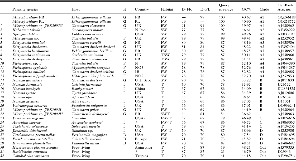
The microsporidian sequences from D. villosus were edited and aligned by eye using BioEdit software (Hall, Reference Hall1999). Alignment with published sequences of microsporidian parasites infecting D. villosus was also performed (GenBank Accession numbers: EF119217, EF119218, EF119219, EF119220, EF119221, EF119222, EF119223). Similarity of the new sequences to those already available in GenBank was determined using BLAST (Table 2). A set of 12 sequences was selected for use in the phylogenetic analysis. This included sequences which were close to parasites from D. villosus, sequences from species that morphologically resembled microsporidia from D. villosus (i.e. Nosema, Pseudonosema cristatellae, Trichonosema pectinatellae, Bryonosema plumatellae) and other sequences chosen to span the diversity of Microsporidia in amphipods. Finally, 3 fungi species were selected as outgroups following Vossbrinck and Debrunner-Vossbrinck (Reference Vossbrinck and Debrunner-Vossbrinck2005) (Table 2). Sequences were aligned with Clustal W (Thompson et al. Reference Thompson, Higgins and Gibson1994) in MEGA 4 software, using default settings (Tamura et al. Reference Tamura, Dudley, Nei and Kumar2007). Only fragments of the sequences that could be unambiguously aligned were used for analysis (1046 bp). Phylogenetic reconstructions included maximum likelihood (ML) processed with PHYML 2.4.4 software (Guindon and Gascuel, Reference Guindon and Gascuel2003) and Bayesian inference processed with MRBA YES v 3.1.2 software (Ronquist and Huelsenbeck, Reference Ronquist and Huelsenbeck2003). For ML and Bayesian analysis, the GTR plus gamma model was used. The model was selected with Modeltest software (Posada and Crandall, Reference Posada and Crandall1998). For the Bayesian approach, we ran the analysis for 500 000 generations, with 1 tree retained every 100 generations. Likelihood stationarity was achieved after 16 000 generations and we excluded this ‘burn-in’ period when constructing a consensus tree. Nodal support was assessed by 1000 bootstrap replicates ML analysis and posterior probabilities for the Bayesian method as estimated from the 3750 trees included after the burn-in.
Sequence similarities of the studied parasite with other microsporidia based on SSU rDNA
(D-FR, D-PL – percentage of identity with studied microsporidian parasites from France and Poland respectively. H, host taxonomic classification; G, Gammaridea; F, Fish; I, Insect; B, Bryozoa. Habitat, FW, Fresh Water; BW, Brackish Water; TSW, Specific zone at the upper limit the sea shore (terrestrial habitat regularly impacted by sea water); T, Terrestrial; FW-T: larvae, Fresh Water but adult, Terrestrial. Clade refers to the clade identified in the phylogenetic tree presented in Fig. 5. Out, outgroup.)

Parasite detection and identification by PCR-RFLP
In order to determine if infection was preferentially associated with host sex and size, 140 D. villosus individuals from Zegrze were sexed under the light microscopy (Nikon SMZ 1500). The 4th coxal plate was measured using Lucia G 4.81 software. This measurement is known to be representative of total size (Bollache et al. Reference Bollache, Gambade and Cezilly2000). Infection status was determined by Microsporidium sp. D diagnostic PCR-RFLP (Wattier et al. Reference Wattier, Haine, Beguet, Martin, Bollache, Musko, Platvoet and Rigaud2007). Microsporidian specific SSU rDNA primers V1f (Baker et al. Reference Baker, Vossbrinck, Maddox and Undeen1994) and 530r (Vossbrinck et al. Reference Vossbrinck, Baker, Didier, Debrunner-Vossbrinck and Shadduck1993) were used to amplify a ~480 bp long PCR fragment, which was then restricted with the enzyme VspI. The Microsporidium sp. D specific restriction profile shows 2 bands of ~400 bp and ~80 bp in length (Wattier et al. Reference Wattier, Haine, Beguet, Martin, Bollache, Musko, Platvoet and Rigaud2007). PCR, restriction and electrophoresis conditions were as described by Wattier et al. (Reference Wattier, Haine, Beguet, Martin, Bollache, Musko, Platvoet and Rigaud2007). In female hosts DNA was extracted separately for muscle tissues and ovaries to detect if parasites preferentially infect ovaries, as previously described for vertically-transmitted microsporidia in other gammarid species (Haine et al. Reference Haine, Brondani, Hume, Perrot-Minnot, Gaillard and Rigaud2004; Slothouber-Galbreath et al. Reference Slothouber-Galbreath, Smith, Terry, Becnel and Dunn2004). In females with embryos laid in the brood pouch, we also extracted DNA from the whole brood to test if parasites could be present in the clutch.
Transmission experiment
Seven infected individuals of D. villosus were collected from the Zegrzynski Reservoir on the Bug River in October 2008. Their infection status was confirmed by examination of muscles for the presence of spores with a light microscope. The infected tissues were stored in a refrigerator at +4°C until the moment of experimental infection. One hundred individuals of D. villosus were collected from the Bug River in November 2008. Animals were maintained in the laboratory for 120 days to exclude any naturally-infected individuals from experiments. The infection status was checked by examination under the light stereoscopic microscope. During that time, gammarids were fed with dry forage (Hikari Tropical Algae Wafers). Forty uninfected individuals were then selected for the experimental infection experiment, and starved for 72 h. Pieces of the body of the 7 infected individuals were then provided as the only source of food for 3 days. After this exposure period, animals were fed with dry fish food pellets for 60 days. All living gammarids were then killed and dissected under a light microscope for examination of microsporidian infection. Control treatment consisted of 40 uninfected animals which were fed with fish food instead of infected tissues during the 3 days of the exposure period.
RESULTS
Macroscopical and light microscopical observations
Some whitish stripes of infected abdominal musculature were macroscopically noticed during observations of infected hosts. Heavy infection causes white colouration of muscles, easily visible through the semi-transparent cuticle (Fig. 1A). In semi-thin sections, the infected fragments of muscle tissue were filled with numerous parasite spores and developmental stages (Fig. 1B). At the later stages of infection, microsporidia could also be found in adipose tissues, gonads and within haemocytes, but were not observed in the gut wall. The free spores in fresh smears were easily recognized as belonging to the phylum Microspora (Fig. 1C). The developmental stages identified in Giemsa-stained smears were diplokaryotic merozoites and sporonts, tetranucleate plasmodia and elongate sporoblasts (Fig. 1E–I). All developmental stages possessed nuclei in diplokaryotic arrangement. Diplokaryotic merozoites were rounded measuring approximately 5×6 μm in size (Fig. 1E). Multiplication was by binary division of diplokaryotic cells (Fig. 1F, H). Rounded diplokaryotic merogonial stages occurred in chains (Fig. 1D, I). The spores were elongate (cucumiform), sometime slightly curved. The size of live spores was 3·82±0·02×2·21±0·01 μm (n=531) and size of Giemsa-stained spores was 3·88±0·02×2·03±0·01 μm (n=493).
(A–I) Light micrographs of the microsporidium Cucumispora dikerogammari n. gen., n. sp. (A) Heavily infected host (Dikerogammarus villosus). Pale-coloured musculature indicates microsporidian infection. (B) Semi-thin section of infected muscle blocks filled with numerous spores and developmental stages. (C) Live spores in fresh smears (Nomarski contrast). (D) Methanol-fixed and Giemsa-stained smears. Diplokaryotic meronts (me) in chain-like disposition, sporont (sp), sporoblasts (sb) and separate spores are visible. (E–I) Giemsa-stained developmental stages: meront (E), merogonial plasmodium with 2 diplokaryons (F), sporont (G), sporogonal plasmodium (H) and groups of meronts (I) are shown.

TEM observations
Description of the developmental stages
Parasites developed in direct contact with the sarcoplasm of muscle cells. The earliest observed stages were rounded merozoites (Fig. 2A). These possessed 2 nuclei in diplokaryotic arrangement, each nucleus measuring 1·5–2·2 μm in diameter. In the region of apposition between the nuclei, the nuclear membranes were markedly electron dense (Fig. 2A–D). The cytoplasm of the early merogonial stages was homogeneously granular (Fig. 2A). Lamellar endoplasmic reticulum was seen in the cytoplasm of late merozoites (Fig. 2B). Most of the merogonial stages occurred in chains (Fig. 2C).
(A–D) Transmission electron micrographs of early developmental stages. (A) Diplokaryotic meront of first generation with cytoplasm containing free ribosomes. (B) Meront of second generation with endoplasmic reticulum inside of cytoplasm (arrowed). (C) Group of meronts presents chain-like structure. (D) Ultrastructure of young sporont showing thickening of plasma membrane (arrowed). Epinuclear plate (ep) and dark clusters of chromatin indicate beginning of nuclear division.

The beginning of sporogony was marked by the structural transformation of the cell wall (Fig. 2D). During late merogonial stages, thickening of the cell surface was accompanied by the appearance of electron-dense granules adhering to the plasmalemma, eventually covering the sporont surface to a depth of about 40 nm (Fig. 3A). The appearance of the centriolar plaque (cp) at the nuclear periphery coincided with the release of early sporonts (Fig. 2D). Few membrane profiles and vacuoles were visible inside of the cytoplasm of the sporont (Fig. 3A). Early diplokaryotic sporonts were rounded to broadly oval in shape (Fig. 2D) while late sporogonial stages were elongated (Fig. 3A). The central diplokaryon with electron-dense nucleoles occupied most of the volume of the sporont (Fig. 3A).
(A–C) Ultrastructure of sporogonial stages. (A) Elongate sporont with nuclei (nu) and electron-dense nucleoli (arrowed). (B) Sporoblast with 2 nuclei (nu) and cytoplasm filled with numerous ribosomes and vacuolar structures indicating extrusion apparatus development. (C) Divided late sporoblast with developing polar filament coils (pf).
Each sporont gives rise by binary fission to 2 diplokaryotic sporoblasts. Sporoblasts and spores lying pairwise were frequently observed on slides and ultrathin sections (Fig. 3C). The cytoplasm of each sporoblast contained 2 nuclei (nu), cisternae of the endoplasmic reticulum, numerous ribosomes and the extrusion apparatus primordia (Fig. 3B, C). The wall of the future spore consists of an electron-transparent endospore about 30 nm thick and a uniform exospore (Fig. 3B). The anchoring disc was initiated as a rounded area, connected to the manubrial part of polar filament at the anterior pole of young spore (Fig. 4A). During spore development, the anchoring disc was transformed to an umbrella-like construction closely connected to the polar filament via a narrow pad-like structure covering the apical part of manubrium (Fig. 4B).
(A–G) Ultrastructure of the spores. (A–B) Longitudinal section of developing anchoring apparatus (ad) and manubrial part of polar filament (pf). (C) Ultra-thin section of infected muscle block containing numerous late sporoblasts and electron-dense spores. (D–G) Ultrastructure of mature spores showing large nuclei (nu), polar filament (pf), bipartite polaroplast (pp1, pp2) and well-developed anchoring disc (ad).
Spore ultrastructure
The mature spores were visible separately or in irregular groups in the muscle tissue. The episporal space was surrounded by a lucent area, outside of the developing sporophorous vacuole wall (Fig. 4C). The spore envelope was composed of the plasmalemma, a 60–80 nm thick electron-transparent endospore and a 20–25 nm wide electron-dense uniform exospore (Fig. 4D, E). The endospore was greatly thinned over the anchoring disc (Fig. 4F, G). In the anchoring disc, strata of varying electron density were seen (Fig. 4G). The anchoring disc was umbrella shaped, with a diameter of 850–900 nm; and a width of 15–30 nm, covering approximately 80% of the anterior polaroplast (Fig. 4F).
The relatively short polaroplast was composed of 2 distinct lamellar parts (Fig. 4F, G). In the anterior zone, the thinnest lamellae were tightly packed within a 100–120 nm wide umbrella-shaped structure measuring 900–1000 nm in diameter (Fig. 4F). The posterior polaroplast was composed of wide lamellae closely packed in a rhomboidal structure, and exhibited a tubular structure filled with an electron-dense substance in transverse section (Fig. 4F).
In longitudinal section, the anterior part of the polar filament (manubrium) measured about 120 nm in diameter and displayed a dense anterior core, surrounded by a structureless zone covered by the plasmalemma (Fig. 4G). The isofilar polar filament was arranged into 6–8 coils in a single layer close to the spore wall (Fig. 4D, E), with a diameter of approximately 100 nm. The angles of tilt of the 3 anterior coils were different from those of the 4–5 posterior coils.
The posterior vacuole was relatively small (not more than 1/6 of spore volume). The voluminous diplokaryon occupied the spore space between the polaroplast and the posterior vacuole and polar filament coils surrounded the nuclei (Fig. 4D). Each nucleus measured about 800 nm in diameter. The cytoplasm was dense and rich in ribosomes, with traces of rough endoplasmic reticulum visible on ultrathin sections (Fig. 4D–F).
Phylogenetic analysis of SSU rDNA sequences
The amplified region of SSU rDNA was 1318 bp long. We identified 2 haplotypes differing by 4 nucleotides: 1 found in the Polish samples and 1 in the French sample (GeneBank Accession numbers GQ258752 and GQ246188, respectively). These sequences were aligned with partial sequences of SSU rDNA of Microsporidium sp. D infecting D. villosus (Wattier et al. Reference Wattier, Haine, Beguet, Martin, Bollache, Musko, Platvoet and Rigaud2007) available in GenBank and they showed 99·7% identity, which indicates the same parasite species. A BLAST search showed that our sequences have 91% identity with an undescribed parasite of Gammarus chevreuxi, named provisionally Microsporidium sp. JES2002G (Table 2). In contrast, our novel sequences showed a low level of identity (67%) with Nosema bombycis, the type species of the genus Nosema. The analysis of GC content confirmed this tendency (Table 2). The GC content is similar among 2 haplotypes of Microsporidium sp. D. and Microsporidium sp. JES2002G and differs with values observed in the case of Nosema spp.
A phylogenetic tree constructed with Bayesian inference analysis grouped the studied microsporidia with Microsporidium sp. JES2002G within 1 clade (A1) (Fig. 5). Outside this clade, the closest relatives of the D. villosus parasites were Kabatana takedai, Spraguea lophii, Microgemma sp., and also the gammarid microsporidia Dictyocoela spp. and Pleistophora spp. in the clade A. Moreover, all of the microsporidian sequences previously ascribed to genus Nosema (Anncaliia algarae) or displaying Nosema-like morphology (P. cristatellae, T. pectinatellae, B. plumatellae) occurred in the separate clades C and D, very distant from the novel sequences of the D. villosus parasites. The maximum likelihood tree showed similar typology, with high support for the 2 new sequences being outside the Nosema clade (bootstrap value 92) and put the novel sequences together with Microsporidium sp. JES2002G on a separate branch (bootstrap value 100) within clade A outside clades A2, A3 and A4.
Microsporidian phylogenetic tree based on a 1046 bp region of SSU rDNA. The 2 haplotypes: Polish and French infecting Dikerogammarus villosus are highlighted in bold.
Prevalence of microsporidia in the host
Molecular screening for the parasite presence by PCR-RFLP showed that 50·0% of D. villosus individuals (n=140) were infected with Microsporidium sp. D. No other RFLP pattern except the one typical for Microsporidium sp. D. was observed. Among the 72 individuals tested positive by the molecular screen, only 21 (29·2%) were symptomatic, probably meaning that symptoms appear late within the host. Taking all animals into account, i.e. adults (n=127) and juveniles (n=13), the probability of being infected increased with the size of the host (Logistic regression: χ2=9·76, d.f.=1, P=0·002) (Fig. 6). Among adult hosts, the probability of being infected was not significantly different between males (57·5%, n=73) and females (48·3%, n=58) (2-tail Fisher's Exact test, P=0·38). Finally, among the 27 infected females where it was measured, 26 (92·6%) were infected in muscles or in both muscles and ovaries, while only 1 was infected in ovaries only. We detected infection in 6 clutches out of 14 breeding infected females.
Size-dependent probability of infection in Dikerogammarus villosus by Cucumispora dikerogammari.
Experimental transmission
Twenty nine D. villosus out of 40 individuals fed with infected tissue survived until the end of the experiment. All of them demonstrated clearly visible symptoms of infection. Light microscopic observations confirmed the presence of numerous spores and other developmental stages of microsporidians inside the somatic tissues of the host. None of the controls displayed any evidence of infection.
DISCUSSION
The taxonomy of Nosema-like species parasitizing amphipods has not been fully defined. Classification of Nosema-like microsporidia using morphological data is very difficult because the description of genus Nosema is practically devoid of diagnostic characters. This genus has been used to collect species with oval or elongate diplokaryotic spores and with development in direct contact with host cell cytoplasm (Sato et al. Reference Sato, Kobayashi and Watanabe1982; Larsson, Reference Larsson1999). However, recent studies using electron microscopic and molecular techniques have revealed that many microsporidian taxa initially included within the genus Nosema are phylogenetically distant from the type species Nosema bombycis and hence should be ascribed to other genera (Silveira and Canning, Reference Silveira and Canning1995; Müller et al. Reference Müller, Trammer, Chioralia, Seitz, Diehl and Franzen2000; Canning et al. Reference Canning, Refardt, Vossbrinck, Okamura and Curry2002; Slamovits et al. Reference Slamovits, Slamovits, Williams and Keeling2004; Franzen et al. Reference Franzen, Fischer, Schroeder, Scholmerich and Schneuwly2005, Reference Franzen, Nassonova, Scholmerich and Issi2006; Sokolova et al. Reference Sokolova, Issi, Morzhina, Tokarev and Vossbrinck2005).
Seven named and 2 unnamed Nosema species have been reported to date in amphipods of Boreal-Atlantic, Ponto-Caspian and Mediterranean origins (Spraque, Reference Spraque1977; Larsson, Reference Larsson1983; Ovcharenko and Kurandina, Reference Ovcharenko and Kurandina1987; Terry et al. Reference Terry, Smith, Bouchon, Rigaud, Duncanson, Sharpe and Dunn1999). However, only Nosema granulosis was completely described, including phylogenetic analysis of the small submit ribosomal RNA gene sequences (Terry et al. Reference Terry, Smith, Bouchon, Rigaud, Duncanson, Sharpe and Dunn1999). Morphological and molecular study of the Nosema sp. infecting the North American amphipod Crangonyx pseudogracilis gave ground to description of a novel genus Fibrillanosema (Slothouber-Galbreath et al. Reference Slothouber-Galbreath, Smith, Terry, Becnel and Dunn2004).
In the present study we show that the previously described Nosema dikerogammari and Microsporidium sp. D are definitely a single parasite species. Morphological analysis (light and electron microscopy) of D. villosus parasites from Poland allowed microsporidia to be identified as the previously described Nosema dikerogammari (Ovcharenko and Vita, Reference Ovcharenko and Vita1996). However, molecular phylogenetic analysis of parasites from the same individual hosts revealed that this microsporidian species could not be included in the genus Nosema sensu stricto. Second, SSU rDNA sequences obtained for parasites infecting D. villosus from France (Microsporidium sp. D) and from Poland have 99·7% identity, sufficiently high to indicate that they are unlikely to represent 2 different species. These parasites were grouped within a clade closely related to Pleistophora parasites (parasites infecting fish and amphipod) and Dictyocoela parasites (infecting amphipods only). Ultrastructural analysis of spores indicated several details that can be used as diagnostic features to differentiate the microsporidium infecting D. villosus from other Nosema-like parasites. These are elongate (cucumiform) spores, sometimes slightly curved; short polaroplast covered with a well-developed anchoring disk; relatively short polar filament turned into several coils with various tilt of anterior turns; thin spore wall and relatively small posterior vacuole, containing posterosome.
We therefore propose to create a new genus Cucumispora for this parasite. Since it was discovered in Dikerogammarus villosus, we propose to call this microsporidium Cucumispora dikerogammari. Given that the undescribed parasite Microsporidium sp. G infecting Gammarus chevreuxi which has a closely-related SSU rDNA sequence should also be placed in Cucumispora. However, in the absence of ultrastructural data, an adequate taxonomic description of this parasite is not yet possible.
Wattier et al. (Reference Wattier, Haine, Beguet, Martin, Bollache, Musko, Platvoet and Rigaud2007) who studied French individuals of D. villosus infected with Cucumispora dikerogammari showed that male and female hosts had an equal probability of being infected. Our data obtained for the Polish population confirmed these results. In females, infection was equally likely in somatic tissues and gonads, and only 1 female was infected in the ovaries only. In addition, symptoms were obvious in muscles, where a rough estimate of up to a million spores could be seen in most infected individuals. These features contrast with those of asymptomatic, vertically-transmitted sex ratio distorting microsporidia found in other amphipod species, where infection is limited almost entirely to the ovaries of females (Bulnheim and Vávra, Reference Bulnheim and Vávra1968; Haine et al. Reference Haine, Brondani, Hume, Perrot-Minnot, Gaillard and Rigaud2004, Reference Haine, Motreuil and Rigaud2007; Mautner et al. Reference Mautner, Cook, Forbes, McCurdy and Dunn2007). However, the presence of parasites in embryos showed that vertical transmission occurs, but since our molecular investigation was made on entire broods, we cannot assess the precise rate of this vertical transmission. The whole brood can be infected, but a single embryo per brood can be infected as well.
In addition, we showed that an efficient horizontal transmission mode of C. dikerogammari can occur through consumption of infected tissue, a mode of transmission similar to that of Pleistophora mulleri infecting Gammarus duebeni celticus (MacNeil et al. Reference MacNeil, Dick, Hatcher, Fielding, Hume and Dunn2003). The importance of this mode of transmission is emphasized by our observation that hosts of large size have a higher probability of being infected than hosts of small size. It is known that individuals of D. villosus are cannibalistic predators (Kinzler and Maier, Reference Kinzler and Maier2003; Kinzler et al. Reference Kinzler, Kley, Mayer, Waloszek and Maier2009) and their main source of infection may therefore be preying upon infected conspecifics. All gammaridean species are known to be omnivorous, often necrophagous and predacious (Kelly et al. Reference Kelly, Dick and Montgomery2002). This makes the microsporidium C. dikerogammari a potential danger for other gammaridean species which co-occur with D. villosus, including the native species in invaded areas. On the other hand, the parasite may function as a factor controlling the population dynamics of its invasive host (Henry and Oma, Reference Henry, Oma and Burges1981; Sweeney and Becnel, Reference Sweeney and Becnel1991). Further ecological studies on this parasite are therefore necessary to understand whether C. dikerogammari should be considered as an emerging disease in Central and Western European rivers.
Description of the genus
Name: Cucumispora n. gen.
Diagnosis: In all developmental stages the nuclei are diplokaryotic and develop in direct contact with the host cell cytoplasm. Merogonic and sporogonic stages divide by binary fission. Each sporont produces 2 elongate sporoblasts which develop into 2 elongate spores with thin spore walls, uniform exospores and isofilar polar filaments arranged in 6–8 coils. The angle of the anterior 3 coils differs from that of subsequent coils. A thin, umbrella-shaped, anchoring disc covers the anterior region of the polaroplast, which has 2 distinct lamellar regions, occupying approximately one fourth of the spore volume. The parasite infects gammaridean hosts and infects primarily muscle tissue but can also occur in other tissues.
Description of the species
Name: Cucumispora dikerogammari (Ovcharenko and Kurandina, Reference Ovcharenko and Kurandina1987).
Synonym: Nosema dikerogammariOvcharenko and Kurandina, Reference Ovcharenko and Kurandina1987
GenBank Accession numbers: GQ246188, GQ258752
Type host: Dikerogammarus villosus (Sowinsky, 1894)
Type locality: Estuarine region of Dnieper River (Ukraine)
Other localities: Dnieper's Reservoirs (Ukraine), Danube River, the Vistula, the Bug, the Oder Rivers; Rhine, Rhône, Seine, Loire, Saone Rivers.
Location in the host: Sarcoplasm of muscle cells (primary site); somatic and adipose tissues, haemolymph and gonads.
Type specimens: Not available. The re-description based on material collected in the Oder River (N51°14′54·79″; E16°50′47·61″) and in the Zegrzynski Reservoir on the Bug River, Poland (N52°27′30·79″; E21°01′04·52″). The paratype slides containing mature free spores were deposited in Protozoan Type Slide Collection at Smithsonian Institution Washington, DC 20560, USA. Other slides were deposited at the Laboratory of the corresponding author at the Institute of Parasitology of the Polish Academy of Sciences, Warszawa 00-818, Poland.
Etymology: Cucumispora after the spore shape, dikerogammari after the host genus.
Diagnosis: Like the genus. The spore sizes averaged 3·82 (2·50−5·03) ± 0·48×2·21 (1·32−3·07) ±0·33 μm. Giemsa stained spores measured 3·74 (2·50−4·78) ±0·39×1·91 (1·21−2·47)±0·24 μm.
Transmission: Horizontal, per os. Transovarial transmission cannot be dismissed because some clutches were infected, but more studies are necessary to confirm the significance of this route of transmission.
ACKNOWLEDGMENT
We thank Urszula Czaplińska (W. Stefański Institute of Parasitology, Polish Academy of Science, Poland) and Michał Grabowski (University of Lodz, Poland) for their help in material collection and technical assistance. The study was supported financially by grants from the Polish Ministry of Science and Higher Education, nos. 2 P04C 138 29 and NN 304081535, and by a post-doctoral grant from the Région Bourgogne (France) to Karolina Bacela.