Introduction
The clinical phenomenon of perilymph fistula describes the condition where patients exhibit episodic vertigo, sudden, progressive or fluctuating sensorineural hearing loss and tinnitus.Reference Kohut1 The underlying aetiology is an abnormal communication between the inner and middle ear, allowing the leak of perilymph.Reference Kohut1, Reference Friedland and Wackym2
Perilymph fistulae occur most commonly in the setting of trauma, following otologic surgery and from other pathological processes involving the middle and inner ear.Reference Friedland and Wackym2 A spontaneous perilymph fistula is an uncommon and contentious diagnosis. One issue is the difficulty in establishing clinical criteria for diagnosis.Reference Vartiainen, Nuutinen, Karjalainen and Nykänen3
With the aid of histological studies of temporal bones, the research community proposed that spontaneous leak occurs at deficiencies or microfissures of the otic capsule.Reference Friedland and Wackym2 The two most described anatomical areas where this occurs are at the fissula ante fenestramReference Harada, Sando and Myers4 and in the region of the round window niche – the posterior semicircular canal ampulla.Reference Okano, Myers and Dickson5
The fissula ante fenestram has been described as a channel running from the middle ear, traversing the otic capsule and entering the inner ear medial to the anterior limit of the oval window.Reference Anson and Donaldson6 The permeable fissula is postulated to be a potential pathway for perilymph leakage.
While histological examination of cadaveric temporal bones has been invaluable, there are inherent problems due to the destructive nature involved in specimen preparation.Reference Uzun, Curthoys and Jones7 Conventional radiology has failed to provide information on otic capsule deficiencies due to limitations in image resolution. X-ray microtomography uses micro-focused X-rays for non-destructive, three-dimensional (3D) imaging with spatial resolution comparable to traditional histology.Reference Uzun, Curthoys and Jones7
Materials and methods
We imaged cadaveric adult human temporal bones with X-ray microtomography using the Xradia Micro-XCT-400 (Carl Zeiss X-ray Microscopy, Inc., Pleasanton, CA, USA) housed in the Australian Centre for Microscopy & Microanalysis. We optimised specimen preparation, developed a standardised scanning protocol for microtomography and used the Avizo Fire software (Visualization Science Group, Merignac Cedex, France) for image visualisation.
Specimen preparation
We obtained fresh frozen temporal bones; we removed extraneous soft tissue and bone external to the otic capsule by microdissection. We performed a cortical mastoidectomy, followed by posterior tympanotomy. This allowed optimal visualisation of the region of the stapes and vestibule. We fenestrated the semicircular canals and performed a cochleostomy at the apical turn to allow perfusion of fixative and stains into the inner ear, thereby improving visualisation of the soft tissue structures of the membranous labyrinth.
Following dissection, we preserved the specimens by one of two methods studied. With the first method, we placed the bones in 4 per cent neutral buffered formaldehyde, followed by polyethylene glycol of an average molecular weight of 400 g/mol (polyethylene glycol 400) in a vacuum chamber for two weeks. Alternatively, we placed the specimens in phosphate-buffered Karnovsky's fixative (3 per cent paraformaldehyde and 0.5 per cent glutaraldehyde) for 5–7 days, and then transferred them to a 0.1 M phosphate buffer solution (pH 7.3). Following this, we placed the specimens in 2 per cent gadolinium chloride or 2 per cent osmium tetroxide for 10 days.
Bones that were PEGylated and unstained only required wrapping with thermoplastic sheeting (Parafilm M; Pechiney Plastic Packaging, Inc., Chicago, Illinois, USA) prior to mounting onto the scanning platform. We suspended the bones that had been preserved with Karnovsky's fixative and stained in the phosphate buffer, and secured them within an airtight container.
Scanning protocol
We performed primary morphological scans using the × 0.5 objective lens of the Micro-XCT-400, capturing images with voxel sizes of approximately 8–10 µ. Once we identified the fissula ante fenestram, we selectively performed a secondary scan using a × 4 objective lens, achieving voxel sizes of approximately 3–4 µ.
We positioned the specimens within the microtomography scanner by selecting the oval window as the central tomographic point. During each scan, we obtained 1800–3000 continuous tomographic projections as we rotated the specimen through 360 ° (at angular increments of 0.12–0.2 °).
We set the X-ray source voltage at 70–100 keV with a consistent power setting of 10 W. We adjusted the voltage and exposure time, which ranged from 10 to 15 s per projection, to maximise dynamic range using the test projections. We obtained stacks of 10 averaged reference images for every 100 tomographic images; we performed noise and ring reduction during image acquisition. We performed image reconstruction with the Xradia software following adjustments for central shift and beam hardening artefacts.
Image visualisation
We performed 3D visualisation and analysis with the Avizo Fire software. We used three main modes: orthogonal slices, volume rendering and isosurface rendering of labelled voxels. We selected oblique over traditional views to improve visualisation of the tortuous fissula. We performed volume rendering with the High Quality Voltex function. We performed isosurface rendering with the Label Field modality.
The Human Research Ethics Committee of our institution approved the study.
Results
Using X-ray microtomography, we were successful in acquiring a data set that demonstrated the morphology of the adult human otic capsule. By analysing the data set, we consistently identified the fissula ante fenestram as a structure arising from the middle ear anterior to the stapes footplate and oval window, traversing the dense bone of the otic capsule and entering the inner ear at the vestibule at its junction with the scala vestibuli. We generated 3D models of the fissula, and stratified them into four broad morphological categories, to create a new proposed classification system for types of fissula ante fenestram: rudimentary pit; partial fissula; complete but occluded fissula; and complete and patent fissula.
Rudimentary pit
Many otic capsules exhibited only a small rudimentary pit or indentation rather than a clearly defined channel where we expected to identify the orifices of the fissula on the tympanic and vestibular surfaces. This may represent fissulae that are almost completely ossified or closed by cartilage.
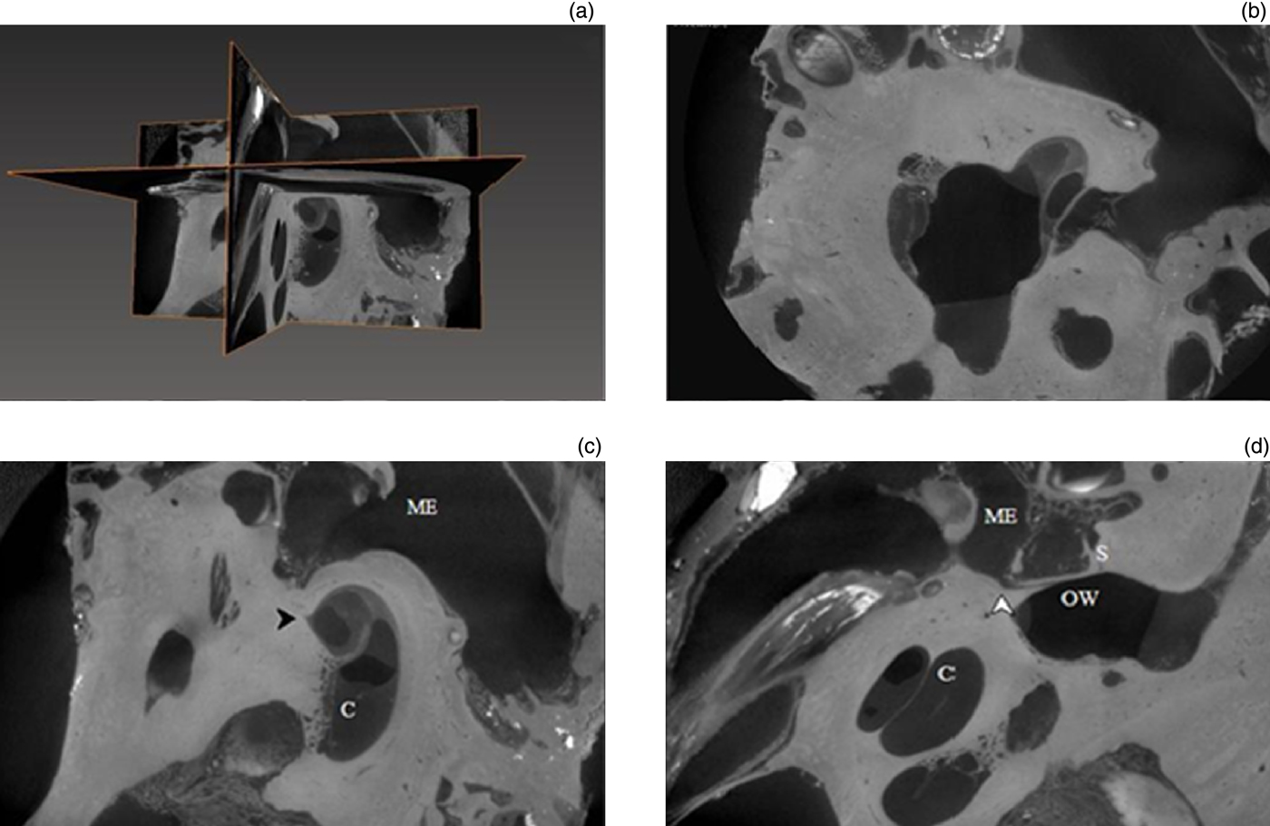
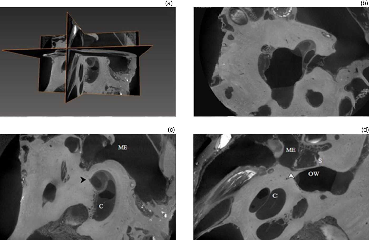
Figure 1 shows orthogonal slices selected from microtomography images of an otic capsule that had a rudimentary pit arising from both the middle- and inner-ear ends of the fissula, without a channel to connect the two pits. In Figure 1(c), a rudimentary pit can be seen at the inner-ear end of the fissula at the vestibule. In Figure 1(d), the pit arises from the middle ear just anterior to the stapes and the oval window.
(a)–(d) X-ray microtomographs of the fissula ante fenestram. As observed in part (c), the inner-ear end of the fissula forms a rudimentary pit or indentation (black arrowhead) within the vestibule, adjacent to the cochlea (C). In part (d), the middle-ear (ME) end of the fissula forms a pit (white arrowhead) anterior to the stapes (S) and oval window (OW).
Partial fissula
We characterised a partial fissula when we could visualise a distinct channel, rather than a pit, within the bone of the labyrinthine capsule. In these specimens, the fissula was not a continuous structure traversing the entire depth of the otic capsule.
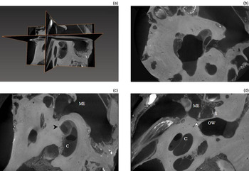
Figures 2, 3 and 4 display the orthogonal slices representing three separate examples of otic capsules with partial fissulae. In Figure 2, the inner-ear end of the fissula is arising from the vestibule and passing through the bone of the otic capsule in the direction of the middle ear. Figure 2(d) demonstrates the partial channel within the otic capsule, with its origin from the middle ear just anterior to the stapes and oval window.
(a)–(d) X-ray microtomographs of the partial fissula ante fenestram. The black arrowheads in parts (b) and (c) indicate the inner-ear end of the fissula ante fenestram; this is a channel arising from the vestibule (V) adjacent to its junction with the scala vestibuli. The fissula passes through the otic capsule towards the middle ear (ME). Part (d) shows the partial channel within the otic capsule; it originates from the middle ear (ME), is anterior to the stapes (S) and oval window (OW) and proceeds towards the cochlea (C).

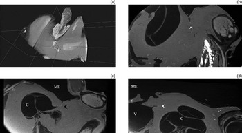
Figure 3 shows another example of a partial fissula ante fenestram. In Figure 3(c), a large portion of the middle-ear end of the fissula can be seen. The channel has a thin, tapered end arising in close proximity to the anterior extent of the oval window, and widens out within the bone of the otic capsule, almost reaching the inner ear. We stained this bone with gadolinium prior to scanning, and there is enhancement within the wall of the fissula, suggesting the presence of a thin, soft tissue lining with a patent core. A small segment of the inner-ear end of the fistula arising at the vestibule can be seen in parts (b) and (d) of Figure 3.
(a)–(d) X-ray microtomographs of the partial fissula ante fenestram. In parts (b) and (d), the black arrowheads indicate the small inner-ear end of the partial fissula, which arises from the vestibule (V) near the hook region of the cochlea (C). The white arrowhead in part (c) indicates the middle-ear (ME) end of the fissula. A significant channel arises in close proximity to the anterior limit of the stapes (S) footplate. Although starting off as a tapered point, the channel expands within the otic capsule and continues towards the vestibule and cochlea (C). The lining of the fissula demonstrates enhancement and preferential uptake of staining with gadolinium surrounding a central patent core.

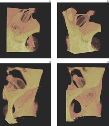
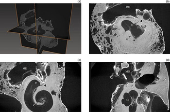
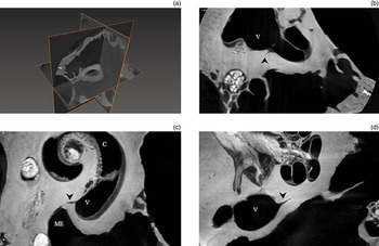
Figure 4 displays a third example of a partial fissula ante fenestram. Figure 4(d) shows the tympanic end arising anterior to the oval window. Parts (b) and (d) of Figure 4 highlight the course of the fissula within the labyrinthine capsule from its origin at the vestibule of the inner ear. Figure 5 demonstrates the volume-rendered model of the fissular region featured in Figure 4.
(a)–(d) X-ray microtomographs of the partial fissula ante fenestram. In parts (b) and (c), the black arrowheads indicate the partial fissula ante fenestram; its inner-ear end arises from the vestibule (V) and continues towards the middle ear (ME). In part (d), the fissula arises in close proximity to the stapes footplate (S) (white arrowhead). Its appearance as a discrete, patent channel within the bone has the irregular appearance of a microfissure running adjacent to it through the otic capsule.

(a)–(d) Volume-rendered, three-dimensional model of the partial fissula ante fenestram. The model shows the relationship between the partial fissula, middle-ear cavity, stapes and vestibule. The fissula ante fenestram appears as a curvilinear cleft within the otic capsule, with its long axis rotating through 90 ° during its course. In this example, the fissula almost reaches its middle-ear orifice, and its inner-ear orifice is widely patent and visible in the vestibule, anterior to the oval window.

Complete but occluded fissula
We determined a complete fissula ante fenestram if we could visualise a definitive channel in its entirety extending from the inner ear to the middle ear. We found that in some examples, the core of the fissula contained connective tissue that had become occlusive at a point along its course. This tissue enhanced on imaging of specimens stained with gadolinium and did not appear to be composed of bone or dense cartilage.
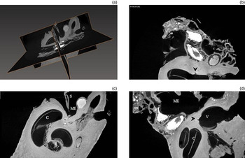
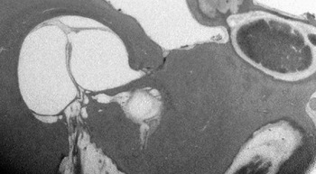
Figure 6 illustrates an example of a complete, but occluded fissula ante fenestram. The complete course of the fissula is best displayed in part (c) of Figure 6, where the fissure is continuous between the inner-ear end at the vestibule and the orifice at the middle-ear surface. There is occlusion of the channel by soft tissue which enhances following staining with gadolinium. Parts (b) and (d) of Figure 6 present the middle-ear end of the fissula as a cleft within the otic capsule. The same microtomography data used in Figure 6 generated Figure 7; however, we altered the contrast settings to better demonstrate the well-defined fissula ante fenestram.
(a)–(d) X-ray microtomographs of the complete but occluded fissula ante fenestram. As observed in part (c), the entire course of the fissula through the otic capsule is a continuous channel (black arrowhead) with openings directly into the vestibule, close to the scala vestibuli of the cochlea (C), and into the middle-ear cavity (ME). The fissula is occluded by connective tissue but not bone or cartilage. Parts (b) and (d) show the cleft-like fissula (white arrowhead) and its relationship to the middle ear (ME), stapes (S), vestibule (V) and cochlea (C).

X-ray microtomograph of a complete fissula ante fenestram.

Complete and patent fissula
The final category in the proposed classification of the morphology of the fissula ante fenestram is the complete and patent fissula, and this is of most interest due to the potential relationship between a patent channel (between the inner and middle ear) and the clinical entity of perilymph fistula.
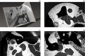
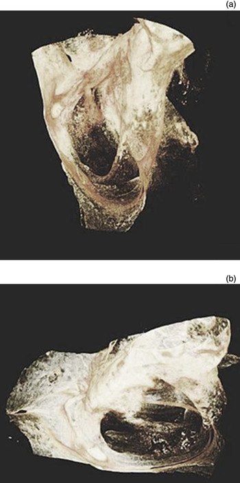
Figure 8, part (c), demonstrates an uninterrupted fissula arising from the vestibule, running a curvilinear course and entering the middle ear cavity anterior to the stapes. Its core does not contain any evidence of bony, cartilaginous or connective tissue obstruction making it a potential pathway for perilymph leakage. Figure 9 shows a volume-rendered model of the fissular region generated from a data set obtained by imaging the specimen shown in Figure 8 with a higher-powered (4 ×) objective lens.
(a)–(d) X-ray microtomographs of the complete patent fissula ante fenestram. In part (c), the black arrowhead indicates the complete fissula within the otic capsule, which begins at the vestibule (V), near the hook region of the cochlea (C), and enters the middle-ear cavity (ME), anterior to the stapes (S). In parts (b) and (d), the arrowheads again highlight the vestibular (black arrowhead) and tympanic (white arrowhead) portions of the fissula.

(a) and (b) Volume-rendered, three-dimensional model encompassing the stapes and oval window, and the region just anterior to it; this contains the tympanic orifice of the fissula ante fenestram.

Figure 10 is a second complete and patent fissula we identified in a separate temporal bone. As with the previous example, there is no obstruction within the core of the channel; note that the overall course and shape of the fissula is similar between specimens.
(a)–(d) X-ray microtomographs of the complete patent fissula ante fenestram. As shown in parts (b)–(d), the completely dehiscent fissula ante fenestram (black arrowheads) forms a communication between the perilymph-filled vestibule (V) in the inner ear and the air-filled middle ear (ME). C = cochlea

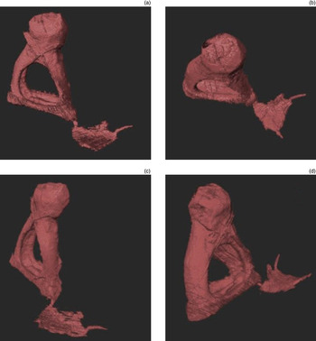
To complete the 3D micromodelling of the fissula ante fenestram using X-ray microtomography, we segmented the fissula as a region of interest by labelling the structure for each individual projection along with the stapes that represented a consistent landmark. We then applied isosurface rendering to these labels. An example of the models is shown in Figure 11.
(a)–(d) Isosurface-rendered, three-dimensional model demonstrating the morphology of the fissula ante fenestram and its anatomical relationship to the stapes.

The models demonstrate the structural morphology of the fissula, which although showing minor variation between specimens in terms of size, had a fairly consistent origin and course. Of importance to otologic surgery, in every otic capsule examined the fissula ante fenestram had a consistent anatomical relationship to the anterior limit of the stapes and oval window. We observed the path of the fissula through the otic capsule rotating along its long axis, and having a wing-like appearance with an expanded middle portion between its narrow ends at the tympanic and vestibular surfaces.
In this preliminary study, we imaged and analysed a total of 15 temporal bones. Seven of the bones contained fissular morphology that could be categorised into the proposed classification system: rudimentary pit; partial fissula; complete but occluded fissula; and complete and patent fissula. The most illustrative examples have been used in the figures.
Discussion
A perilymph fistula is a clinical entity where an abnormal communication between the fluid-filled inner ear and air-filled middle ear allows the leak of perilymph.Reference Kohut1, Reference Friedland and Wackym2
The majority of cases of perilymph fistula occur following trauma,Reference Minor8 surgery such as stapedectomy or cochlear implantation, or from infection, cholesteatoma, granuloma and neoplasm.Reference Friedland and Wackym2 In contrast, the diagnosis of spontaneous perilymph fistula is less common and remains contentious.Reference Friedland and Wackym2, Reference Shea9 Much of the debate has been due to incomplete understanding of the anatomical and pathophysiological bases of perilymph fistulae. The identification and description of a pathway of perilymph leakage was therefore considered of potential research interest.
The postulated pathogenesis of fistulae involves perilymph leakage at developmental deficiencies or microfissures within the labyrinthine capsule.Reference Friedland and Wackym2, Reference Harada, Sando and Myers4, Reference Okano, Myers and Dickson5 This hypothesis has been supported by findings at exploratory tympanotomy, and by predictive histological temporal bone studies.Reference Kohut, Hinojosa, Howard and Ryu10 The frequent identification of perilymph accumulation at specific sites, especially anterior to the oval window and at the floor of the round window niche, in patients with hearing loss and vertigo have led to the conclusion that perilymph fistulae occur most often at the fissula ante fenestramReference Harada, Sando and Myers4 and at the fissure of the round window niche – the posterior semicircular canal ampulla.Reference Okano, Myers and Dickson5 In a series of over 200 cases of exploratory tympanostomies, Seltzer and McCabeReference Seltzer and McCabe11 reported that over half of the identified fistulae occurred in the region of the oval window, and within this group 67 per cent were located anteriorly, at the fissula ante fenestram.
With the established clinical data, histological temporal bone studies have been driven to identify evidence of otic capsule microfissures. Researchers led by KohutReference Kohut, Hinojosa, Howard and Ryu10, Reference Kohut, Hinojosa and Ryu12 concluded that histological otic capsule patencies could be anticipated on the presence of fistula symptoms during life,Reference Kohut, Hinojosa, Howard and Ryu10 and conversely, the presence of a histologically identifiable capsule patency led to a fourfold increased chance of having a clinical disorder of hearing or balance during life.Reference Kohut, Hinojosa and Ryu12
While histological examination of temporal bones has been invaluable, there are inherent problems with the destructive nature of specimen processing, including decalcification, embedding and sectioning, and the dehydration effects of alcohol-based preservatives and stains.Reference Uzun, Curthoys and Jones7 Some researchers have questioned the validity of observed otic capsule dehiscence and attributed them to artefact.
X-ray microtomography allows non-destructive examination, with objective lenses that micro-focus photon beams to create projections at a resolution of image voxel size less than 1 µ. This is comparable to that seen in traditional histology and significantly more powerful than clinical high-resolution computed tomography.Reference Uzun, Curthoys and Jones7 The stacked serial microtomography projections allow tracking and 3D modelling of long, tortuous structures, such as the fissula ante fenestram, which may not be possible with individual histological sections due to the potential loss of information between sections.
The method of specimen preparation used for this study allowed the entire otic capsule to remain intact. The specimens had minimal chemical processing with preservatives and stains that do not alter the compositions of the bony and soft tissues,Reference Anniko and Lundquist13 and the structures within the otic capsule remained in their natural anatomical configurations allowing direct extrapolation to surgical anatomy.
This study found that both forms of primary fixation (formaldehyde with polyethylene glycol, or Karnovsky's fixative) were effective in preserving both osseous and soft tissue structures of the temporal bones. The PEGylated bones could not be stained but had the advantage of being easy to handle and able to be stored without special precautions. The Karnovsky's fixative allowed post-fixation staining, but the specimens required careful handling with protective equipment and suspension in buffered solution within airtight containers for storage. Considerations were made as to the characteristics and thickness of the walls of the containers so as not to interfere with or degrade image quality.
With its known uses in traditional light and electron microscopy techniques, osmium tetroxide was selected due to its preservation of and deposition in delicate membranous structures in the inner ear, even in dense surrounding bone, due to its affinity for double-bond configurations in lipid. Gadolinium was used as a contrast agent, similar to its use in clinical radiography, due to its ability to attenuate X-rays. Again, this provided additional enhancement of the soft tissue against the bony components.
To date, the understanding of the morphology of the fissula ante fenestram has been provided almost exclusively from histological examination. The fissula has been described as an appendage of the perilymphatic labyrinth, forming a slit-like channel traversing the bony otic capsule between the oval window and cochlea.Reference Anson and Donaldson6 Within the microtomography data set from this study, the fissula ante fenestram was consistently identified at the location described by the histological data – the middle-ear (or tympanic) end begins posteroinferior to the cochleariform process, just anterior to the oval window, and the inner-ear (or vestibular) end lies at the junction of the vestibule with the scala vestibuli.Reference Dawes, Pearman and Kochilas14–Reference Anson and Martin17
In Anson and Cauldwell'sReference Anson and Cauldwell16 description of the fissula ante fenestram, they concluded that ‘variations in fissular shape and extent are multifold’ (p. 671); however, there has not been any established classification of fissular morphology. From the data in this study, a new classification has been proposed, based on the extent and patency of the fissula ante fenestram. Four categories have been stratified: rudimentary pit; partial fissula; complete occluded fissula; and complete patent fissula. Anson and MartinReference Anson and Martin17 observed in some specimens the occurrence of incomplete fissulae. They also reported in one specimen there being no distinct fissula, but an indentation of connective tissue at the expected sites of the tympanic and vestibular orifices. Examples of these findings are present in our microtomography data, and have been classified as rudimentary pits and partial fissulae depending on their extent.
The 3D models generated from complete fissula ante fenestram captured on microtomography demonstrate an irregular curvilinear cleft with narrow, tubular ends and an expanded middle portion on the same axis as the base of the stapes. Consistent with existing data,Reference Anson and Cauldwell16, Reference Anson and Wilson18, Reference Wilson19 the long axis of the fissula was seen to rotate through 90 ° during its course through the otic capsule, with the middle-ear end elongating in the horizontal plane before curving around and descending vertically to its vestibular termination.
The composition of the core of the fissula ante fenestram in adults has been widely debated. PerozziReference Perozzi20 first described it as being composed of dense cartilage. BastReference Bast15 and Anson and MartinReference Anson and Martin17 reported the fissula as being composed centrally of loose reticular and vascular connective tissue that became denser at the periphery, with hyaline cartilage at the orifices of the fissula. In a later report, Anson and CauldwellReference Anson and Cauldwell16 described the core of the fissula ante fenestram as being composed of necrotic cartilage and loose vacuolated mesenchyme with plugs of fibroblastic tissue. WilsonReference Wilson19 proposed that the connective tissue core became progressively replaced by cartilage or bone with age. He thought the process was dependent on blood supply from the middle ear and, importantly, that it could be interrupted.
Kohut et al.Reference Kohut, Hinojosa and Ryu12 examined the temporal bones of patients who in life had surgically proven perilymph fistula, and found loose connective tissue with large intercellular spaces forming the entire core of the fissula ante fenestram and extending to the endothelial lining of the vestibule. They compared this appearance to that of the perilymph-filled spiral ligament, and described the absence of bony or cartilaginous obstruction along the extent of the patent fissula.
From microtomography examination of the fissula ante fenestram, it can be demonstrated that a complete fissula may be occluded by loose connective tissue, with evidence of preferential uptake in specimens stained with gadolinium. The ends of the fissulae were widely patent, which is consistent with the report from Kohut et al.Reference Kohut, Hinojosa and Ryu12 Moreover, examples of fissula ante fenestram that were free from bony, cartilaginous or connective tissue obstruction from the inner to middle ear were also observed in the microtomography analysis. We postulate that these could be permeable to perilymph and present a prospective pathway for perilymph fistula.
A significant question remains regarding the potential relationship between a patent microfissure and the clinical entity of perilymph fistula. Microfissures are common histopathological findings, with Harada et al.Reference Harada, Sando and Myers4 reporting a 25 per cent prevalence of fissures in over 300 temporal bones examined, yet the clinical diagnosis of spontaneous perilymph fistula is uncommon. This suggests that the relationship is not straightforward – small patencies may be impermeable, leakage can be inactive or sporadic, and fistulae with slow leaks may remain asymptomatic. Therefore, there are likely other pathological factors that precipitate fistula formation and associated symptoms.Reference Friedland and Wackym2, Reference Cole21, Reference Grundfast and Bluestone22
• X-ray microtomography allows non-destructive, three-dimensional imaging and modelling of the delicate structures within the otic capsule
• We used microtomography to characterise the morphology of the fissula ante fenestram and then proposed a new classification system that includes a rudimentary pit, partial fissula, complete occluded fissula and complete patent fissula
• The core of the fissula ante fenestram appears to be composed of connective tissue that may be permeable to perilymph
• This finding supports the postulated theory of the fissula ante fenestram being a potential site for perilymph leakage
One hypothesis is that leakage through microfissures occurs with sudden and transient increases in perilymph pressure. Transmission of fluctuating cerebrospinal fluid pressures to the perilymphatic space may be a contributing factor.Reference Cole21 In our analysis, one of the examined specimens contained both a complete patent fissula ante fenestram and an abnormally wide cochlear aqueduct.
A limitation of the presented research on the fissula ante fenestram is the lack of clinical and demographic data available on the patients from whom the temporal bones were harvested as they were de-identified prior to processing. Therefore, no comment can be made on whether the patencies were observed in patients with fistula symptoms during life and at what age these were present. However, improvement and refinement of the understanding of the pathophysiological mechanism underlying perilymph fistulae has implications on clinical diagnosis and management.
Conclusion
X-ray microtomography is an effective method of temporal bone examination that allows non-destructive, 3D modelling of otic capsule deficiencies, in particular the morphology of the fissula ante fenestram and its anatomical relationship with the stapes, oval window and vestibule. Microtomography as a research tool has significant advantages over traditional histological examination, including avoidance of the artefactual effects of specimen dissection and processing.
With this work, we propose a new structural classification of the fissula ante fenestra in adults, one that is based on extent and patency of the channel. The fissula can range from rudimentary pit to complete channel extending from the inner ear at the junction of the vestibule and scala vestibuli to the middle ear just anterior to the oval window. We have shown the core of the fissula to be composed of connective tissue, rather than bone or dense cartilage, and there were examples of widely patent fissulae with permeable cores.
The presence of a dehiscent channel through the otic capsule provides support to the model of perilymph leakage at the site of the fissula ante fenestram. Greater understanding of the anatomical and pathophysiological mechanisms underlying spontaneous perilymph fistulae may lead to improvements in accuracy of diagnosis, planning of surgical repair and post-operative outcomes for hearing and vestibular function.