I. INTRODUCTION
Thermolysis of hydrated and/or organometallic compounds is a well-known preparation route for new derivatives, intermediate or anhydrous forms of inorganic compounds, possibly metastable, usually obtained as fine powders. The full crystallographic characterization of such materials can be difficult, requesting expertise in the most up to date powder diffraction methodologies. Many crystal structures of new fluoride compounds that were prepared according to such routes or to more classical solid-state approaches, were determined ab initio from the powder diffraction data. The list includes fluoride compounds with a binary chemical formulation such as SF6 (Cockroft and Fitch, Reference Cockcroft and Fitch1988), τ-AlF3 (Le Bail et al., Reference Le Bail, Fourquet and Bentrup1992), ReF7 (Vogt et al., Reference Vogt, Fitch and Cockroft1994), and C6F10 (Smrcok et al., Reference Smrcok, Mach and Le Bail2013). Such discoveries of new binary compounds are especially interesting owing to their rarefaction: the ReF7 crystal structure being published in Science, τ-AlF3 being later “post-predicted” together with other yet unknown possible AlF3 polymorphs (Le Bail and Calvayrac, Reference Le Bail and Calvayrac2006). New ternary compounds are logically a bit more frequently produced, some crystal structures being also solved from the powder diffraction data, such as Li2TbF6 (Laligant et al., Reference Laligant, Le Bail, Férey, Avignant and Cousseins1988), β- and γ-BaAlF5 (Le Bail et al., Reference Le Bail, Férey, Mercier, De Kozak and Samouël1990b), polymorphs of CsAlF4 (Bentrup et al., Reference Bentrup, Le Bail, Duroy and Fourquet1992), β-Ba3AlF9 (Le Bail, Reference Le Bail1993) a complex one with 29 independent atoms, ThZr2F12 (Taoudi et al., Reference Taoudi, Mikou and Laval1996), Ba2ZrF8 (Le Bail and Laval, Reference Le Bail and Laval1998), θ-KAlF4 (Le Bail, Reference Le Bail2009a), and NaAlF4 (Le Bail, Reference Le Bail2009b), to give a few examples, and there is no place here for citing quaternary compounds. In the same A2MF5 family (A I = K, Rb, Cs, and NH4; M III = Al, Cr, V, Fe, and Ga) as the title compounds, α- and β-(NH4)2FeF5 (Fourquet et al., Reference Fourquet, Le Bail, Duroy and Moron1989) were the results of the thermal decomposition of (NH4)2FeF5•H2O, their structures were also solved from the powder diffraction data. On continuing such studies, the thermal dehydration of K2FeF5•H2O was found to lead to two new K2FeF5 forms, one of them being characterized structurally ab initio in this paper by powder diffraction methodologies having demonstrated their efficiency during several recent blind tests (Le Bail et al., Reference Le Bail, Cranswick, Adil, Altomare, Avdeev, Cerny, Cuocci, Giacovazzo, Halasz, Lapidus, Louwen, Moliterni, Palatinus, Rizzi, Schilder, Stephens, Stone and van Mechelen2009).
II. EXPERIMENTAL
The new K2FeF5 forms are the products of monohydrate thermolysis, which was reported by Bentrup (Reference Bentrup1991) to lead to an orthorhombic form at 135 °C. It is shown here that this orthorhombic form (noted III) is not the high-temperature solid-state one (Le Bail et al., Reference Le Bail, Desert and Fourquet1990a) belonging to the Pbcn space group (noted form-I), nor the hydrothermally synthesized form (Fourquet and Duroy, Reference Fourquet and Duroy1993) crystallizing in the Pbam space group (noted form-II), both these known phases being characterized from single crystal data. Moreover, a phase transition from form-III to a fourth form is observed at 412 °C, which finally undergoes a phase transition to the normal Pbcn HT form-I at 580 °C. The powder diffraction patterns of K2FeF5 forms III and IV could be recorded at room temperature. Note that form-II transforms at 190 °C irreversibly to K3FeF6 + KFeF4 (Fourquet and Duroy, Reference Fourquet and Duroy1993).
III. INDEXING, STRUCTURE DETERMINATIONS, AND RIETVELD REFINEMENTS
A. K2FeF5 form-III
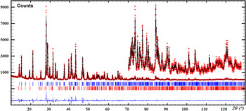
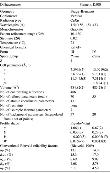
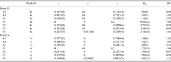
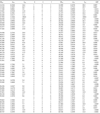
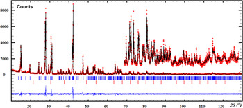
At the identification stage by search-match procedures applied to the powder diffraction pattern recorded at room temperature, by using the ICDD-PDF database, the best match was from K2VO2F3 (see PDF entry 04-009-3298; PDF, 2012) (Ryan et al., Reference Ryan, Mastin and Reisfeld1971). Rb2CrF5 (Jacoboni et al., Reference Jacoboni, De Pape, Poulain, Le Marouille and Grandjean1974) belongs to the same crystal structure type. Thus, the Rietveld (Reference Rietveld1969) refinement, using the FULLPROF software (Rodriguez-Carvajal, Reference Rodriguez-Carvajal1993) was made starting from the Rb2CrF5 atomic coordinates (Pnma space group), and readily converged to satisfy R values. In order to improve the sample crystallinity, it was maintained at 400 °C just before the form-III → form-IV transition, nevertheless, a small amount of form-IV had to be considered in the final Rietveld refinement (Figure 1), with fixed atomic coordinates, after the form-IV structure was solved. The Rietveld refinement details, the atomic coordinates, selected geometric parameters, and X-ray diffraction data are shown in Tables I–IV, respectively.
(Color online) Rietveld plot for K2FeF5 form-III (CuKα). The second phase corresponds to a 6.1(2)% contribution of form-IV starting to appear. A third phase remains unidentified, showing a main peak at 23.2° (2θ).

Experimental and Rietveld refinement details for the K2FeF5 forms III and IV.

Fractional atomic coordinates and isotropic displacement parameters (Å2) at 23 °C for K2FeF5 forms III and IV in the Pnma and C2/m space groups, respectively; BV is the bond-valence calculated according to Brese and O'Keefe (Reference Brese and O'Keefe1991).

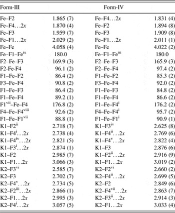
Selected interatomic distances (Å) and angles (°) for the K2FeF5 forms III and IV at 23 °C.

Symmetry codes – Form-III : (i) −x+1, y + 1/2, and −z + 1; (ii) x − 1/2, −y + 1/2, and −z + 1/2; (iii) −x + 1, y − 1/2, and −z + 1; (iv) x − 1, y, and z; (v) x − 1, −y + 1/2, and z; (vi) x − 1/2, −y + 1/2, and −z + 3/2; (vii) −x + 3/2, −y, and z + 3/2; (viii) x + 1/2, y, and −z + 1/2; (ix) −x + 3/2, −y − 1, and z + 3/2; Form-IV : (i) x, −y, and z; (ii) −x + 1/2, y − 1/2, and −z + 1; (iii) −x + 1/2, y + 1/2, and −z + 1; (iv) −x + 1, y, and −z + 1; (v) x + 1/2, y − 1/2, and z + 1; (vi) −x, y, and −z + 1; (vii) x, y, and z + 1; (x) x, −y, and z + 1.
X-ray diffraction data for K2FeF5 form-III at 23 °C (CuK α1).

B. K2FeF5 form-IV
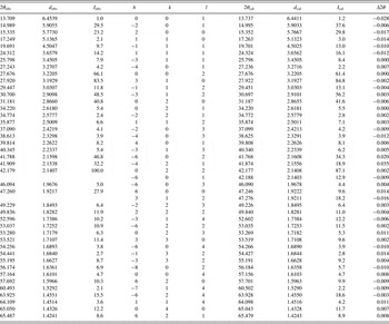
The search-match process was negative for that sample quenched at 500 °C. Indexing was realized by using the McMaille software (Le Bail, Reference Le Bail2004), leading to a C-centered monoclinic cell. The Figures of Merit (FoMs) were M 20 = 55.9 (de Wolff, Reference de Wolff1968), F 20 = 79.5 (0.0043, 58) (Smith and Snyder, Reference Smith and Snyder1979), and McM 20 = 169.9 (Le Bail, Reference Le Bail, Clearfield, Reibenspies and Bhuvanesh2008). The cell volume was similar to that of form-III. On using Le Bail fitting (Le Bail, Reference Le Bail2005) for evaluating the reliability of the indexing and for intensity extraction, the C2/m, C2, or Cm space groups were found to be the most probable. The structure solution was carried out in direct space by using the ESPOIR software (Le Bail, Reference Le Bail2001), moving in the cell the appropriate number of K, Fe, and F atoms by a Monte Carlo process. A satisfying solution allowing us to recognize [FeF6] octahedra was obtained in the C2/m space group, which was confirmed by a Rietveld refinement using FULLPROF (Figure 2). Traces of KFeF3 were identified indicating some Fe3+ → Fe2+ disproportionation; another impurity in a very small proportion could not be defined. The Rietveld refinement details, the atomic coordinates, and selected geometric parameters are shown in Tables I–III, respectively, and the X-ray diffraction data are in Table V.
(Color online) Rietveld plot for K2FeF5 form-IV (CuKα). The second phase corresponds to 3.4(1)% of the cubic KFeF3 phase. A third phase remains unidentified, showing weak reflections between ~15° and 20° (2θ).

X-ray diffraction data for K2FeF5 form-IV at 23 °C (CuK α1).

IV. STRUCTURE DESCRIPTION AND CORRELATIONS WITH RELATED COMPOUNDS
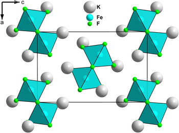
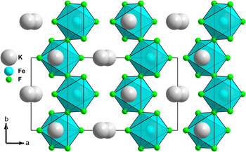
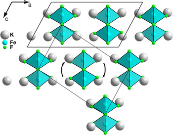
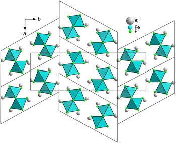
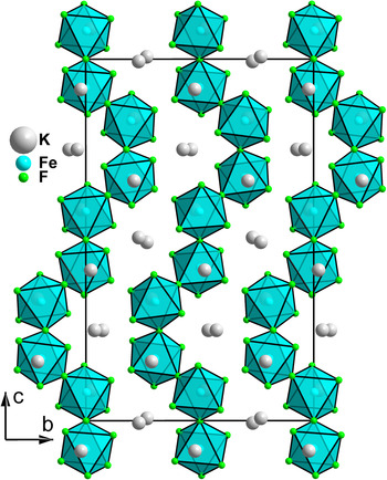
The form-III structure (Figure 3) is very common since it is adopted by Rb2 M IIIF5 (M III = Cr, Fe, and Ga) and Cs2FeF5, as well as by some oxyfluoride compounds. MF6 octahedra are connected by cis-corners to form infinite (FeF5) n 2n− zigzag chains. Calculated bond valences reported in Table II for both forms III and IV are close to the expected values (maximum difference attaining 12%), indicating that the unrestrained Rietveld refinements from laboratory powder data have led to medium quality results. The bridging Fe–F distance (2.029 Å in form-III and 2.011 Å in form-IV) is longer than the terminal ones 1.864 < Fe–Fterm < 1.960 Å in form-III and 1.831 < Fe-Fterm < 1.909 Å in form-IV; the bridging Fe–F–Fe angle is 180° in both forms. The same cis-chains of the FeF6 octahedra are the characteristic of K2FeF5 form-II hydrothermally synthesized (Fourquet and Duroy, Reference Fourquet and Duroy1993) but the mutual orientation of the chains is different leading to a doubling of the longer axis (b II = 23.0897 Å ~2 × c III = 11.544 Å), the two other cell parameters being very similar (Table VI). All of the potassium ions are tenfold coordinated in both forms II and III, with a clear cut of the K–F distances. In form-IV all of the potassium ions are also tenfold coordinated (they have coordination 9, 10, and 11 in form-I) and the same zigzag cis-connected (FeF5) n 2n− chains (Figure 4) appear to present again a different mutual orientation (Figure 5). A topotactic reaction hypothesis looks highly plausible to explain the form-III → IV transition : it is sufficient to rotate half the chains accompanied by the potassium ions nearest neighbors by 90° along the b-axis (arrows in Figure 5), and to translate the chains by 1/2 b in order to make form-III appear back from form-IV (but since the reaction is irreversible, the inverse moves have to be considered). Polymorphic transitions were also proposed to be topotactic and a similar displacive mechanism was suggested for the α, β, and γ-BaAlF5 phases characterized by the same cis-connected octahedral chains, some of these structures being also solved from the powder data (Le Bail et al., Reference Le Bail, Férey, Mercier, De Kozak and Samouël1990b). K2FeF5 form-II (Figure 6) may be described as containing kink-folded blocks related to form-IV, and if the junction zone between these blocks is examined, one recognizes similarities with form-III (Figure 3). All of the four K2FeF5 forms are characterized by cis-connected octahedral chains, and their V/Z ratios at room temperature (Table VI) are almost identical. The periodicity is of N = 2 octahedra for forms II, III, and IV, corresponding to the shortest cell parameter close to 5.7 Å (Table VI), whereas form-I shows N = 8 (Figure 7) so that the phase transition IV → I at 580 °C looks to require a much larger atomic displacement amplitude than for the III → IV transition. A different organization with N = 4 was established for α-(NH4)2FeF5 (Fourquet et al., Reference Fourquet, Le Bail, Duroy and Moron1989), also produced by dehydration and solved from the powder data, so that the current number of structure types for the A2MF5 compounds presenting cis-connected octahedral chains is five. Concerning K2FeF5, only form-I is yet characterized magnetically, however the three other forms should logically share the same characteristic one-dimensional antiferromagnetic model (Chelkowski and Wijn, Reference Chelkowski and Wijn1995). Other A2 MF5 structural types not considered here, characterized by trans-connections leading to linear octahedral chains, are described elsewhere (Babel and Tressaud, Reference Babel, Tressaud and Hagenmuller1985).
(Color online) Projection along the short b-axis of the structure of K2FeF5 form-III.

(Color online) View of the structure of K2FeF5 form-IV along the c axis, showing the infinite cis-connected octahedral chains. A view of form-III along its a-axis would be quite similar.

(Color online) Projection of the structure of K2FeF5 form-IV along the b axis, to be compared to Figure 3 in order to recognize how the rotation of half of the chains (see the arrows) and their translation of 1/2 b would allow us to rebuild the form-III structure (thin lines).

(Color online) Projection of the structure of K2FeF5 form-II along the c axis, to be compared to Figure 5 in order to recognize kink folded blocks of the form-IV monoclinic cell (thin lines) which reproduce also the form-III arrangement at their junctions.

(Color online) Projection of the structure of K2FeF5 form-I along the a axis, showing a N = 8 periodicity along the octahedral chains, to be compared to Figure 4 showing N = 2 for form-III (and also for forms II and IV).

Summary of the crystallographic details of the four K2FeF5 forms.

V. CONCLUSIONS
Two additional K2FeF5 forms are disclosed: one belonging to the Rb2FeF5 type and the other establishing a new A2MF5 structural type. With four different forms, K2FeF5 appears to be the richest polymorphic composition of all of the A2 I M IIIF5 (A I = K, Rb, Cs, Tl, and NH4; M III = Al, Cr, Mn, Fe, and Ga) series. All of these forms are showing cis-connected (FeF5) n 2n− octahedral chains. The role of powder diffraction in the elucidation of the crystal structures of such compounds obtained by thermolysis is absolutely pivotal since the possibility of suitable single crystal growth is generally excluded.
ACKNOWLEDGMENTS
The author thanks H. Duroy and J.L Fourquet (both retired) for the preparation of the samples and the recording of the powder diffraction data, twenty years ago.
SUPPLEMENTARY MATERIALS AND METHODS
The supplementary material referred to in this article can be found online at http://www.journals.cambridge.org/pdj.