


An appropriate balance between protein synthesis and degradation is essential for the maintenance of skeletal muscle mass. Protein degradation in particular plays an important role in normal muscle growth(Reference Goldspink1,Reference Goldspink and Goldspink2) and is primarily regulated by the ubiquitin–proteasome system(Reference Jagoe and Goldberg3–Reference Lecker, Goldberg and Mitch5). The suppression of ubiquitin–proteasome system-dependent protein degradation has been reported to contribute to both the maintenance of and increases in skeletal muscle mass(Reference Tawa, Odessey and Goldberg6–Reference Baptista, Leal and Artioli8).
The rate-limiting step of ubiquitin–proteasome system is the regulation of E3 ubiquitin ligases, which determine substrate specificity within the system(Reference Hershko and Ciechanover9). In skeletal muscle, two muscle-specific E3 ubiquitin ligases, muscle atrophy F-box (Atrogin-1/MAFbx) and muscle RING finger 1 (MuRF-1), play central roles in the control of protein breakdown(Reference Bodine, Latres and Baumhueter10,Reference Sandri, Sandri and Gilbert11) . Atrogin-1/MAFbx expression is transcriptionally upregulated during muscle atrophy and is regulated by forkhead boxO (FoxO) transcription factors(Reference Sandri, Sandri and Gilbert11), and MuRF-1 targets major myofibrillar proteins, such as myosin heavy chain for ubiquitination and subsequent degradation(Reference Bodine, Latres and Baumhueter10).
Rice bran, a byproduct of the rice milling process, comprises several cellular layers, including the pericarp, tegmen and aleurone, and is rich in fibre(Reference Shi, Mazza and Maguer12). The outer layer of rice bran mainly comprises the pericarp, which is richer in lignin than the inner layer, and the amounts of pectic substances, hemicellulose and α-cellulose present in the inner and outer layers are similar(Reference Shibuya, Nakane and Yasui13).
Multi-break milling systems have recently become prevalent in the rice industry. In such systems, multiple milling machines are used to remove more bran from the rice kernels after the initial break. In a previous study, we collected the outer bran fraction of rice (OBFR) from the first and second breaks of a commercial quadruple-break milling system and found that feeding a diet containing this fraction altered the concentrations of metabolites in the cecal contents, plasma and livers of rats(Reference Kawaguchi, Nishikoba and Shimamoto14) and prevented lipid accumulation by enhancing faecal lipid excretion(Reference Ijiri, Nojima and Kawaguchi15).
In the present study, we collected the inner bran fraction of rice (IBFR) from the third and fourth breaks of a commercial quadruple-break milling system. First, we examined the effects of dietary supplementation with either IBFR or OBFR on the growth performance and tissue weight of rats and found that IBFR consumption increased the skeletal muscle weight of the rats, possibly through the suppression of muscle protein breakdown. Then, based on these findings, we further investigated the mechanisms of this effect by feeding rats a protein-deficient (PD) diet, which induces muscle protein catabolism(Reference Sugawara, Ito and Nishizawa16), and supplemented their diet with either an aqueous extract (AE) or a hexane extract (HE) of IBFR. To determine their effects on the metabolism of the rats, we performed an untargeted metabolomic analysis using GC–MS and a targeted quantitative analysis of amino acids using HPLC with ortho-phthalaldehyde derivatisation and fluorescence detection. Moreover, we conducted a peptidomic analysis of the AE in IBFR to assess whether peptides are involved in metabolic alterations.
Materials and methods
Preparation of outer bran fraction of rice and inner bran fraction of rice
OBFR and IBFR were collected from a commercial rice milling plant (Shokkyo Co., Ltd) operating a quadruple-break milling system. In this study, two rice milling machines (RB40A, SATAKE Corporation), each equipped with two internal milling units, were connected in series. OBFR and IBFR were collected from the first and second machines, respectively. OBFR corresponds to approximately the 100–95 % fraction of brown rice by weight, whereas IBFR corresponds to approximately the 95–90 % fraction.
Preparation of experimental diets
To determine the effects of feeding IBFR or OBFR on the growth performance and tissue weights of rats, three experimental diets were prepared: (1) an AIN-93G-based control diet, (2) a diet containing 5 % (w/w) OBFR and (3) a diet containing 5 % (w/w) IBFR. The protein components of the three diets were adjusted to be equal. The chemical compositions (moisture, protein, fat, carbohydrate and ash) of OBFR and IBFR are shown in Supplementary Table S1. The ingredients and nutrient contents of the three diets are shown in Supplementary Table S2.
In Experiment 2, to determine whether the effects of IBFR could be ascribed to its hydrophilic or hydrophobic components, AE and HE were prepared as follows. To prepare AE, 100 g of IBFR was extracted with 300 mL of water by homogenising (NP-60, SATAKE MultiMix Corporation) for 3 min and then centrifuged at 5900 × g for 10 min at 4 °C. The supernatant was lyophilised and used as AE. For extraction of HE, 100 g of IBFR was extracted with 500 mL of n-hexane at room temperature for 2 h, with gentle stirring, then filtered, and the residue was re-extracted twice. The filtrates were then collected, vacuum-dried in a rotary evaporator at 50 °C, redissolved in 140 mL of corn oil and used as the HE. The chemical composition (moisture, protein, fat, carbohydrate and ash) of the AE and HE are shown in Supplementary Table S1. Four experimental diets were prepared: (1) an AIN-93G-based control diet, (2) a PD diet, (3) the PD diet containing 1 % AE of IBFR and (4) the PD diet containing 7 % HE of IBFR in maize oil. The PD diet was formulated according to Sugawara et al. (2009)(Reference Sugawara, Ito and Nishizawa16) with slight modification. The protein contents of the PD and AE and HE-containing diets were adjusted to be equal (0·38 %), and the compositions of the AE and HE of IBFR in each diet were adjusted to be equivalent to 5 % IBFR in Experiment 1. The ingredients and nutrient contents of the four diets are shown in Supplementary Table S2.
Experiment 1
Eighteen male Sprague–Dawley rats (7 weeks old; approximately 200 g) were purchased from Japan SLC Co., Ltd. They were individually housed in wire-bottomed aluminium cages (152 × 201 × 170 mm) in a temperature-controlled room (25 °C) and provided with water and the control diet ad libitum for 1 week prior to the experiment. The rats were monitored daily, and no signs of pain, suffering or distress were observed. All the procedures were performed according to the guidelines of the Animal Care and Use Committee of Kagoshima University, and the study was approved by this committee (approval number A16011).
At 8 weeks of age, the rats were randomly assigned to one of the three diet groups (n 6 rats per group) and fed the experimental diets for 21 d. Their body weights were recorded weekly, and their individual food intake was measured daily. At the end of the feeding period, they were anesthetised with carbon dioxide and euthanised by cervical dislocation. Their hearts, livers, kidneys, abdominal fat depots and soleus muscles were then collected and weighed. Blood samples were collected in heparinised tubes and centrifuged at 5900 × g for 10 min at 4 °C, and then plasma was collected and stored at −30 °C until analysed.
Experiment 2
Twenty-four male Sprague–Dawley rats (4 weeks old; approximately 45 g) were obtained from Japan SLC Co., Ltd. They were individually housed in the wire-bottomed cages at 25 °C and provided with water and the control diet ad libitum for 1 week prior to the experiment. Their health and welfare were checked daily, and no signs of distress were noted. All the procedures were performed according to the guidelines of the Animal Care and Use Committee of Kagoshima University, and the study was approved by this committee (approval number A21015).
At 5 weeks of age, the rats were randomly divided into four groups (n 6 rats per group) and fed one of the four experimental diets for 7 d. Their body weights were measured at the beginning and end of the experiment, and their individual food intake was recorded daily. At the end of this period, the rats were anesthetised with carbon dioxide and euthanised by cervical dislocation. Their hearts, livers, kidneys, abdominal fat depots and soleus muscles were then collected and weighed. Blood samples were collected in heparinised tubes and centrifuged at 5900 × g for 10 min at 4 °C, and the plasma was stored at −30 °C until analysed.
RNA extraction and quantitative real-time PCR
Soleus muscles were homogenised in ISOGENII (Nippon Gene), and RNA was extracted in accordance with the manufacturer’s instructions. Quantitative real-time PCR was performed as previously described(Reference Kawaguchi, Nishikoba and Shimamoto14). In brief, complementary DNA was synthesised from 60 ng of RNA per 10 µL of reaction solution using a PrimeScript RT Reagent Kit (RR036A, Takara). The samples were incubated at 37 °C for 15 min, 85 °C for 5 s and then 4 °C for 5 min. Gene expression levels were measured by real-time PCR using the 7300 Real-Time PCR system (Applied Biosystems) and SYBR Select Master Mix (Applied Biosystems). The thermal cycling conditions were 50 °C for 2 min, 95 °C for 2 min and then 45 cycles of 95 °C for 15 s, 55 °C for 15 s and 72 °C for 1 min. The following primers were used: Atrogin-1/MAFbx (5’-CCACATGTGGGTGTATCGAATGG-3’ and 5’-GCCTCTGCATGATGTTCAGTTGTAA-3’), MuRF-1 (5’-GGGAACGACCGAGTTCAGACTATC-3’ and 5’-GGCGTCCAAACTTGTGGCTCA-3’), FoxO1 (5’-CTTCCCGCACAGTGTCAAGA-3’ and 5’-AGGGGTGAAGGGCATCTTTG-3’), FoxO3a (5’-CGGACAAACGGCTCACTTT-3’ and 5’-TCGGCTCTTGGTGTACTTG-3’), insulin-like growth factor-1 (IGF-1) (5’-TGTACTGTGCTCCGCTGAAG-3’ and 5’-AGTGTACTTCCTTCTGAGTCTTGG-3’), myosin heavy chain 1 (MHC-1) (5’-ACAGAGGAAGACAGGAAGAACCTAC-3’ and 5’-GGGCTTCACAGGCATCCTTAG-3’), actinalpha1 (ACTA1) (5’-TGCCTGCTATGTATGTGGCTATTCA-3’ and 5’-CTCATAGATGGGCACGTTGTGG-3’) and ribosomal protein S18 (18S) (5’-AAGTTTCAGCACATCCTGCGAGTA-3’ and 5’-TTGGTGAGGTCAATGTCTGCTTTC-3’). Because there were no significant differences in the 18S cycle threshold values of the groups, 18S was used as the reference gene. The expression levels of target genes are shown as ratios relative to the values for the control group of rats.
Determination of free amino acid concentrations
An analysis of the free amino acid concentrations in the plasma samples was performed using a pre-column technique with HPLC, as previously reported(Reference Azuma, Hirao and Hayakawa17). The analysis was performed by the HPLC system NexeraX2 (Shimadzu Co., Ltd) with a Kinetex 2·6 µm column (EVO C18; 100 × 3·0 mm). A gradient elution was performed using mobile phase A (17 mM potassium dihydrogen phosphate, 3 mM dipotassium hydrogen phosphate) and mobile phase B (distilled water/acetonitrile/methanol, 15:45:40, v/v/v) according to the following program: 0–1·5 min, 11 % B; 1·5–6 min, linear increase from 11 % to 22 % B; 6–8 min, from 22 % to 30 % B; 8–10·5 min, from 30 % to 53 % B; 10·5–12·5 min, 53 % B; 12·5–13 min, from 53 % to 100 % B; 13–17 min, 100 % B; 17–17·5 min, linear decrease from 100 % to 11 % B, followed by a re-equilibration step of 5·5 min under the initial conditions. Pre-column derivatisation was performed in the HPLC system using 45 µL, 22 µL and 7·5 µL of mercaptopropionic acid, ortho-phthalaldehyde and sample, respectively. The mixture was left to stand for 2 min, then 5 µL of fluorenylmethyl chloroformate was added and mixed in, and the mixture was left to stand for a further 2 min. Finally, 1 µL of the mixture was injected into the column. The flow rate and column temperature were set to 0·85 mL/min and 35 °C, respectively. An RF-20Axs high-sensitivity fluorescence detector with channel 1 set up with excitation and emission wavelengths of 350 nm and 450 nm, and channel 2 set up with excitation and emission wavelengths of 266 nm and Em 305 nm, respectively. The plasma concentrations of the amino acids are expressed in µmol/L.
Sample preparation for GC–MS/MS analysis
GC–MS/MS analysis was performed according to Shimamoto et al.(Reference Shimamoto, Nakamura and Tomonaga18). In brief, 50 µL of aliquots of plasma were each suspended in 250 µL of methanol/chloroform/water (5:2:2), with 5 µL of 1 mg/mL 2-isopropylmalic acid as an internal standard. The samples were then mixed in a shaker at 1200 rpm at 37 °C for 30 min and then centrifuged at 16 000 × g at 4 °C for 5 min. Next, 225 µL of the supernatant was mixed with 200 µL of distilled water and vortex-mixed, and then the mixture was centrifuged at 16 000 × g at 4 °C for 5 min. Subsequently, 250 µL of the supernatant was dried under vacuum using a centrifugal evaporator (RD-400; Yamato Scientific), after cooling at −80 °C for 10 min. Methoxyamine hydrochloride in pyridine (20 mg/mL, 40 µL) was then added to the tubes, and they were vortex-mixed and then shaken at 1200 rpm at 30 °C for 90 min in the dark to allow oximation. N-methyl-N-trimethylsilyltrifluoroacetamine (20 µL) was then added to each tube, and the contents were vortex-mixed. To prepare trimethylsilyl derivatives, the tubes were shaken at 1200 rpm at 37 °C for 45 min in the dark.
GC–MS/MS analysis and data processing
GC–MS/MS analysis was performed using a GCMS-TQ8050 (Shimadzu Corporation). A 30 m × 0·25 mm (internal diameter) BPX-5 column (SGE) with a 0·25-µm film thickness was used, according to the method described in the Smart Metabolites Database (Shimadzu).
Data processing was performed using the Smart Metabolites Database (Shimadzu), MS-DIAL version 5.2(Reference Lai, Tsugawa and Wohlgemuth19) for SCAN mode, and the MRMPROBS program version 3.64(Reference Tsugawa, Kanazawa and Ogiwara20) for MRM mode. For the identification, the peaks in SCAN mode recorded for the 45–600 m/z mass range were automatically detected by MS-DIAL using the peak detection option of a minimum peak height of 2000. A data quality check was conducted using thresholds of −10 < RI < 10, dot production > 0·8 and presence > 0·6, and the remaining data were then manually checked. For the quantification, data obtained by MRM mode was analysed by MRMPROBS. Ultimately, 165 metabolites were identified in the plasma samples. The relative quantities of the metabolites were calculated using the peak areas for each metabolite relative to that of the internal standard (2-isopropylmalic acid) and are expressed as a percentage of that of an arbitrary control.
Peptidomic analysis and data processing
Peptidomic analysis for comprehensive peptide profiling was performed by Creative Proteomics Inc. Samples were prepared from the aqueous fraction of IBFR as described in Section 2·2 and subsequently shipped to the company for analysis. For peptide sequencing, the samples were analysed using an Ultimate 3000 nano UHPLC system coupled to an Orbitrap Q Exactive HF mass spectrometer (Thermo Fisher Scientific). The precursor mass tolerance was set to ±5 ppm.
In silico peptide digestion was performed using Rapid Peptides Generator (RPG) version 2.0.5 (https://gitlab.pasteur.fr/nmaillet/rpg)(Reference Maillet21). Peptide sequences were input into the tool, and analysis was conducted using pepsin-pH1.3, trypsin and chymotrypsin-high in sequential mode.
Statistical analysis
Statistical comparisons were performed using Dunnett’s test (Experiment 1) and Tukey’s test (Experiment 2), and all the data are presented as mean and standard error of the mean. These analyses were performed using R (https://www.R-project.org/, accessed on 16 February 2025)(22). To explore the metabolic pathways affected by the PD or AE diets, pathway analysis was performed using pathway-associated metabolite sets in MetaboAnalyst 6.0(Reference Pang, Lu and Zhou23). Differences were considered to be statistically significant at P < 0·05.
Results
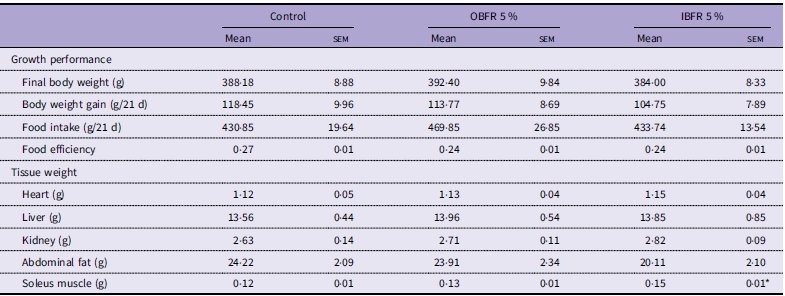
Table 1 shows the effects of supplementation with either OBFR or IBFR on the growth performance and tissue weights in rats of the Experiment 1. Feeding the OBFR diet did not affect the body weight gain and feed intake of the rats during the 21-d feeding period. It also had no significant effects on the weights of the individual organs or tissues (liver, heart, kidney, abdominal fat and soleus muscle). In contrast, although feeding the IBFR diet did not affect the growth performance of the rats during the experimental period, it significantly increased the weights of the soleus muscles compared with their counterparts fed the control diet. However, it did not affect the weights of the other tissues and organs.
Effects of feeding OBFR and IBFR on growth performance and tissue weights of rats

Table 1. Long description
Image fully described in caption.
OBFR, outer bran fraction of rice bran; IBFR, inner bran of rice bran.
Values are expressed as mean and SEM (n 6 rats per group).
* Significantly different (P < 0.05).
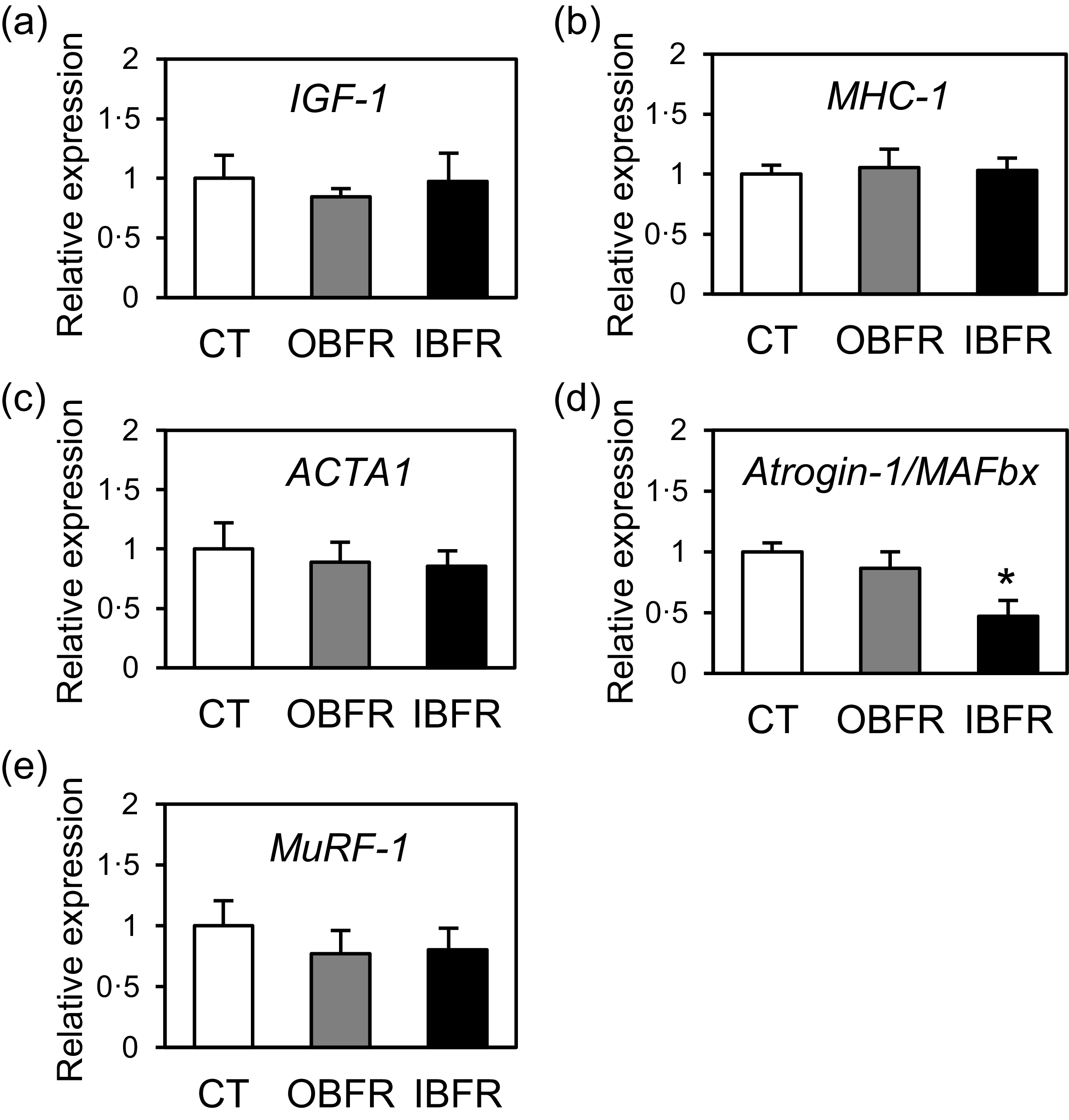
Figure 1 shows the effects of supplementation with either OBFR or IBFR on expression of genes encoding IGF-1, MHC-1, ACTA1, Atrogin-1/MAFbx and MuRF-1 in the soleus muscle of the rats. Neither the OBFR diet nor the IBFR diet affected IGF-1 mRNA expression (Figure 1a). In addition, neither diet affected the expression of MHC-1 or ACTA1 (Figure 1b and c). In addition, the OBFR diet did not affect the mRNA expressions of Atrogin-1/MAFbx and MuRF-1 (Figure 1d and e). In contrast, the IBFR diet significantly reduced the mRNA expression of Atrogin-1/MAFbx in the soleus muscle of the rats, but it did not affect the expression of MuRF-1 (Figure 1d and e).
Effects of the IBFR-containing diet on the mRNA expression of IGF-1 (a), MHC-1 (b), ACTA1 (c), Atrogin-1/MAFbx (d) and MuRF-1 (e) in the soleus muscle of rats. The results of the quantitative real-time PCR were normalised to the 18S mRNA levels and expressed relative to the mean value of the control rats fed an AIN-93G-based diet. Values are expressed as means ± sem (n 6 rats per group). *P < 0·05 (v. control rats). CT, control; OBFR, outer bran fraction of rice; IBFR, inner bran fraction of rice; IGF, insulin-like growth factor; MHC, myosin heavy chain; ACTA, actin alpha; MuRF-1, muscle ring finger; 18S, ribosomal protein S18.

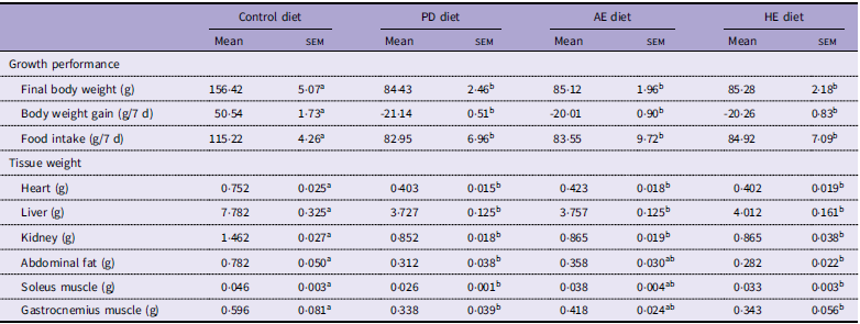
Table 2 shows the effects of the AE or HE of IBFR on the skeletal muscle of the PD diet-fed rats of the Experiment 2. Compared with rats fed the AIN-93G-based control diet, those fed the PD diet showed less body weight gain and lower food intake at the end of the 7-d feeding period (Table 2). Feeding the PD diet also resulted in lower weights of the liver, heart, kidney, abdominal fat and soleus muscle. Similarly, feeding the HE diet reduced the body weight, food intake and tissue weights of the rats (Table 2). In contrast, feeding the AE diet markedly reduced several parameters (body weight, food intake and the weights of liver, heart and kidney), but the weights of the soleus and gastrocnemius muscles did not significantly differ from those of rats fed the AIN-93-based control diet (Table 2).
Effects of supplementation with AE and HE on growth performance and tissue weight in rats

Table 2. Long description
Image fully described in caption.
AE, aqueous extract; HE, hexane extract; PD, protein-deficient.
Values are expressed as mean and sem (n 6 rats per group). Means with the same superscript letter within rows are not significantly different at P < 0.05.
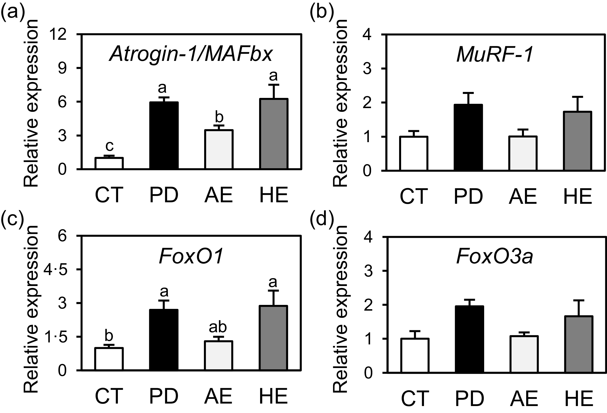
The mRNA expression of Atrogin-1/MAFbx in the soleus muscles of rats fed the PD diet was significantly higher than that of rats fed the AIN-93-based control diet (Figure 2a). Although the differences were not significant, the mRNA expression levels of MuRF-1, FoxO1 and FoxO3a were also higher in the soleus muscles of rats fed the PD diet (Figure 2b–d). The expression levels of Atrogin-1/MAFbx and FoxO1 in the soleus muscles were significantly higher in rats fed the HE diet than in those fed the AIN-93-based control diet (Figure 2a and c). Moreover, the mRNA expression levels of MuRF-1 and FoxO3a tended to be higher in rats fed the HE diet (Figure 2b and d). In contrast, the mRNA expression of Atrogin-1/MAFbx in the soleus muscles of rats fed the AE diet was significantly lower than that of rats fed the PD or HE diets, although it was higher than that of rats fed the AIN-93-based control diet (Figure 2a). However, the mRNA expression level of FoxO1 in the soleus muscles of the rats fed the AE diet was comparable to that of the rats fed the AIN-93-based control diet (Figure 2c). Furthermore, the expression pattern of MuRF-1 and FoxO3a mRNA was similar to that of Atrogin-1/MAFbx mRNA, albeit that the differences were not statistically significant (Figure 2b and d).
Effects of the AE- or HE-containing diet on the mRNA expression of Atrogin-1/MAFbx (a), MuRF-1 (b), FoxO1 (c) and FoxO3a (d) in the soleus muscle of rats. The results of the quantitative real-time PCR were normalised to the 18S mRNA levels and expressed relative to the mean value of the control rats fed an AIN-93G-based diet. Values are expressed as means ± sem (n 6 rats per group). Means in the same graph with different superscript letters are significantly different (P < 0·05). CT, control; PD, protein-deficient; AE, aqueous extract of IBFR; HE, hexane extract of IBFR; IBFR, inner bran fraction of rice; MuRF-1, muscle ring finger; FoxO, forkhead boxO; 18S, ribosomal protein S18.

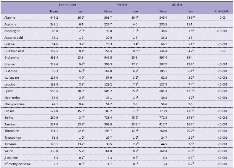
The plasma concentrations of glycine, serine, histidine and N τ-methylhistidine in rats fed the PD diet were significantly higher than those of the rats fed the AIN-93-based control diet (Table 3). In addition, the plasma concentrations of alanine, asparagine, cystine, isoleucine, leucine, lysine, methionine, proline, threonine, tryptophan, tyrosine, valine and β-alanine were significantly lower than those of the rats fed the AIN-93-based control diet. In contrast, plasma concentrations of cystine and β-alanine in rats fed the AE diet were significantly higher than those in rats fed the PD diet, whereas the N τ-methylhistidine concentration was lower (Table 3).
Effects of supplementation with AE on plasma amino acids of rats fed the PD diet

Table 3. Long description
The data in the table show the plasma concentrations of individual amino acids in rats fed three experimental diets: the control, PD, and AE diets.
AE, aqueous extract; PD, protein-deficient.
Values are expressed as mean and sem (n 6 rats per group). Means with the same superscript letter within rows are not significantly different at P < 0.05.
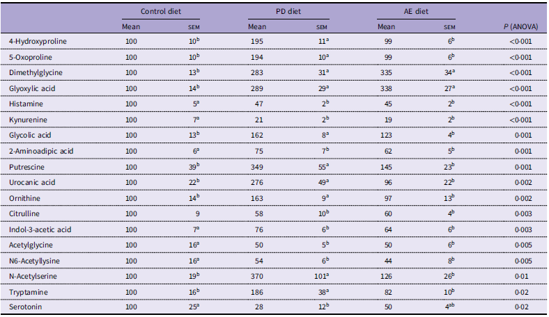
The untargeted GC–MS-based metabolomic analysis identified a total of 144 metabolites in the plasma of the rats, excluding the amino acids quantified by the targeted analysis (Supplementary Table S3). Of these, fifty-eight metabolites were significantly affected by the diet fed: the levels of twenty-five were increased by the PD diet v. the AIN-93-based control diet, and the levels of thirty-three were reduced (Supplementary Table S4). In accordance with the results of the targeted quantitative analysis, the levels of many of the amino acids and their metabolites, such as 4-hydroxyproline, kynurenine, ornithine, citrulline and serotonin, were significantly affected by the PD diet (Table 4). Additionally, the plasma levels of both ornithine and putrescine, precursors of polyamines, were increased by the PD diet (Table 4).
Altered plasma levels of amino acids and their metabolites in PD diet-fed rats

Table 4. Long description
The data in the table show changes in plasma levels of amino acids and their metabolites altered by AE in PD diet-fed rats, expressed relative to the control group set at 100.
PD, protein-deficient; AE, aqueous extract.
Values are expressed as mean and sem (n 6 rats per group). Means with the same superscript letter within rows are not significantly different at P < 0.05.
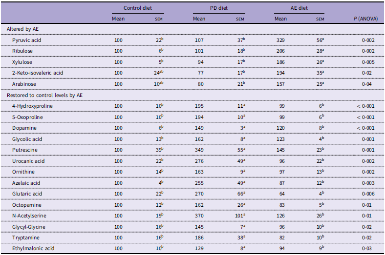
Despite being based on the PD diet, feeding the AE diet significantly affected the plasma levels of nineteen metabolites: five were increased and fourteen were reduced by the AE diet v. the PD diet (Table 5). Fourteen of these metabolites, including 4-hydroxyproline, 5-oxoproline, putrescine, urocanic acid, ornithine and cystine, were restored to levels comparable to those of rats fed the AIN-93G control diet. In addition, six of the metabolites, including pyruvic acid, 2-keto-isovaleric acid and β-alanine, were higher than those of rats fed either the control or PD diet (Tables 4 and 5).
Plasma metabolites altered by AE supplementation in PD diet-fed rats

AE, aqueous extract; PD, protein-deficient.
Values are expressed as mean and sem (n 6 rats per group). Means with the same superscript letter within rows are not significantly different at P < 0.05.
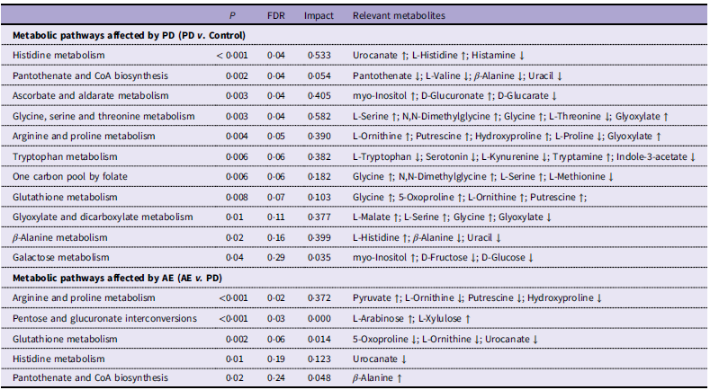
The pathway analysis showed that eleven metabolic pathways were influenced by the PD diet: six of these were involved in amino acid metabolism, and the other five were involved in the metabolism of either carbohydrates or vitamins (Table 6). Among the enriched pathways, tryptophan and histidine metabolism showed particularly notable alterations: the plasma tryptamine level was higher, whereas those of tryptophan, kynurenine and serotonin were lower (Tables 3 and 4). With respect to histidine metabolism, the plasma histidine, urocanic acid and N τ-methylhistidine levels were higher, whereas histamine level was lower. The pathway analysis showed that feeding the AE diet affected five metabolic pathways: arginine and proline metabolism, pentose and glucuronate interconversions, glutathione metabolism, histidine metabolism and pantothenate and CoA biosynthesis (Table 6). Although the effects of the PD and AE diets were compared with different control groups (PD v. AIN-93G normal diet and AE v. PD diet), both were shown to influence glutathione metabolism, arginine and proline metabolism and histidine metabolism. The PD diet affected the levels of metabolites related to the γ-glutamyl cycle: it increased the 5-oxoproline level and reduced the cystine level. Conversely, the AE diet restored their plasma levels to those comparable with the control diet-fed rats (Table 5).
Metabolic pathways significantly affected by the PD diet and AE supplementation in the plasma of rats

PD, protein-deficient; AE, aqueous extract.
Upward (↑) and downward (↓) arrows indicate increases and decreases in metabolite levels, respectively.
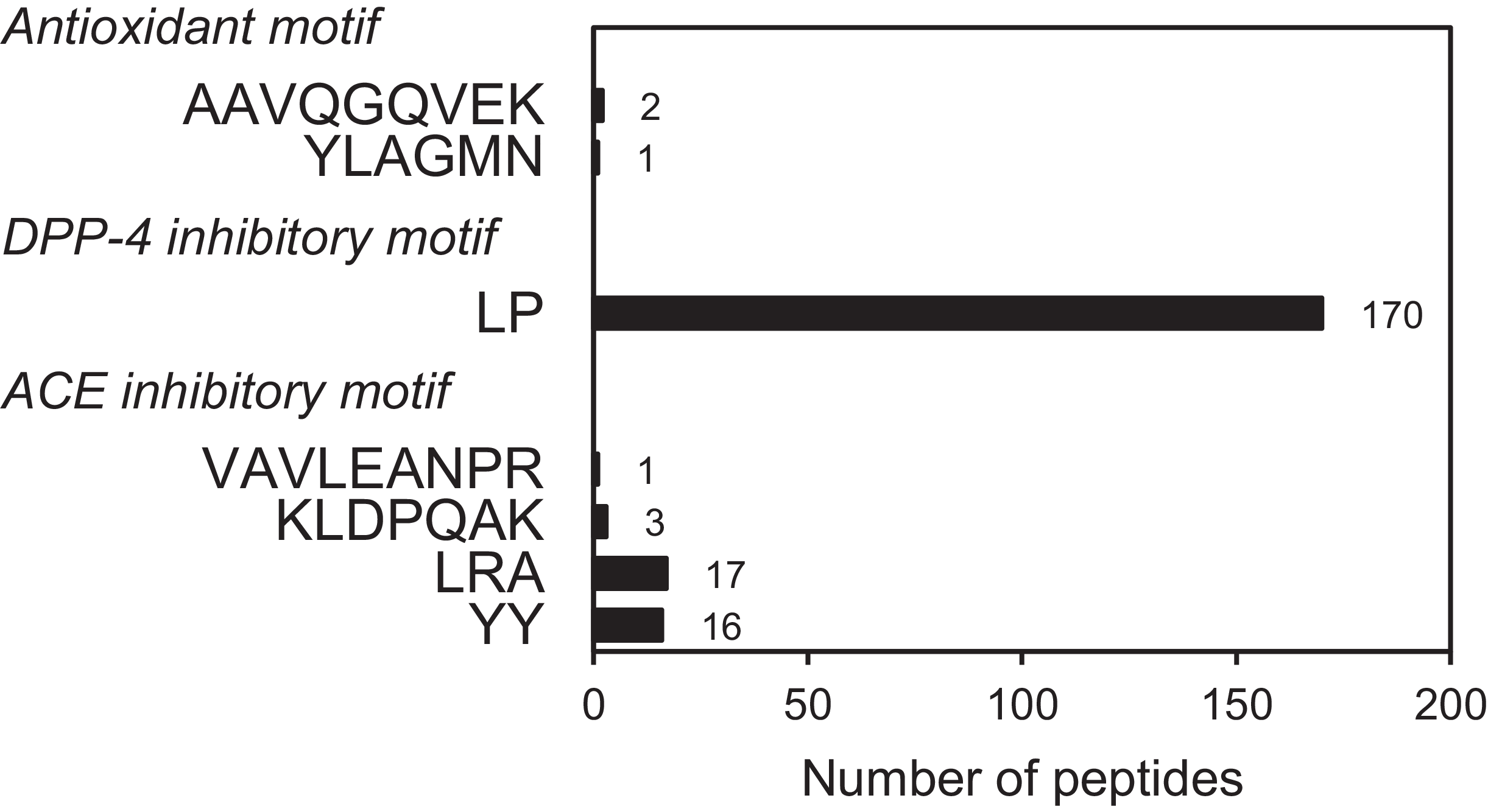
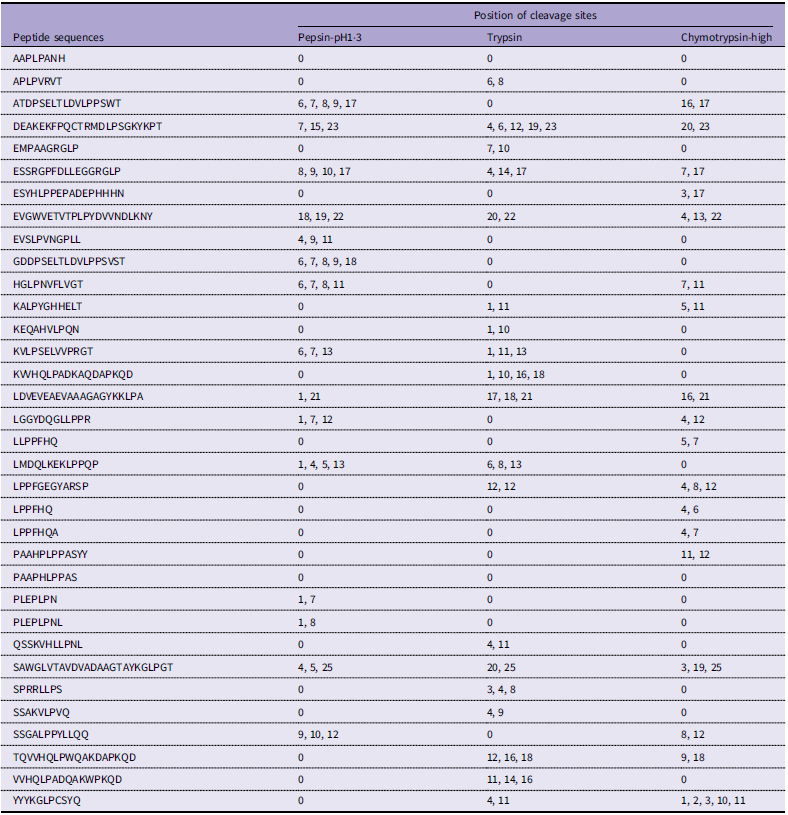
To explore peptides from water-soluble molecules in the AE of IBFR, a peptidomic analysis was performed. In total, peptidomic analysis of AE identified a total of 1222 peptides (Supplementary Table S5). AE peptides contained sequences previously reported in rice bran-derived peptides to exhibit antioxidant, dipeptidyl peptidase-4 (DPP-4) inhibitory and angiotensin-converting enzyme (ACE) inhibitory activities (Figure 3). Specifically, three peptides contained internal sequences with the antioxidant motifs ‘AAVQGQVEK’ and ‘YLAGMN’, whereas 170 peptides contained internal sequences corresponding to the DPP-4 inhibitory dipeptide ‘LP’ (Supplementary Table S6)(Reference Wattanasiritham, Theerakulkait and Wickramasekara24–Reference Hatanaka, Uraji and Fujita26). In addition, thirty-seven peptides contained sequences corresponding to the ACE inhibitory peptides, such as ‘VAVLEANPR’, ‘KLDPQAK’, ‘LRA’ and ‘YY’ (Supplementary Table S6)(Reference Uraipong and Zhao27,Reference Shobako, Ogawa and Ishikado28) . They are expected to undergo digestion by gastric and intestinal enzymes, such as pepsin, trypsin and chymotrypsin. Accordingly, we predicted the effects of these enzymes on the candidate peptides using the RPG tool. As shown in Table 7, thirty-four peptides containing ‘LP’ were considered to retain the ‘LP’ sequence after gastrointestinal digestion.
Counts of AE-derived peptides containing sequence identical to antioxidative, DPP-4 inhibitory and ACE inhibitory peptides. The number of peptides is shown for each sequence. AE, aqueous extract; ACE, angiotensin-converting enzyme; DPP-4, dipeptidyl peptidase-4.

Candidate bioactive peptides containing LP sequences identified in the AE of IBFR

Table 7. Long description
The data in the table show the cleavage sites in potential bioactive peptides containg LP sequences identified in AE after digestion with pepsin, trypsin, and chymotrypsin.
LP, Leu-Pro; AE, aqueous extract; IBFR, inner bran of rice bran.
Discussion
In Experiment 1, feeding the IBFR diet under normal-protein conditions increased soleus muscle weight and reduced Atrogin-1/MAFbx mRNA expression, without affecting IGF-1 or MuRF-1 mRNA expression (Figure 1a, d and e). Sacheck et al. (2004) reported that the mRNA expression of Atrogin-1/MAFbx linearly and positively correlates with the overall rate of muscle protein degradation, but there is no correlation between MuRF-1 mRNA expression and the overall rate of protein breakdown(Reference Sacheck, Ohtsuka and McLary29). These results suggest that consumption of the IBFR diet reduced muscle protein degradation by suppressing Atrogin-1/MAFbx mRNA expression, likely independent of IGF-1, thereby increasing skeletal muscle weight in rats. However, Yoshida et al. (2010) reported that IGF-1 prevents skeletal muscle wasting and inhibits Atrogin-1/MAFbx expression(Reference Yoshida, Semprun-Prieto and Sukhanov30). Although a direct correlation between IGF-1 and Atrogin-1/MAFbx was not confirmed in the present study, a potential connection could still be suggested for future research based on findings reported by Yoshida et al. (Reference Yoshida, Semprun-Prieto and Sukhanov30)
It was unclear which components of IBFR are responsible for its effects on skeletal muscle. To gain further insight into the effects of IBFR on the regulation of muscle protein degradation, we fed rats a PD diet, an approach that is commonly used to study skeletal muscle proteolysis(Reference Sugawara, Ito and Nishizawa16,Reference Kobayashi, Somoto and Mitsuyama31) . We then evaluated the effects of the AE or HE of IBFR on the skeletal muscle of the PD diet-fed rats. In agreement with previous studies of the effects of a PD diet(Reference Jacobson32–Reference Warren, Benedito and Skinner35), we confirmed that feeding the PD diet inhibited growth performance and decreased the weights of the liver, heart, kidney, abdominal fat and soleus muscle. However, the AE diet ameliorated the skeletal muscle loss induced by the PD diet and exhibited an mRNA expression pattern for FoxO1 and Atrogin-1/MAFbx similar to that of the control diet (Figure 2a and c). On the other hand, feeding the HE diet did not ameliorate the skeletal muscle loss. These results suggest that the water-soluble molecules in the AE of IBFR may be involved in the suppression of muscle protein degradation, possibly via a reduction in FoxO1 and Atrogin-1/MAFbx mRNA expression in the skeletal muscle of rats.
To investigate why the AE diet alleviated the PD diet-induced muscle protein degradation in the rats, we performed a targeted quantitative analysis of the amino acids and an untargeted metabolomic analysis of the plasma metabolites of rats fed the AIN-93-based control diet, the PD diet or the AE diet.
The pathway analysis showed that the PD diet affected tryptophan metabolism (e.g. tryptophan and tryptamine) and histidine metabolism (e.g. histidine and urocanic acid) (Table 6). Tryptamine, produced from tryptophan by the gut microbiota(Reference Marcobal, Kashyap and Nelson36), was elevated by the PD diet, suggesting that it may reflect microbial changes. The observed pattern of histidine metabolites, such as elevated urocanic acid levels, suggests enhanced protein degradation owing to insufficient dietary protein supply, in contrast to histidinemia, which is associated with reduced urocanic acid levels(Reference Taylor, Levy and McInnes37). In addition, the increased level of 4-hydroxyproline, a marker of collagen degradation, implied that skin homeostasis may also have been affected.
The pathway analysis also revealed that the AE diet shared three common pathways, including glutathione metabolism (e.g. 5-oxoproline and cystine) (Tables 3 and 5). These similar effects suggest that these pathways may be involved in the attenuation of muscle protein degradation identified in the AE diet-fed rats. Protein malnutrition has been reported to result in lower glutathione levels and changes in myofibrillar structures(Reference Dubick, Heng and Rucker38,Reference Oumi, Miyoshi and Yamamoto39) . Because glutathione is a major antioxidant in skeletal muscle, the redox status of the tissue is thought to be involved in protein degradation and atrophy(Reference Ji, Fu and Mitchell40,Reference Powers, Kavazis and DeRuisseau41) . The AE supplementation restored the 5-oxoproline and cystine levels altered by the PD diet to values comparable to those observed in rats fed the control diet. These results suggest that the AE supplementation could ameliorate the perturbations in glutathione metabolism induced by PD diet. Glutathione depletion leads to the accumulation of reactive oxygen species, which activate FoxO proteins(Reference Maher42,Reference Klotz, Sánchez-Ramos and Prieto-Arroyo43) . FoxO transcription factors regulate the expression of Atrogin-1/MAFbx (Reference Sandri, Sandri and Gilbert11,Reference Stitt, Drujan and Clarke44) . One possible explanation is that the positive effects of the AE diet on glutathione metabolism contributes to the alleviation of muscle protein degradation through the attenuation of reactive oxygen species-mediated FoxO activation.
The AE diet suppressed the increase in N τ-methylhistidine concentration induced by the PD diet (Table 3). Because N τ-methylhistidine is an index of muscle protein degradation(Reference Nagasawa, Yoshizawa and Nishizawa45), these results indicate that AE supplementation attenuates the enhanced muscle protein degradation in rats fed a PD diet. Furthermore, we identified fourteen metabolites altered by the PD diet and restored to control levels by the AE diet (Table 5). Notably, changes in ornithine and putrescine levels suggest a possible impairment of polyamine synthesis under the PD diet rather than effects on urea cycle metabolites, which remained unchanged. The regulation of polyamine has been shown to be related to hypertrophy and atrophy in skeletal muscle(Reference Lee and MacLean46,Reference Tabbaa, Ruz Gomez and Campelj47) and particularly in FoxO1 overexpression-induced muscle atrophy(Reference Oyabu, Sato and Kawaguchi48). Polyamines (spermidine and spermine) have been reported to suppress the expression of Atrogin-1/MAFbx in C2C12 cells(Reference Yoshioka, Onishi and Yasui49). Therefore, these improvements in plasma polyamine levels may contribute to the AE diet-mediated suppression of muscle protein degradation. To further elucidate the mechanisms underlying the effects of AE on PD diet-induced muscle proteolysis, additional studies of these metabolites and related enzymes in skeletal muscle are required.
To explore peptides from water-soluble molecules in the AE of IBFR, a peptidomic analysis was performed. We found that AE contained 210 peptides harbouring antioxidant, DPP-4 inhibitory and ACE inhibitory motif (Figure 3). It has been well known that soya glycinin contains the ‘DIYNP’ sequence, which is structurally similar to a ubiquitin ligase Casitas B-cell lymphoma-b inhibitor (Cblin), a peptide reported to prevent muscle atrophy(Reference Nakao, Hirasaka and Goto50,Reference Abe, Kohno and Yama51) . This peptide sequence has been reported to attenuate muscle protein degradation through suppression of Atrogin-1/MAFbx and MuRF-1 expression (Reference Abe, Kohno and Yama51). However, none of the identified peptides from the AE of IBFR contained the Cblin-like sequence ‘DIYNP’. The AE peptides containing ‘LP’ sequence may suppress Atrogin-1/MAFbx expression by inhibition of DPP-4, since its inhibition has been reported to attenuate skeletal muscle mass loss in diabetic patients(Reference Bouchi, Fukuda and Takeuchi52). DPP-4 rapidly inactivates glucagon-like peptide-1 (GLP-1). GLP-1 receptor signalling has been shown to induce phosphorylation of FoxO1 via phosphatidylinositol-3 kinase (PI3K)/Akt pathway(Reference Kimura, Okouchi and Fujioka53,Reference Shao, Nie and Chen54) . Therefore, DPP-4 inhibition by these peptides may enhance GLP-1 signalling, leading to attenuation of FoxO1 and Atrogin-1/MAFbx expression.
Additionally, AE peptides with ‘LRA’ and ‘YY’ sequences may contribute to attenuating muscle wasting. This is because in rodents, angiotensin II enhances protein degradation in skeletal muscle, accompanied by inducing the expression of Atrogin-1/MAFbx (Reference Yoshida, Semprun-Prieto and Sukhanov30,Reference Brink, Price and Chrast55) , and its inhibition attenuates muscle protein degradation(Reference Sanders, Russell and Tisdale56). Therefore, these peptides may reduce Atrogin-1/MAFbx expression through ACE inhibition.
Subsequently, the potential cleavage sites of these peptides by gastric and intestinal enzymes were predicted. Thirty-four peptides were estimated to retain the ‘LP’ sequence, a DPP-4 inhibitory motif. Pro-containing di- and tripeptides are known to exhibit resistance to gastrointestinal enzymes(Reference Ohsawa, Satsu and Ohki57). Moreover, this sequence has also been reported to be resistant to digestion by brush border membrane enzymes, including postproline endopeptidase and aminopeptidase(Reference Morita, Chung and Freeman58). In contrast, most peptides with bioactive sequences appear to be degraded by gastrointestinal enzymes (Supplementary Table S6). However, the peptides of ‘LRA’ and ‘YY’ from thermolysin-treated rice bran remain 81 % and 82 %, respectively, even after digestion with pepsin and pancreatin(Reference Shobako, Ogawa and Ishikado28). Furthermore, the digestibility of rice proteins extracted using alkali or α-amylase, major industrial processing, has been reported to be approximately 6–16 % lower in vitro and 10–12 % lower in vivo than that of casein in rats(Reference Yang, Chen and Xu59). Therefore, not all peptides are completely degraded, allowing the generation of bioactive peptides. These results suggest that the effects of AE may be partially mediated by peptides with the potential to attenuate muscle protein degradation. These AE-derived peptides may exert suppressive effects on protein degradation.
In conclusion, supplementation with IBFR reduces Atrogin-1/MAFbx mRNA expression and muscle protein degradation in rats. In addition, water-soluble components of IBFR reduce Atrogin-1/MAFbx mRNA expression by reducing FoxO1 mRNA expression in rat skeletal muscle. These effects appear to be accompanied by improvements in glutathione metabolism and polyamine synthesis. Furthermore, dietary AE supplementation could ameliorate the redox imbalance and modulate polyamine metabolism under conditions of protein malnutrition, potentially through bioactive peptides generated during AE digestion.
Supplementary material
For supplementary material/s referred to in this article, please visit https://doi.org/10.1017/S0007114526108216
Acknowledgements
The authors thank Mark Cleasby, PhD, from Edanz (https://jp.edanz.com/ac) for editing a draft of this manuscript.
This study was partially supported by grants provided to D. Ijiri from Kiyokawa Akio Ikuei Shogaku Foundation and S. Shimamoto from the Uchida Energy Science Promotion Foundation (no. R04-1-055).
S. Shimamoto, A. Ohtsuka and D. Ijiri designed the study. M. Kamimura, S. Shimamoto, S. Tomonaga and D. Ijiri contributed to the methodology. M. Kamimura, S. Shimamoto, K. Hirao, A. Katafuchi, M. Kawaguchi and K. Nakamura performed the analysis and collected the data. Y. Yamauchi and Y. Fujita provided resources. M. Kamimura, S. Shimamoto, K. Hirao, K. Nakamura, A. Katafuchi and D. Ijiri prepared the original draft of the manuscript. S. Shimamoto, S. Tomonaga, Y. Fujita, A. Ohtsuka and D. Ijiri reviewed and edited the manuscript. A. Ohtsuka supervised the study. D. Ijiri administered the project. All authors have read and agreed to the published version of the manuscript.
No potential conflict of interest was reported by the authors.
The data underlying this article will be shared on reasonable request to the corresponding author.