INTRODUCTION
Leishmania is a protozoan parasite, endemic in 98 countries worldwide, responsible for a spectrum of diseases in humans (World Health Organization, 2010). Out of the 2 million new cases notified every year, about 1·5 million are cutaneous or mucocutaneous leishmaniasis. The human-infective species are classified into 2 subgenera on the basis of their location in the vector's intestine: Leishmania (Leishmania) and Leishmania (Viannia). Leishmania braziliensis is one of the representatives of the L. (Viannia) subgenus, and one of the major causative agents of cutaneous or mucocutaneous leishmaniasis in wide areas of Central and South America.
In higher eukaryotes, the nucleosome constitutes the chromatin structural unit, and is basically composed of DNA wrapped around an octamer of core histones. Another histone, the linker histone or histone H1, packs the chromatin by interacting with the nucleosomal DNA and the histone octamer (Woodcock et al. Reference Woodcock, Skoultchi and Fan2006). Histone H1 is formed by a globular domain flanked by 2 unstructured regions at both the N-and the C-terminal ends. The latter presents numerous basic residues that counteract the negative charges of DNA (Hendzel et al. Reference Hendzel, Lever, Crawford and Th'ng2004).
The genus Leishmania and other lower eukaryotes, present a particular histone H1 that lacks the central globular domain, a feature that has been related to the imperfect condensation of chromatin in chromosomes during cell division (Hecker et al. Reference Hecker, Betschart, Bender, Burri and Schlimme1994). Despite that, Leishmania H1 is apparently functional since it is expressed differently at different developmental stages (Noll et al. Reference Noll, Desponds, Belli, Glaser and Fasel1997) and an increase in its expression levels has been related to chromatin compaction (Zangger et al. Reference Zangger, Mottram and Fasel2002; Masina et al. Reference Masina, Zangger, Rivier and Fasel2007). A relationship between H1 expression levels and infectivity has also been found in L. major (Papageorgiou and Soteriadou, Reference Papageorgiou and Soteriadou2002; Masina et al. Reference Masina, Zangger, Rivier and Fasel2007). Since the completion of the Leishmania Genome Project, histone H1 variants have been described in L. infantum (JPCM5), L. major (Friedlin), L. mexicana (MHOM/GT/2001/U1103) and L. braziliensis (MHOM/BR/75/M2904). These H1 variants show important divergences in their sequence, although no phylogenetic analysis of these important genes has been performed so far in Leishmania species.
A conflictive point in the characterization of histone H1 proteins has traditionally been the determination of their ability to self-interact both in the absence and in the presence of DNA, with authors showing conflicting results in different models (Thomas et al. Reference Thomas, Rees and Finch1992; Carter and Van Holde, Reference Carter and Van Holde1998; Salvati et al. Reference Salvati, Conforti, Conte, Matassa, Fucci and Piscopo2008). Besides, most of these investigations have been performed using the H1 globular domain, thus comparative analysis with Leishmania histone H1 is unfeasible. These long-standing controversies render the analysis of the structural and functional characteristics of Leishmania H1 an unresolved issue for the scientific community.
MATERIALS AND METHODS
Parasites
The parasites used in this study belong to the following species: L. braziliensis (MOHN/PE/95/LQ-2), L. amazonensis (MHOM/BR/77/LTB0016), L. major (LM 252), L. infantum (MHOM/ES/1990/BCN-143), and L. tropica (MON58/LEM2578). Promastigotes of all these species were grown in Tobie´s medium, containing 10% (v/v) of HIFBS (heat-inactivated fetal bovine serum) at 22°C. Experimental cultures were initiated at 0·5×106 promastigotes and harvested at the desired density.
Protein extracts
All Leishmania total protein extracts were made of 2×10⁷ promastigotes in the late-logarithmic phase, washed twice in cold PBS. The pellet was sonicated in lysis buffer (150 mm Tris-HCl, pH 7·4, 50 mm NaCl, 0·05% (v/v) NP-40), and then centrifuged at 18 000 g for 1 h at 4°C.
Parasitic Histone H1 was specifically isolated by perchloric acid extraction from nuclei as described (Noll et al. Reference Noll, Desponds, Belli, Glaser and Fasel1997). All protein extracts were quantified using MicroBCA protein Assay (Pierce).
rLbH1 purification and antibody production
The gene encoding L. braziliensis H1 was subcloned on plasmid pET29b(+) (Novagen) in-frame with the C-terminal His6-tag, after PCR with specific oligonucleotides (pETH1-Rev, TGATCTCGAGCTTCTTCGCCGC; pETH1-For, CTCTACCATATGTTCGCTAACTCC) using as template clone 3.3 (Martínez et al. Reference Carmelo, Martínez, González, Piñero, Patarroyo, Del Castillo and Valladares2002) (GenBank Acc. no: AF131892), to generate pETH1 plasmid. Recombinant L. braziliensis H1 protein (rLbH1) was generated as a C-terminal His6-tagged protein in E. coli BL21, after co-transformation with RIG-plasmid to improve expression levels (Baca and Hol, Reference Baca and Hol2000). rLbH1 was solubilized by sonication in binding buffer (BB: 20 mm Tris-HCl, pH 8, 0·5 m NaCl, 5 mm imidazole). The cleared lysate was purified by affinity chromatography using a Ni-NTA column, in BB with increasing imidazole concentration, and was maintained at −80°C in storage buffer (50 mm HEPES-NaOH, pH 8, 50 mm NaCl, 50% (v/v) glycerol). Both purity and integrity were assessed by Coomassie blue-stained SDS-PAGE electrophoresis. Mass spectrometry was also performed in order to accurately determine the actual molecular mass of rLbH1, since its particular charge (net charge +34, deduced isoelectric point: 12·44), induces an abnormal protein migration in SDS-PAGE (Panyim and Chalkley, Reference Panyim and Chalkley1971).
Two New Zealand white rabbits were immunized with 500 μg of purified rLbH1in CFA (Complete Freund's Adjuvant, Sigma-Aldrich). Three subsequent 350 μg boosts in IFA (Incomplete Freund's Adjuvant, Sigma-Aldrich) were injected at 10-day intervals, until good anti-rLbH1 antibody levels were detected by Western blot analysis. All procedures involving animals were conducted in accordance with the European Guidelines, approved by the Bioethical Committee of the University of La Laguna, Spain.
Phylogenetic analysis
All sequences identified as Histone H1 or putative Histone H1 from L. (Leishmania) major (LmjF), L. (Leishmania) infantum (LinJ), L. (Leishmania) mexicana (LmxM) and L. (Viannia) braziliensis (LbrM) were retrieved from the Leishmania Genome Database (www.genedb.org) and aligned using ClustalW as implemented in MEGA 4 software (Tamura et al. Reference Tamura, Dudley, Nei and Kumar2007). The same software was used to construct a Neighbor-joining tree with p-distances. Trypanosoma brucei H1 sequence (Tb11.42.0005) obtained also from the same source was used as outgroup.
Mass spectrometry analysis
Matrix-assisted laser desorption ionization/time-of-flight (MALDI-TOF) mass spectra were performed in an Autoflex (Bruker-Daltonics) spectrometer, at the University of La Laguna Proteomic Service. Briefly, 0·5 μl of protein (10 μ m stock) was mixed with 0·5 μl of CHCA matrix (α-cyano-4-hydroxycinnamic acid) (AcN:H20 (7:3) in 2% (v/v) TFA), and allowed to evaporate at room temperature prior to mass spectral analysis.
DNA substrates
Three different DNA substrates were used for binding assays: a biotinylated A+T rich ds-oligonucleotide called H1-DB (oligo 1: GCGCAAAATATTGAAAACGCC; oligo 2: GCGGCGTTTTCAATATTTTGC) (Bharath et al. Reference Bharath, Ramesh, Chandra and Rao2002); a PCR-amplified 219 bp fragment of L. braziliensis RNA-Polimerase II gene using oligonucleotides RNApol-For: 5′CGCCTATGGTGCCTGGTC3′ and RNApol-Rev: 5′CTCACCGACATCACCAGC3′; and plasmid pGT-RNApol II (3,234 bp), resulting from cloning this PCR-amplified 219 bp fragment in pGemT-easy vector (Promega). Plasmid pGT-RNApol II was purified using Qiagen Spin Miniprep Kit (Qiagen), and linearized by complete digestion with NotI (Roche Diagnostics).
Further DNA purification was performed by phenol-chloroform extraction and ethanol precipitation, followed by examination by electrophoresis in 1% (w/v) agarose gels with ethidium bromide staining and UV visualization. DNA concentration was determined using Nanodrop 1000 spectrophotometer (Thermo).
Chemical cross-linking
Chemical cross-linking was performed using 10 μ m protein and 10 mm dimethyl pimelimidate (DMP, Sigma-Aldrich) or 0·2% (v/v) glutaraldehyde as cross-linking agents, in cross-linking buffer (50 mm HEPES-KOH, pH 8, 5 mm KCl, 5 mm MgCl, 10 mm NaCl) (Carmelo et al. Reference Carmelo, Barillà, Golovanov, Lian, Derome and Hayes2005).
UV cross-linking
UV cross-linking was performed incubating 10 μ m protein in the presence or absence of 10 μ m H1-DB oligonucleotide in cross-linking buffer at 25°C for 20 min, and then irradiated on ice for 1 h, at a distance of 1·5 cm from the 254 nm UV lamp. Reaction was stopped by boiling in SDS-PAGE loading buffer and was visualized by SDS-PAGE.
Electrophoretic mobility shift assays (EMSA)
Plasmid pGT-RNApol II (0·5 μg) or the PCR-amplified 219 bp fragment of L. braziliensis RNA-Polymerase II gene (0·7 μg) were incubated with protein at 25°C in binding buffer (50 mm HEPES-KOH, pH 8, 5 mm KCl, 5 mm MgCl, 10 mm NaCl, 2·5% (v/v) glycerol) for 20 min (Carmelo et al. Reference Carmelo, Barillà, Golovanov, Lian, Derome and Hayes2005). Reactions were visualized by electrophoresis in 0·7% (w/v) agarose gels (10 V/cm) and run at room temperature in TBE buffer with ethidium bromide staining.
Confocal microscopy
L. braziliensis promastigotes (5×10⁶) were washed twice using cold PBS and fixed in 500 μl of PLP (2% (w/v) paraformaldehyde, 37·5 mm sodium phosphate pH, 7·4, 60 mm L-lysine, 100 mm sodium m-periodate). Cells were then permeabilized using PBS-0·5% (v/v) Triton X-100 for 5 min, and blocked by incubation in blocking buffer (1% (w/v) BSA in PBS) for 1 h. All incubations were performed in a humid chamber at 37°C. Primary antibody staining was performed using anti-rLbH1 diluted in blocking buffer (1/200 (v/v)) for 1 h, and then removed by extensive washing. Incubation with secondary antibody (dilution 1/250 (v/v), FITC-conjugated anti-rabbit, Sigma-Aldrich) was performed similarly. Parasite DNA was stained for 5 min at room temperature with 5 μg/ml DAPI (4′, 6-diamidine-2-phenyl indole, Pierce). Counterstaining using Evan's blue was performed where necessary. Cover-slips were mounted with Prolong Antifade Kit (Molecular Probes). Microscopical analysis of the samples was performed using a Leica DMI6000 confocal microscope using a FITC filter (medium wavelength 530 nm, max. 490 nm).
RESULTS
Phylogenetic relationship in Leishmania H1 genes
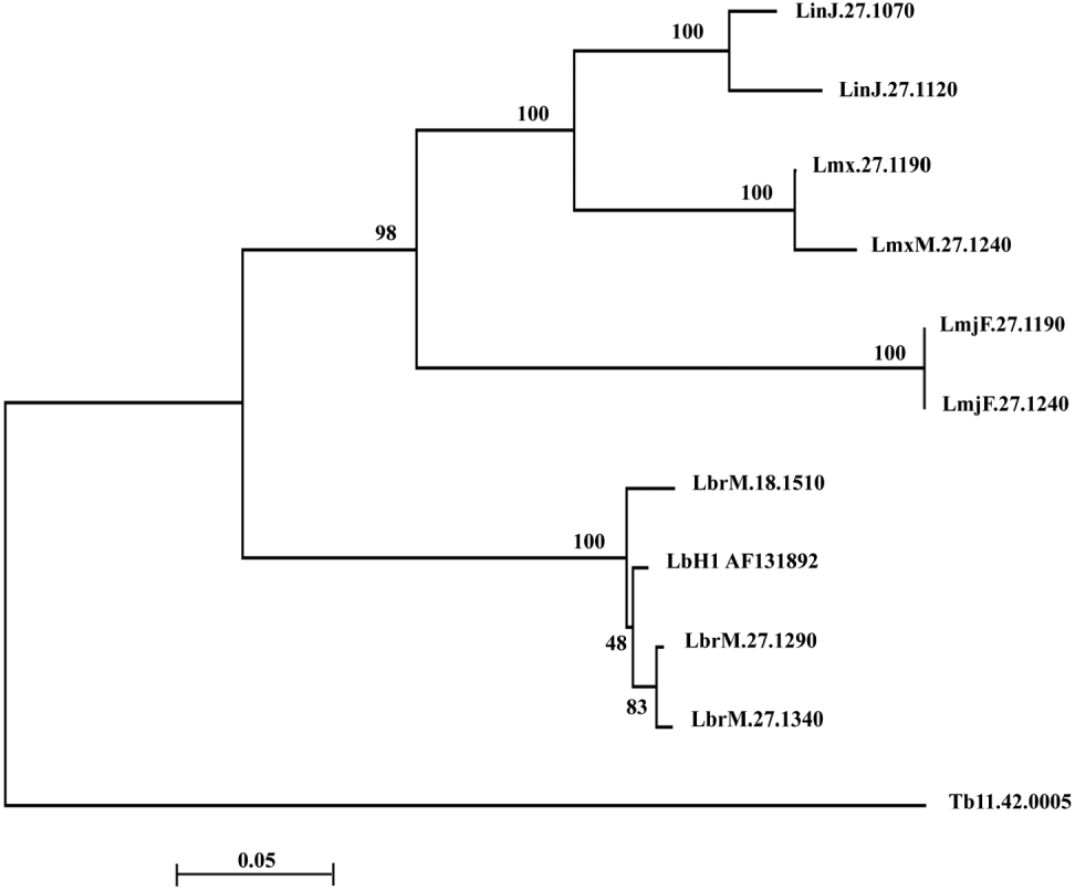
The completion of the Leishmania Genome Project exposed a collection of sequences termed Histone H1 or putative Histone H1. Although the alignment of these sequences proved to be challenging due to the differences in sequence among them, 2 main clusters could be identified by phylogenetic analysis (Fig. 1). One of them presents 2 almost identical copies in each L.(Leishmania) species (LmjF.27.1190 and LmjF.27.1240, 96 aa; LinJ.27.1070, 111 aa and LinJ.27.1120, 102 aa; LmxM.27.1190, 100 aa and LmxM.27.1240, 102 aa). This cluster showed a high bootstrap value (98%), suggesting a common ancestor for all of them. In contrast, L. braziliensis presents a group of 3 closely related genes (LbrM.27.1290, LbrM.27.1340 and LbrM.18.1510), including the gene coding for the L. braziliensis H1 protein analysed in this manuscript (AF131892) (Fig. 1). This cluster is only distantly related to the former, reflecting the sequence divergences found for the L. braziliensis gene, compared to the L. (Leishmania) species analysed. This phylogenetic analysis of H1 variants in Leishmania revealed that L. (Viannia) braziliensis orthologues are quite divergent to the other species included in this study. Phylogenies based on amino acid sequences agreed with the nucleotide-based trees, although with lower resolution power, so the latter were chosen for this analysis.
Phylogenetic analysis of Leishmania Histone H1 genes. Neighbor-joining tree of Histone H1 and putative Histone H1 genes from L. (Leishmania) major (LmjF), L. (Leishmania) infantum (LinJ), L. (Leishmania) mexicana (LmxM) and L. (Viannia) braziliensis (LbrM). The sequences were retrieved from Leishmania Genome Database (www.genedb.org) and aligned using ClustalW as implemented in MEGA 4 software. Bootstrap support of the branches is given in percentages at the internodes. The tree is drawn to the scale at the bottom, expressed as distance per nucleotide, and rooted with a Trypanosoma brucei H1 (Tb11.42.0005) sequence as outgroup.

Identification and immunolocalization of L. braziliensis Histone H1
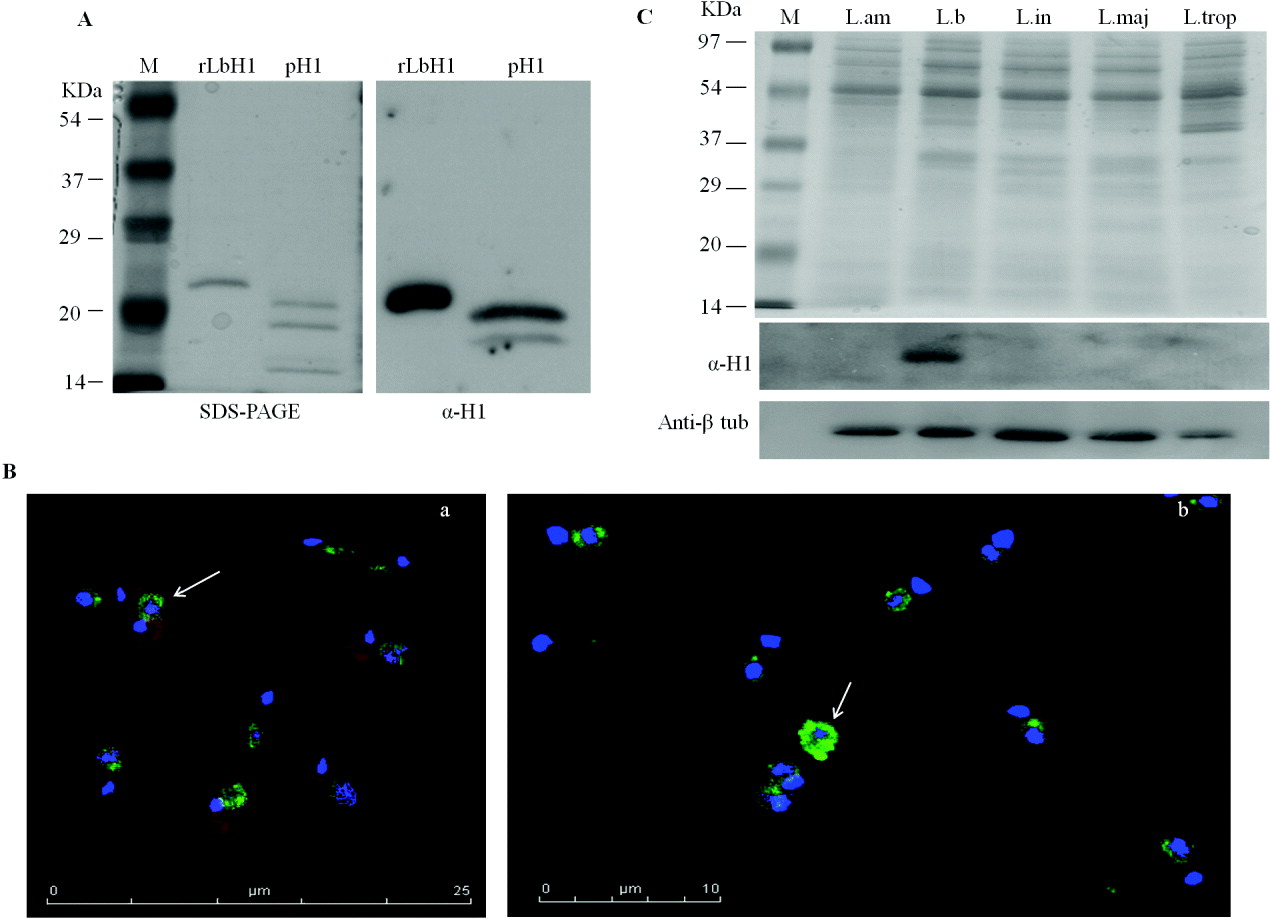
The antibody generated against recombinant L. braziliensis Histone H1 was assayed in Western-blot against the same protein used for immunization (rLbH1) and also against histone H1 extracted from parasite nuclei using perchloric acid extraction (pH1). Fig. 2A shows SDS-PAGE and Western-blot using rabbit polyclonal antiserum. Anti-rLbH1 (α-H1) antibody was able to strongly recognize rLbH1 and 2 of the 3 bands obtained using perchloric acid extraction, none of which was detected by the pre-immune serum (data not shown). These results demonstrate that the anti-rLbH1 antibody recognizes histone H1 in L. braziliensis, detecting a band with a strong signal, but also a minor band with very low intensity, probably related to the presence of protein variants, as observed in the phylogenetic tree.
Identification and immunolocalization of Leishmania braziliensis Histone H1. (A) Coomassie blue-stained (right) SDS-PAGE (15% acrylamide) and Western-blot (left) using anti-rLbH1 (α-H1). M, molecular weight marker; rLbH1: 0·5 μg recombinant L. braziliensis Histone H1; pH1: 1 μg of perchloric acid H1 extraction. (B) Confocal immunofluorescence microscopy of L. braziliensis promastigotes stained using anti-rLbH1 (green), DAPI (blue) and Evan's blue (red) (a–b). White arrows indicate the ring-like H1 deposits. (C) Coomassie blue-stained SDS-PAGE (12% acrylamide) and Western blot using anti-rLbH1 (α-H1) and β-tubulin antibody (anti-β-tub) (Sigma-Aldrich) against Leishmania species: L.am, L. amazonensis; L.br, L. braziliensis; L.in, L. infantum; L.maj, L. major; L.trop, L. tropica.
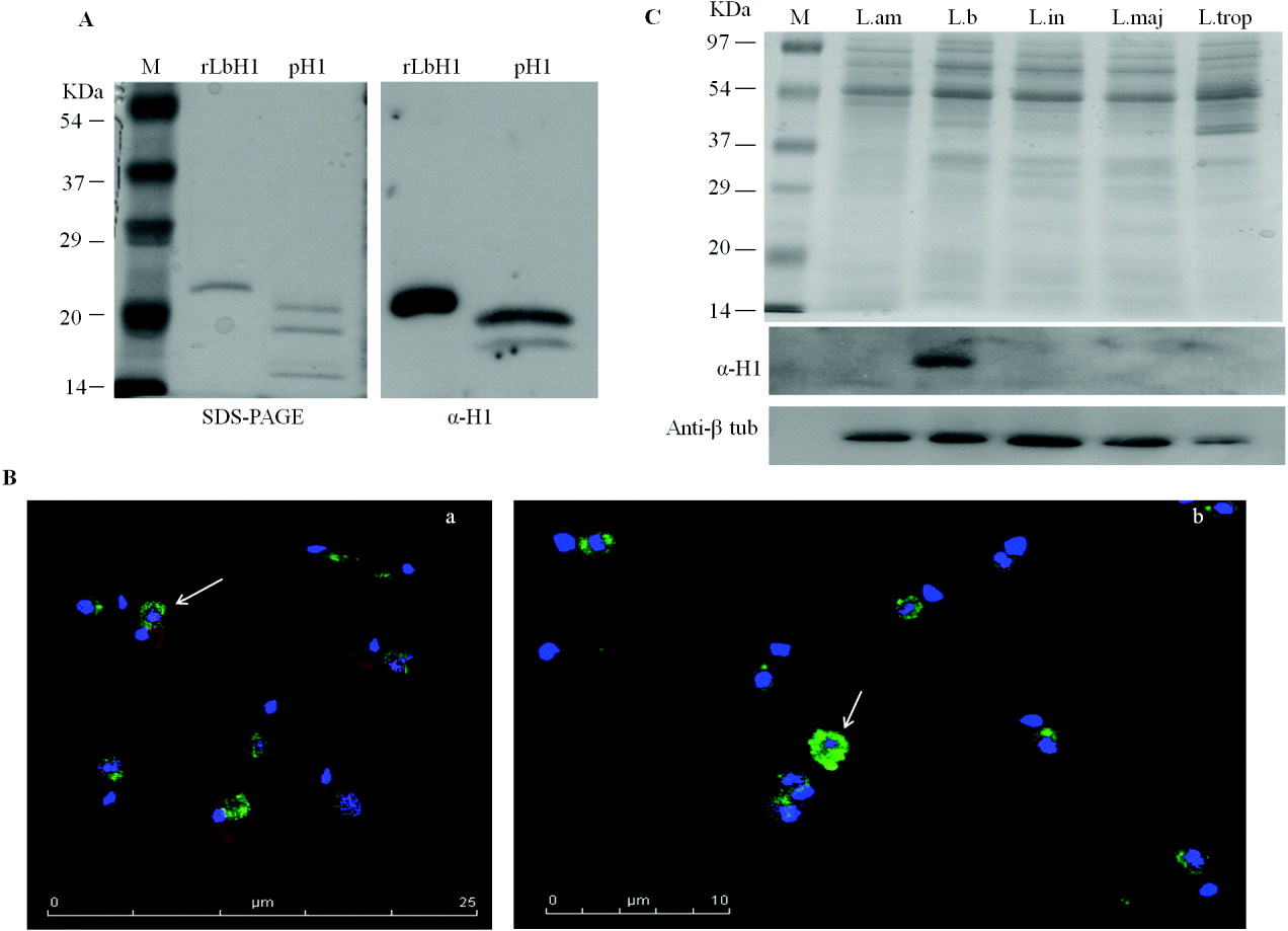
In order to confirm this finding, immunolocalization experiments were performed using confocal microscopy on L. braziliensis promastigotes. Figure 2B(a) shows that LbH1 was located only at the parasitic nuclei, co-localizing with nuclear but not kinetoplast DAPI staining. As shown in Fig. 2B(b), L. braziliensis H1 nuclear staining was not uniform, showing a particular ring localization with empty nuclear spaces (Fig. 2B) resulting from condensation of histone H1 around the nuclear membrane. A similar finding was previously observed for 2 Old World Leishmania species, L.major and L.donovani (Noll et al. Reference Noll, Desponds, Belli, Glaser and Fasel1997; Smirlis et al. Reference Smirlis, Boleti, Gaitanou, Soto and Soteriadou2009).
Surprisingly, anti-rLbH1 antibody was unable to detect a band in any of the other Leishmania species assayed in Western-blot (L. amazonensis, L. infantum, L. major and L. tropica) (Fig. 2C). For this experiment, all Leishmania species were grown until late-log phase (1×10⁷ parasites/ml), and then protein extracts were purified. Similar amounts of protein (6 μg) were loaded in each lane of the SDS-PAGE, and then blotted using anti-rLbH1 and anti-β-tubulin (Sigma Aldrich) antibodies (Fig. 2C). As shown in Fig. 2C, despite loading the same amount of protein in each lane, as shown by the anti-β-tubulin immunodetection, the anti-rLbH1 antibody only detects a band in the L. braziliensis protein extract, but not in any of the other Leishmania species tested. This result contrasts the observations of other authors (Papageorgiou and Soteriadou, Reference Papageorgiou and Soteriadou2002), but is in agreement with the genetic distance observed between L.(Viannia) braziliensis H1 and other L. (Leishmania) spp. H1 genes, as reflected in the tree (Fig. 1).
Leishmania braziliensis Histone H1 is a monomeric protein
In order to determine the aggregation status of rLbH1in solution, chemical cross-linking was applied using 2 different cross-linkers. The first one is DMP, a homobifunctional imidoester with spacer arm length of 9·2 Å that possesses 2 identical groups that can react with primary amine groups to form stable covalent bonds. Incubation of rLbH1 with DMP over a range of different temperatures revealed a single species, showing a small change in the migration of the monomeric protein (Fig. 3A). In contrast, when BSA was used as a control in the same experiment, a band matching the molecular weight of a BSA dimer was observed on SDS-PAGE. MALDI-TOF spectra revealed a 14 087 Da mass, compatible with the monomeric rLbH1bound to 5–6 DMP molecules (DMP molecular weight: 259 Da) (Fig. 3B). Similarly, a negligible 28 466 Da mass was detected, that might result from the formation of a minor dimeric species, but was undetectable in Coomassie-stained gels. Given the fact that DMP has a long reaction distance (9·2 A), a second experiment was performed using glutaraldehyde as cross-linker, a reagent that allows for a much closer molecular interaction and flexibility. Glutaraldehyde cross-linking showed considerable blurring of the monomeric protein, and interestingly, the formation of a minor second species, whose molecular weight cannot be determined by SDS-PAGE given the aberrant migration of the protein (Fig. 3C). MALDI-TOF spectra revealed a 16 230 Da mass, compatible with the monomeric rLbH1bound to 34–35 glutaraldehyde molecules (glutaraldehyde molecular weight: 100.12 Da) (Fig. 3D). The other mass observed was 32 649 Da, probably matching the dimeric species revealed in Coomassie-stained gels. The formation of that minor dimeric species observed under conditions that induce the formation of a strong BSA dimer (data not shown), suggests that the rLbH1 dimer is probably an artefact derived from the fact that rLbH1 has 35 possible sites (35 lysines) where a small flexible molecule such as glutaraldehyde can bind, and not a consequence of protein oligomerization. Consequently, rLbH1 has proven to be a monomeric protein in solution, although some minor aggregation has been observed under these particular reaction conditions.
Analysis of the aggregation state of rLbH1 in solution. (A) Coomassie blue-stained SDS-PAGE of the DMP-cross-linking reactions of rLbH1 and BSA. M, molecular weight marker; -, reaction without DMP; 4°, 25°, 37°, DMP cross-linking reactions incubated at those temperatures; +, reaction with DMP. (B) MALDI-TOF spectrum of rLbH1 DMP-cross-linking reaction. (C) Coomassie blue-stained SDS-PAGE of rLbH1glutaraldehyde-crosslinking reactions. (D) MALDI-TOF spectrum of glutaraldehyde-cross-linking reaction.

DNA binding induces polymerization of L. braziliensis Histone H1 in vitro
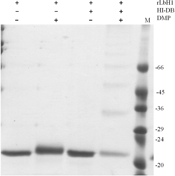
In order to investigate whether DNA is able to induce polymerization of L. braziliensis Histone H1 in vitro, a series of experiments was performed in which a ds-oligonucleotide was incubated with the protein. Eukaryotic histones H1 do not show any binding specificity against any particular DNA sequence, although some affinity of these proteins with A+T-rich sequences has been shown in vitro (Churchill and Suzuki, Reference Churchill and Suzuki1989). Recombinant rLbH1 was incubated with H1-DB oligonucleotide in binding buffer for 20 min, and then DMP was added as a cross-linker. SDS-PAGE of these reactions revealed the presence of a series of bands indicative of the formation of several defined complexes between rLbH1 monomers upon binding to DNA (Fig. 4), since DMP does not induce any protein–DNA cross-links. The precise molecular mass of those bands could not be determined due to the anomalous migration of rLbH1 in SDS-PAGE, and also MALDI-TOF analysis was unable to resolve the stoichiometry of those complexes. Nevertheless, the fact that at least 4 discrete bands were clearly visible on the gel proves that L. braziliensis H1 is able to polymerize when a DNA substrate is present in the reaction.
Analysis of the aggregation state of rLbH1 in the presence of DNA. Coomassie blue-stained SDS-PAGE of rLbH1 DMP-cross-linking reactions in the presence of H1-DB oligonucleotide. M, molecular weight marker.

Leishmania braziliensis Histone H1 is able to form complexes with DNA in vitro
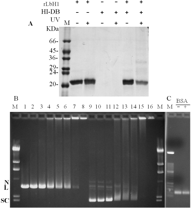
In order to further investigate this interaction, rLbH1 was incubated with H1-DB in binding buffer for 20 min, and then the binding reactions were irradiated with 254 nm UV light on ice for 1 h before SDS-PAGE. UV irradiation induces cross-linking between proteins and nucleic acids in close contact (Pellé and Murphy, Reference Pellé and Murphy1993). Subsequent boiling in SDS-PAGE buffer will cause dissociation of any non-specific complexes. Figure 5A reveals the formation of at least 4 different complexes between rLbH1 and H1-DB after UV cross-linking, therefore demonstrating its DNA-binding ability.
Leishmania braziliensis H1 DNA-binding in vitro. (A) Coomassie blue-stained SDS-PAGE of the UV-cross-linking reactions of rLbH1 in the presence of H1-DB oligonucleotide. M, molecular weight marker. (B) Agarose gel electrophoresis of DNA-binding reactions using 0·5 μg of supercoiled (SC) or linearised (L) pGT-RNApol II plasmid in the presence of increasing molar ratios of rLbH1. N, relaxed circular plasmid; M, λ/Hind III molecular weight marker; lanes 1 and 9: no rLbH1 added; lanes 2 and 10: 25/1 molar ratio; lanes 3 and 11: 50/1 molar ratio; lanes 4 and 12: 100/1 molar ratio; lanes 5 and 13: 200/1 molar ratio; lanes 6 and 14: 200/1 molar ratio; lanes 7 and 15: 300/1 molar ratio; lanes 9 and 16: 500/1 molar ratio. (C) Agarose gel electrophoresis of DNA-binding reactions using 0·7 μg of a PCR-amplified 219 bp fragment of L. braziliensis RNA-Polymerase II, incubated with bovine serum albumin (BSA); M: 100 bp Molecular Weight Marker XIV (Roche Diagnostics); -, reaction without BSA; +, reaction with 1000/1 molar ratio of BSA/DNA.

With the aim to analyse this interaction further, the binding activity was evaluated against a long DNA fragment, pGT-RNApol II, in both supercoiled (SC) and linear form (L). Incubation of 0·5 μg linear pGT-RNApol II (L) with increasing molar ratios of rLbH1 (25/1 to 500/1, Fig. 5B) induced the formation of a smear along the lane, with progressive disappearance of the linear DNA band and resulting in complete DNA aggregation in the wells (Fig. 5B, lanes 1–8). In contrast, when the supercoiled template (SC) was used (Fig. 5B, lanes 9–16), a small but appreciable shift was observed as soon as 100/1 molar ratio (Fig. 5B, lane 12), revealing the formation of a defined retarded rLbH1/DNA complex (Fig. 5B, lanes 12–14) ending up with full DNA-protein aggregation in the wells starting from 300/1 ratio (Fig. 5B, lane 15). Interestingly, the small amount of contaminating relaxed circular plasmid (Fig. 5B, N) was also retarded. Since the sequence of the DNA was identical in both experiments, it is clear that some structural aspect is playing a role in the H1-DNA interaction that is reflected in the higher affinity of histone H1 for supercoiled DNA compared to linear DNA. In order to rule out the possibility of DNA shift or aggregation in the presence of any unrelated protein, bovine serum albumin (BSA) was incubated with the 219 bp fragment of L. braziliensis RNA-Polymerase II gene at a 1000/1 molar ratio, and then electrophoresed as described. Figure 5C shows that BSA was unable to induce any shift in the Leishmania-specific DNA fragment even at that high molar ratio, thereby demonstrating the specificity of the rLbH1-DNA interaction.
DISCUSSION
In this study, a histone H1 from L. braziliensis was identified and partially characterized. Numerous histone H1 genes from different Leishmania species can be found on the databases, showing remarkable differences in their length and sequence. In addition, the sequencing of the genome of 4 Leishmania species in the last few years, has unlocked a collection of new genes, whose characteristics are still unknown for most of them.
Regarding Leishmania H1 genes, the Genome Database includes several H1 and/or putative H1 genes, showing differences in sequence, length and amino acid composition. The phylogenetic relationship of these genes suggests the presence of 2 main clusters, one of them grouping H1 genes from the L. (Leishmania) species sequenced, and a distant one that includes the L.(Viannia) braziliensis orthologue. The H1 described here (AF131892) is included in the group of 3 closely related L. braziliensis paralogues, sharing only a very old ancestor with the other species’ genes, a feature that is probably a consequence of its separation from the Old World Leishmania species and its subgenus classification (Peacock et al. Reference Peacock, Seeger, Harris, Murphy, Ruiz, Quail, Peters, Adlem, Tivey, Aslett, Kerhornou, Ivens, Fraser, Rajandream, Carver, Norbertczak, Chillingworth, Hance, Jagels, Moule, Ormond, Rutter, Squares, Whitehead, Rabbinowitsch, Arrowsmith, White, Thurston, Bringaud, Baldauf, Faulconbridge, Jeffares, Depledge, Oyola, Hilley, Brito, Tosi, Barrell, Cruz, Mottram, Smith and Berriman2007).
The structural and functional analyses of Leishmania histones H1 proteins have traditionally been considered challenging, due to the difficulty in obtaining good quantities of purified protein under non-denaturing conditions (Carmelo et al. Reference Carmelo, Martínez, González, Piñero, Patarroyo, Del Castillo and Valladares2002). The protocol described here allowed overexpression and purification of rLbH1 under non-denaturing conditions. The protein identity was confirmed by immunoblot using a polyclonal antiserum against the recombinant protein that was able to detect it in nuclear extracts, as well as using immnofluorescence microscopy. As observed for Old World Leishmania species L. donovani and L. major (Noll et al. Reference Noll, Desponds, Belli, Glaser and Fasel1997; Smirlis et al. Reference Smirlis, Boleti, Gaitanou, Soto and Soteriadou2009), L. braziliensis H1 nuclear staining is particularly directed towards the nuclear rim, suggesting its implication in nuclear envelope reorganization processes (Smirlis et al. Reference Smirlis, Boleti, Gaitanou, Soto and Soteriadou2009), and nuclear surface-microtubule-organization (Nakayama et al. Reference Nakayama, Ishii, Hotta and Mizuno2008). The actual significance of the ring H1 distribution observed remains unclear, but given the fact that chromatin does not condense into chromosomes, and nuclear membrane remains intact during mitosis in Leishmania, it seems clear that Histone H1 must be playing a role in chromatin and nuclear organization whose nature must be further investigated.
A surprising result was the species-specific nature of the anti-rLbH1 antibody that recognized L. braziliensis H1 only in Western-blots but not the other Leishmania species tested. This observation is clearly related to the low sequence identity observed between this protein and other Leishmania species’ histones H1 (Martinez et al. Reference Masina, Zangger, Rivier and Fasel2002), reflecting the evolutionary separation of L.(Viannia) braziliensis and L.(Leishmania) spp., estimated between 20 and 100 million years (Momen and Cupolillo; Reference Momen and Cupolillo2000; Kerr, Reference Kerr2006).
L. braziliensis Histone H1 has proven to be monomeric in solution. The aggregation state of linker histones in the absence of DNA has long been subject to controversy. Some authors have shown little, if any, self-association of the globular domains of linker histones H1 and H5 (Thomas et al. Reference Thomas, Rees and Finch1992), in contrast to other groups that have proven by different means the ability of linker histones to self-associate (Carter and Van Holde, Reference Carter and Van Holde1998; Salvati et al. Reference Salvati, Conforti, Conte, Matassa, Fucci and Piscopo2008). Most of these studies have been performed on the protein globular domain, that is essential for the association with DNA in eukaryotes, but very little attention has been focused on the full-length protein or the C- and N-terminal tails. Histone H1 or H1-like proteins from trypanosomatid protozoa show particularly divergent sequences compared to their higher eukaryote counterparts, reflected by a much lower molecular mass and the lack of the central globular domain (Noll et al. Reference Noll, Desponds, Belli, Glaser and Fasel1997; Papageorgiou and Soteriadou, Reference Papageorgiou and Soteriadou2002). Those differences have been related to the lack of chromatin condensation observed in these organisms during cell division that renders Leishmania H1 as an interesting model for the analysis of the aggregation state of histones in solution and in the presence of DNA. At low salt concentration (10 mm Na+), L. braziliensis H1 was predominantly monomeric in solution, as shown by SDS-PAGE and mass spectroscopy analysis. In contrast, the presence of DNA has been demonstrated to induce the formation of a series of defined protein-protein and protein-DNA complexes, particularly when a short DNA fragment was assayed. An interesting explanation for this behaviour could be that the binding to DNA is able to induce the folding of L. braziliensis H1, basically a disordered stretch of positively-charged amino acids, into an ordered structure, that would be essential for its activity.
The binding of rLbH1 to a long DNA fragment in either supercoiled or linearised form also revealed a particular DNA-binding activity. The linear DNA fragment was progressively aggregated by the titration with rLbH1, until the aggregates were too large to enter the gel at around 500/1 protein/DNA molar ratio. In contrast, the supercoiled substrate suffered an appreciable shift starting at a 100/1 molar ratio, indicating the formation of a defined complex, until the increase in protein concentration resulted in full DNA-protein aggregation. This different behaviour of rLbH1 is comparable to that observed by Ellen and van Holde (Reference Ellen and van Holde2004) that suggested that the plectonemic structure of supercoiled DNA could provide multiple sites for histone to connect, and therefore its preferential binding.
From the analysis of all these results, it seems clear that the structural and functional characteristics of Leishmania Histone H1 are still unresolved issues that raise interesting questions for the scientific community. The determination of protein structure both in solution and bound to DNA, and the fine-mapping of H1-DNA interactions will focus our attention in the future.
ACKNOWLEDGEMENTS
We would like to thank Dr Hernandez-Fernaud for his kind help with MALDI-TOF spectra.
FINANCIAL SUPPORT
This work has been funded by Fondo de Investigaciones Sanitarias (B.V., grant number PI081984), Red de Investigación de Centros de Enfermedades Tropicales (RICET)-Instituto de Salud Carlos III (B.V., grant number RD06/0021/0005) and Ministerio de Ciencia y Tecnología (A.O., grant number AGL2007-60123/GAN), Spain.