Introduction
Laryngeal squamous cell carcinoma (SCC) is one of the most common malignancies of the head and neck.Reference Jemal, Siegel, Ward, Hao, Xu and Thun1 Locoregional aggressiveness of these malignancies contributes to a five-year death rate of about 35 per cent.Reference Jemal, Siegel, Ward, Hao, Xu and Thun1 Significant functional sequelae, mainly involving speech and swallowing, often afflict long-term survivors, resulting in a heavy social burden.
Despite advances in our understanding of the molecular mechanisms of tumour development, together with technical and methodological progress in head and neck oncology, the prognosis of laryngeal SCC has not improved in the past 30 years. Indeed, the documented 24.5 per cent reduction in mortality rate from 1990 to 2004 in the USA is essentially due to decreased incidence, subsequent to the success of primary prevention, particularly the campaign against cigarette smoking.Reference Jemal, Siegel, Ward, Hao, Xu and Thun1 The 5-year disease-specific survival of laryngeal cancer in the USA changed from 67 per cent in 1977 to 64 per cent in 2003, and, together with adenocarcinoma of the uterine body, it remains the only major human cancer that has shown no significant improvement in survival rate over the past 30 years.Reference Jemal, Siegel, Ward, Hao, Xu and Thun1 These data are even more striking when compared with those reported for oral SCC, another head and neck cancer, whose five-year survival rate rose from 53 to 60 per cent in the same period.
Several explanations for this trend have been suggested.Reference Almadori, Bussu, Cadoni, Galli, Paludetti and Maurizi2 For example, many authors cite the increasing push towards surgical and non-surgical function-preserving treatments.Reference Almadori, Bussu, Cadoni, Galli, Paludetti and Maurizi2–5 Non-surgical function-preserving strategies are often effective and may allow a better quality of life in cured patients.6, Reference Forastiere, Goepfert, Maor, Pajak, Weber and Morrison7 Problems arise in the case of recurrence; for instance, surgical salvage treatment is often associated with higher complication and lower survival rates. Therefore, great efforts have been made in the last few decades to identify predictive chemoradiosensitivity factors and prognostic factors, in order to determine high-risk patients and guide therapeutic choices.
Despite the multiplicity of clinical prognosticators proposed, the only consistent clinical predictors for disease control and disease-specific survival in laryngeal SCC remain tumour (T) and, to a greater extent, nodal (N) stage.Reference Licitra, Bernier, Grandi, Locati, Merlano and Gatta8–Reference Spector, Sessions, Haughey, Chao, Simpson and El Mofty10 This is probably because of the extreme biological heterogeneity of laryngeal SCCs,Reference Bernier, Bentzen and Vermorken11, Reference Haddad and Shin12 which contributes to a lack of consistency in treatment planning.
Molecular markers have given us a breakthrough in this heterogeneity.Reference Almadori, Bussu, Cadoni, Galli, Paludetti and Maurizi2, Reference Belbin, Singh, Barber, Socci, Wenig and Smith13, Reference Leethanakul, Patel, Gillespie, Shillitoe, Kellman and Ensley14 In particular, increasing attention has been focused on the epidermal growth factor receptor (EGFR) because of its important role in the regulation of cell proliferation, differentiation, development and oncogenesis.Reference Mendelsohn15, Reference Arteaga16 Overexpression of EGFR and its ligands, epidermal growth factor and/or transforming growth factor α, has been reported in about 90 per cent of head and neck SCCs, including laryngeal SCC. This overexpression is associated with tumour progression, poor disease-free and overall survival, increased risk of disease recurrence and metastasis, and, finally, resistance to chemotherapy and radiotherapy.Reference Kalyankrishna and Grandis17–Reference Dassonville, Formento, Francoual, Ramaioli, Santini and Schneider24 Unfortunately, evaluation of molecular markers is not included in most accepted international guidelines,5 and therefore does not contribute to clinical decision-making processes at present. There is no consensus or standard, even for detection methods; hence, data from various centres cannot be merged and compared to reach an acceptable level of evidence.
Research on molecular mechanisms also indicates potential targets for therapy and, again, EGFR may be considered one of the most interesting; indeed, several drugs acting on the EGFR pathway have been and are currently being tested (in the clinical phase) on head and neck SCC patients. Cetuximab, an anti-EGFR monoclonal humanised antibody, is one of the targeted therapies approved for treatment of head and neck SCC. Cetuximab interacts with the extracellular binding site of EGFR to block ligand stimulation.Reference Mendelsohn and Baselga25 Although cetuximab has shown only modest clinical efficacy in monotherapy, its radiosensitising effect has been proven in clinical trials, where it increased the efficacy of radiotherapy in patients with locally advanced head and neck SCCs,Reference Bonner, Harari, Giralt, Azarnia, Shin and Cohen26 and a clinical effectiveness has been reported in patients with recurrent or metastatic diseases in combination with cisplatin-based chemotherapy as well.Reference Vermorken, Trigo, Hitt, Koralewski, Diaz-Rubio and Rolland27–Reference Burtness, Goldwasser, Flood, Mattar and Forastiere29
Although the molecular mechanisms of action of anti-EGFR drugs, and in particular of cetuximab, have been studied extensively, they are still not fully understood, and the best modality for the clinical use remains to be further defined.
The present study aimed to add relevant data in this field, using Hep-2 laryngeal cells as an in vitro model of laryngeal SCC. We investigated the effects of the administration of cetuximab alone or in combination with cisplatin (the drug most used in the treatment of laryngeal SCC), in relation to different administration modalities and time courses.
Materials and methods
Reagents
Cetuximab (2 mg/ml sterile solution) was obtained from Merck Serono (Darmstadt, Germany). Cisplatin was purchased from Sigma Chemicals (St Louis, Missouri, USA), and the drug stock solution (1 mg/ml) was prepared by dissolving the substance in ethanol and storing it at −20°C. All of the reagents where then diluted to working concentrations in incubation medium.
Cell line and culture conditions
The human larynx squamous carcinoma cell line Hep-2 (purchased from Istituto Zooprofilattico, Parma, Italy) was cultured in modified Eagle's medium with Earle's salts containing non-hydrolysable glutamine (Biochrom, Berlin, Germany), and supplemented with: 10 per cent fetal bovine serum (Life Technologies, Paisley, Scotland, UK), non-essential amino acids, sodium pyruvate, 100 U/ml penicillin and 100 µg/ml streptomycin. It was then stored at 37°C in a 5 per cent carbon dioxide (CO2) and 95 per cent oxygen (O2) humidified atmosphere. Cell viability was determined by trypan blue dye exclusion.
Microscopic analysis of apoptosis
The morphological features of apoptotic degeneration were analysed using fluorescence microscopy with Hoechst 33258 nuclear dye.Reference Forloni, Chiesa, Smiroldo, Verga, Salmona and Tagliavini30 Cells were plated at a density of 2.5 × 10⁶ cells per 35 mm dish; each dish had glass coverslips coated with 1 mg/ml poly-d-lysine. After exposure to experimental treatments, cells were washed with phosphate buffered saline and fixed in 4 per cent paraformaldehyde in phosphate buffered saline for 30 minutes at room temperature. Cells were then washed three times with phosphate buffered saline and incubated for 10 minutes in a humidified atmosphere of 5 per cent CO2 and 95 per cent O2 at 37°C with the DNA-binding dye Hoechst 33258 (0.5 µg/ml in phosphate buffered saline). After washing with water, coverslips were mounted onto glass slides and photographed with a fluorescence microscope (Zeiss Axiovert 25), with excitation at 360 nm, at × 400 magnification.
Cells were considered viable if their chromatin was diffuse and evenly distributed throughout the nucleus, whereas cells with bright, condensed nuclei were counted as positive for apoptosis. Each condition was represented in three dishes per experiment. Normal and apoptotic cells were counted from three fields per dish in a fixed pattern.
Statistical analysis
The data were analysed by one-way analysis of variance, followed by the post-hoc Newman–Keuls test for multiple comparisons among group means, using Prism software (GraphPad, San Diego, California, USA). Differences were considered statistically significant if p < 0.05. All results are presented as the mean ± standard error of the mean of at least three different experiments performed in triplicate, unless otherwise specified.
Results
Cetuximab inhibited cell growth
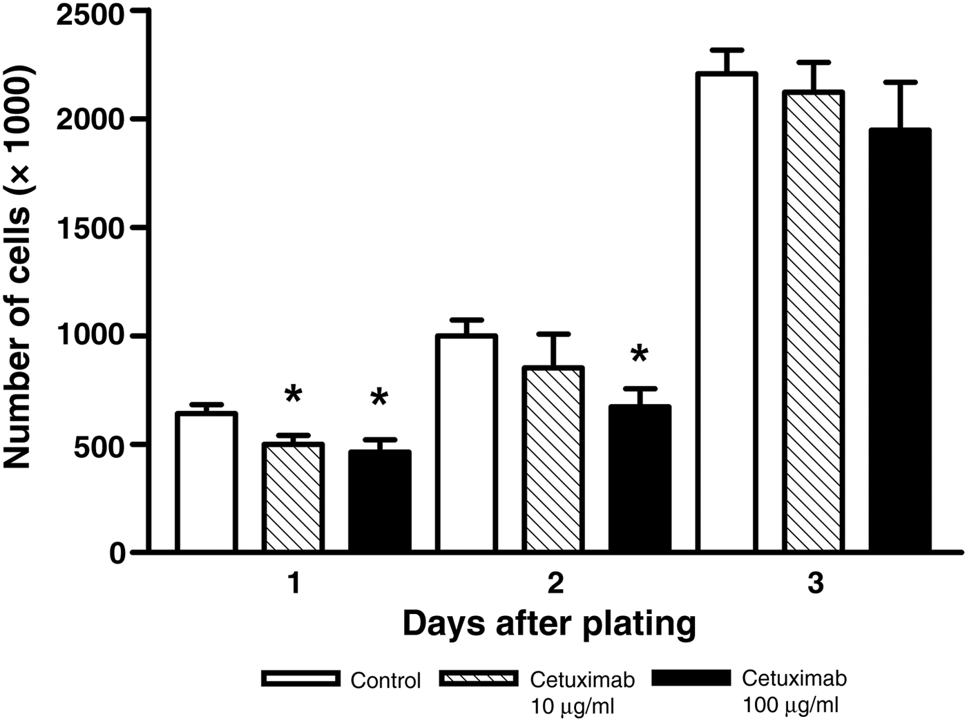
Hep-2 cells were cultured in 60 mm Petri dishes (200 000 cells/dish) and incubated with 10–100 µg/ml cetuximab 1 day after plating, without replacing the drug during the time course of the experiment. Cells were then counted at daily intervals during a 3-day period of culture.
The results, illustrated in Figure 1, showed slight but significant decreases in cell proliferation after 1 day with 10 µg/ml cetuximab and after 1 and 2 days with 100 µg/ml cetuximab, with respect to the untreated control. However, this effect was transient, with a near-complete recovery of proliferative ability after 3 days. As 100 µg/ml cetuximab was the more effective dose, this concentration was used for all subsequent experiments.
Effect of cetuximab on Hep-2 cell proliferation. Cells were incubated with medium alone (control), or with medium containing 10 or 100 µg/ml cetuximab for 24 hours, and proliferation was evaluated for 72 hours. Results are from three independent experiments (*p < 0.05 vs control).

We then compared the effect of 100 µg/ml cetuximab on cell proliferation: cetuximab was added once 1 day after plating or every 24 hours for 3 days. The results showed that when administered every 24 hours, cetuximab was able to maintain its antiproliferative effect at day 3 of culture (Figure 2a).
Effect of cetuximab on Hep-2 cell proliferation. (a) Cells were incubated with medium alone (control) or with medium containing 100 µg/ml cetuximab for the indicated number of days. Results are from three independent experiments (*p < 0.05 vs control, **p < 0.05 vs cetuximab administered only once and vs control). (b) & (c) Staining with Hoechst 33258 fluorescent dye: cells were incubated with medium alone (control) (b) or with medium containing 100 µg/ml cetuximab (c) for 3 days.
The reduction in proliferation rate could be explained by either reduced proliferation or cell death. Therefore, the cells were treated with medium alone or with 100 µg/ml cetuximab added every 24 hours for 3 days. Hoechst 33258 fluorescent dye staining was used to investigate whether the EGFR antibody caused apoptosis. Apoptotic bodies were not observed in significant numbers in either the cetuximab-treated or control cells, suggesting that cytotoxicity was not contributing to the reduction in proliferation rate; this reduction appeared to be at least in part caused by decreased proliferation (Figures 2b and 2c).
Cisplatin induced apoptosis
Starting 1 day after plating, Hep-2 cells were treated with 1-hour exposure to 25 µg/ml cisplatin followed by a wash out (a ‘single high-dose shot’), or with continuous exposure to 2.5 µg/ml cisplatin for 24 hours. Apoptosis in the two different treatment modalities was then evaluated. The effects of the single high-dose shot were comparable with those of continuous exposure (data not shown). After this preliminary experiment, cisplatin was always administered as a single high-dose shot, to simulate the apoptotic stimulus of a radiotherapy session.
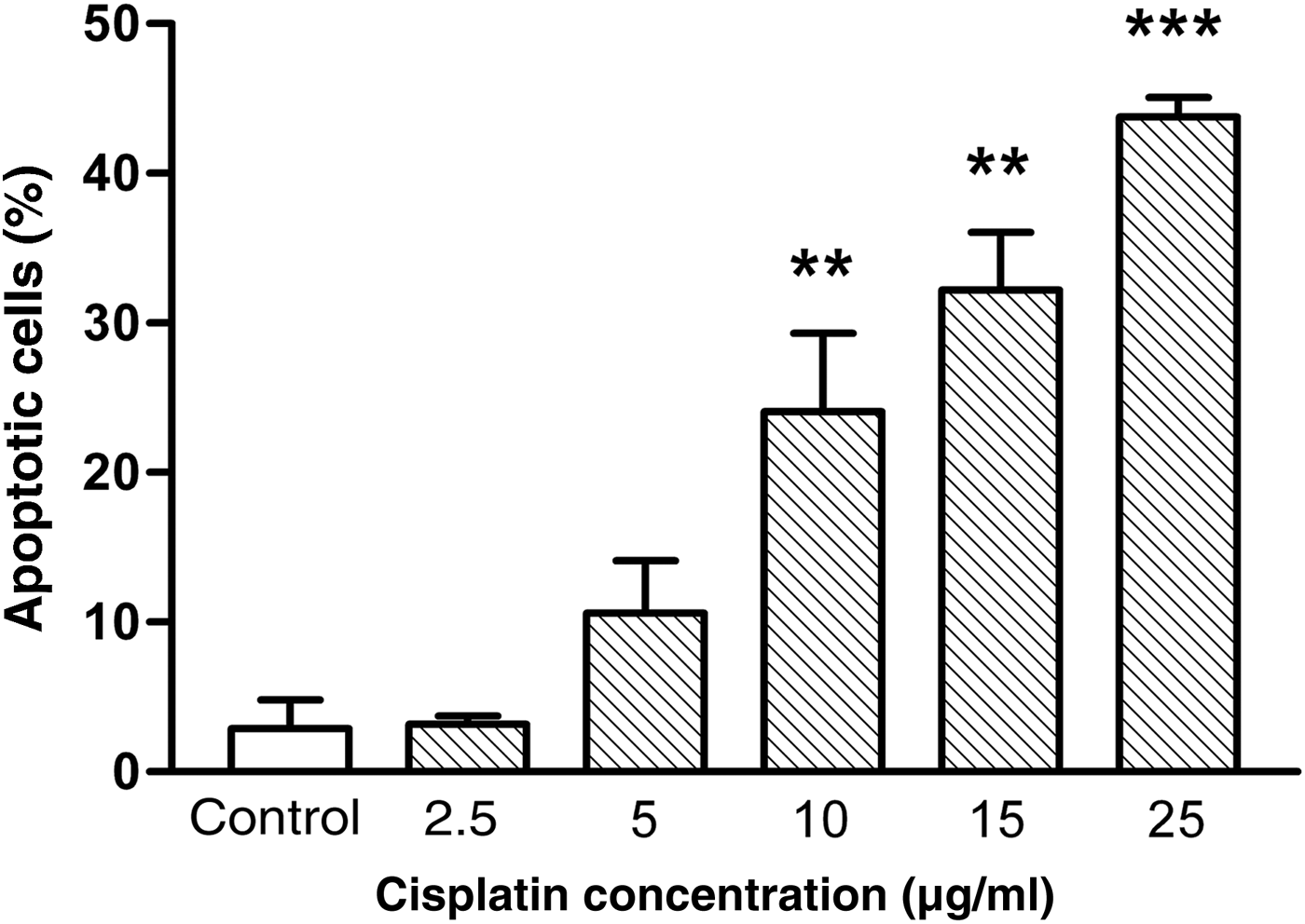
Hep-2 cells were exposed to graded concentrations of cisplatin (2.5–25 µg/ml) and apoptosis was evaluated after 24 hours. Cisplatin induced a concentration-dependent decrease in cell viability, with a statistically significant effect starting at 10 µg/ml (24.1 per cent of apoptotic cells, p < 0.01 vs control), and a maximal effect at 25 µg/ml (43.8 per cent of apoptotic cells, p < 0.001 vs control) (Figure 3), as quantified by microscopic analysis with Hoechst 33258 stain. Degenerated cells exhibited the typical features of apoptosis,Reference Wyllie, Kerr and Currie31, Reference Bursch, Oberhammer and Schulte-Hermann32 including chromatin condensation and fragmentation. In contrast, control cultures showed no change in the number of viable cells, with less than 5 per cent apoptotic cells (Figure 3).
Concentration dependence of the effect of cisplatin on Hep-2 cell viability. Cells were exposed to concentrations of cisplatin ranging from 2.5 to 25 µg/ml for 1 hour (‘single high-dose shot’), and apoptosis was evaluated after 24 hours. Results are from three independent experiments (**p < 0.01, ***p < 0.001 vs control).
Cetuximab enhanced cisplatin pro-apoptotic effect
In order to investigate whether cetuximab modulated the pro-apoptotic effect of cisplatin, Hep-2 cells were incubated with cetuximab for 24 hours (10 or 100 µg/ml). A submaximal concentration of cisplatin (10 µg/ml) was then added to cultures as a single high-dose shot, in the presence or absence of cetuximab. Apoptosis was measured after 24 hours (Figure 4).
(a) Effect of cetuximab and cisplatin on Hep-2 cell viability. Cells were incubated with cetuximab for 24 hours, and subsequently cisplatin was added for 1 hour as stated in the text. Apoptosis was measured after 24 hours. Results are from three independent experiments (***p < 0.001 vs control, °°p < 0.01 vs cisplatin, ##p < 0.01 vs cisplatin plus cetuximab 10 µg/ml). (b) – (g) Staining with Hoechst 33258 fluorescent dye: (b) control; (c) 10 µg/ml cisplatin; (d) 10 µg/ml cetuximab; (e) 100 µg/ml cetuximab; (f) 10 µg/ml cisplatin plus 10 µg/ml cetuximab; (g) 10 µg/ml cisplatin plus 100 µg/ml cetuximab. Arrows indicate typical apoptotic nuclei with fragmented or condensed chromatin.
Cisplatin induced a significant decrease in cell viability (25.5 per cent of apoptotic cells, p < 0.001 vs control), whereas cetuximab alone had no effect whatsoever on cell apoptosis. In the presence of 10 µg/ml cisplatin and 100 µg/ml cetuximab, the percentage of apoptotic cells was greater than that obtained with cisplatin alone (40.7 per cent of apoptotic cells, p < 0.01 vs cisplatin alone). These results indicate that combination treatment with cetuximab significantly increased the pro-apoptotic effect of cisplatin.
Discussion
In the present study, we investigated the effects of the administration, with different modalities, of cetuximab (a monoclonal humanised anti-EGFR antibody), alone or combined with cisplatin (a well-known cytotoxic drug), on the laryngeal cancer cell line Hep-2.
When administered alone, cetuximab was able to inhibit cell proliferation, but did not act as a cytotoxic drug, and did not induce apoptosis. Moreover, inhibition of proliferation tended to be overcome by Hep-2 cells after 48 hours. This antiproliferative activity is consistent with the hypothesised role for cetuximab in inhibiting repopulation,Reference Krause, Ostermann, Petersen, Yaromina, Hessel and Harstrick33 which may be one of the reasons for its proven efficacy on head and neck SCCs when used in combination with radiotherapy.Reference Bonner, Harari, Giralt, Azarnia, Shin and Cohen26
• Cetuximab, an epidermal growth factor receptor inhibitor, is one of the therapies approved for head and neck squamous cell carcinoma
• Its molecular mechanisms of action are not fully understood and the best modality for its clinical use remains unclear
• Cetuximab administered alone could inhibit cell proliferation, but did not act as a cytotoxic drug or induce apoptosis; inhibition of cell proliferation was overcome by Hep-2 cells after 48 hours
• Such results may prompt a re-evaluation of cetuximab administration
• When used in combination with a submaximal concentration of cisplatin, EGFR antibody significantly enhanced cisplatin-induced apoptosis
The recovery from the proliferative block by cancer cells after treatment with cetuximab was at least partially avoided by replacing the drug every 24 hours during the time course of the experiments. The recovery of proliferative ability may be at least in part due to the increasing number of activated receptors over the time course, which may in turn be mediated by an increased autocrine secretion of an agonist (such as epidermal growth factor or transforming growth factor α) via an overexpression of EGFR on the cell surface or via degradation of the drug. However, we cannot exclude the activation of alternative cellular pathways that do not need EGFR activation to promote proliferation. The molecular mechanisms underlying this phenomenon remain to be evaluated.
The recovery of proliferative ability by cancer cells observed 24 hours after treatment may be relevant from a clinical point of view. The current administration protocol of cetuximab in our institution, as in many others, consists of intravenous cetuximab 1 week before radiotherapy, at a loading dose of 400 mg per square metre of body surface area over a period of 120 minutes. This is preceded by a test dose of 20 mg, and followed by weekly 60-minute infusions during the course of radiation therapy (usually for 7 weeks in our institution), as described by Bonner et al. Reference Bonner, Harari, Giralt, Azarnia, Shin and Cohen26 Our results may suggest a re-evaluation of the need for a loading dose before starting irradiation, which may cause some resistance in advance, and of the weekly schedule of concomitant administration, which may be too widely spaced.
Many important apoptotic pathways activated by cisplatin and by irradiation in laryngeal SCC cells are probably shared, and may rely on p53 and related proteins.Reference Mao, Hong and Papadimitrakopoulou34–Reference Bradford, Wolf, Carey, Zhu, Beals and Truelson37 In fact, it is well known that the response to cisplatin predicts radiosensitivity in the clinical setting.6 In our experimental model, when we used lower concentrations of cisplatin, leaving it in the medium for a longer time, we were trying to simulate cisplatin monochemotherapy (mainly utilised in palliation), in which cisplatin is administered as an intravenous infusion for 6–8 hours. On the other hand, when we treated the cells with a higher concentration of cisplatin for a short time, washing out the medium after 1 hour, our intention was to reproduce the strong, short apoptotic stimulus of a single irradiation session (which usually lasts from 5 to 20 minutes in the case of intensity-modulated radiation therapy). The apoptotic effects of cisplatin alone were about the same regardless of whether we treated cells with a high concentration hit for 1 hour or with a lower concentration for a longer time. In the former modality, the apoptotic effect of submaximal concentrations of cisplatin was strongly increased by the concomitant administration of anti-EGFR antibody, as it probably is in vivo during radiotherapy. The molecular pathways underlying such synergy, which is likely to be at least as relevant as the inhibition of repopulation in mediating the clinical effects of anti-EGFR antibodies, remain to be further elucidated. However, the inhibition of binding may render cells more vulnerable to cisplatin-induced apoptosis. In fact, the activation of the STAT-3 pathway by the EGFR is known to strongly inhibit several apoptotic pathways.Reference Song and Grandis38, Reference Quadros, Peruzzi, Kari and Rodeck39
In conclusion, cetuximab inhibited Hep-2 cell proliferation in a time-dependent manner. This effect was not associated with the induction of apoptosis. When used in combination with a submaximal concentration of cisplatin, the EGFR antibody significantly enhanced cisplatin-induced apoptosis. The in vitro increase in the effects of pro-apoptotic agents induced by cetuximab (which has limited effectiveness alone as an anticancer agent) is very interesting with regard to its concurrent use with a regional treatment such as radiotherapy for a disease, like laryngeal SCC, which mainly has a locoregional history. In fact, toxicity would be substantially limited to the irradiated field. Moreover, clinical experience confirms a favourable systemic toxicity profile, different from cisplatin, for patients treated with cetuximab and radiotherapy, whose most significant adverse side effects are usually local or cutaneous, and only rarely systemic or severe, if we exclude hypersensitivity or infusion reactions.
Acknowledgement
This work was supported in part by Grant D.1.-2006 from the Università Cattolica del Sacro Cuore, within its programme of promotion and diffusion of scientific research.