Background
Coronary artery anomalies are diverse lesions which can be associated with the risk of myocardial ischaemia and sudden cardiac death. Ischaemia may result from abnormal ostia, acute angulation, or compression due to an intramural, interarterial, or intramyocardial course of the affected vessel. In patients with the evidence of impaired coronary blood flow or high-risk lesions, surgical intervention may be indicated to improve myocardial perfusion and mitigate risk of sudden cardiac death.
A single left coronary artery giving rise to an interarterial right coronary artery is an exceedingly rare anatomic variant with few case reports in the literature. Reference Lipton, Barry, Obrez, Silverman and Wexler1–Reference Swaminath, Panikkath, Strefling, Rosales, Narayanan and Wischmeyer6 We report a Division I collegiate athlete who presented with ischaemic symptoms and significantly compromised coronary perfusion due to this anatomy who underwent transection and reimplantation of the anomalous right coronary artery.
Surgical technique
The operation was performed via median sternotomy on cardiopulmonary bypass. The patient was cooled to 28°C. The proximal coronary branches were dissected. The right coronary artery shared a common wall with the aorta involving the intercoronary commissure. Following cross-clamping and antegrade cardioplegia, an oblique aortotomy was performed extending into the non-coronary cusp. The right coronary artery was ligated and divided at its take-off from its common wall with the aorta. A neo-ostium was created in the right coronary sinus of Valsalva with 3.5 mm aortic punch, and the right coronary artery was reimplanted with continuous posterior and interrupted anterior fine suture. Cardiopulmonary bypass time was 142 minutes and cross-clamp time was 71 minutes.
Clinical vignette
An 18-year-old female athlete with mitral valve prolapse and 10-year history of exertional chest pain presented for further workup of worsening symptoms during competitive sports. She was subsequently referred to our institution’s Coronary Artery Anomalies Program and completed standardised comprehensive multimodal evaluation Reference Molossi, Agrawal and Mery7 consisting of echocardiogram, CT with coronary angiography, exercise stress testing, dobutamine stress cardiac MRI, and cardiac catheterisation supplemented by coronary angiography, fractional flow reserve, instantaneous wave-free ratio, and intravascular ultrasound.
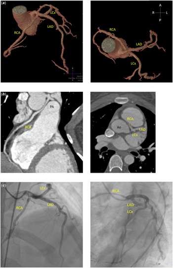
CT demonstrated a single non-stenotic coronary ostium in the left sinus of Valsalva giving rise to a left main coronary artery which trifurcated proximally into the left circumflex, left anterior descending, and right coronary arteries (Fig 1). The 3.5 cm interarterial segment of the right coronary artery had 60–70% narrowing on intravascular ultrasound and significantly decreased dobutamine stress instantaneous wave-free ratio of 0.78 (normal >0.85). Exercise stress testing showed no ischaemic changes. Stress cardiac MRI had no perfusion defects or delayed myocardial enhancement. Biventricular systolic function was low-normal with mildly dilated chambers, and there was mild mitral valve prolapse without regurgitation. Given her symptoms and haemodynamically significant coronary compression at the interarterial portion of the right coronary, she was put forward for surgical intervention by transection and reimplantation.
Preoperative imaging. ( a ) Three-dimensional reconstructions from CTA of preoperative coronary anatomy, viewed from a rotated coronal plane (left) and axial plane (right), demonstrating the single coronary artery originating from the left coronary sinus and trifurcating into an RCA, LAD, and left circumflex. ( b ) Representative oblique (left) and axial (right) cuts from preoperative CTA demonstrate the compressed interarterial course of the RCA. ( c ) Anatomy was confirmed on coronary angiography during cardiac catheterisation and significant RCA compression was noted based on invasive haemodynamic measures. Ao = aorta; CTA = computed tomography with coronary angiography; LAD = left anterior descending artery; LCx = left circumflex coronary artery; RCA = right coronary artery.

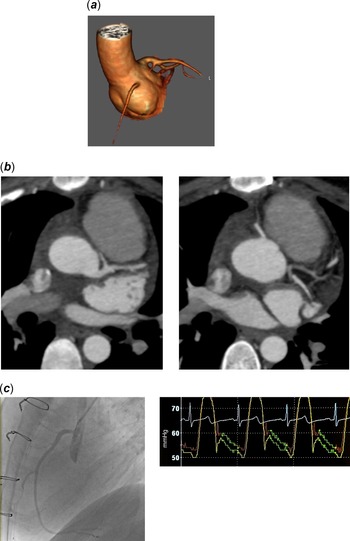
She was discharged home 3 days post-operatively with brief readmission for post-cardiotomy syndrome managed with NSAIDs and diuretics. Holter monitoring was notable for isolated episode of non-sustained four-beat ventricular tachycardia and frequent premature ventricular contractions (the latter also seen pre-operatively). At 3-month post-operative follow-up, bilateral coronaries were patent on CT (Fig 2) with prograde flow on echocardiogram. Stress fractional flow reserve and instantaneous wave-free ratio were normal, and there was no significant luminal diameter change throughout the reimplanted coronary. Stress cardiac MRI confirmed no rest or inducible perfusion defects, wall motion abnormalities, or delayed enhancement. Exercise stress testing was likewise without the evidence of ischaemia. Electrocardiogram showed improving non-specific ST changes. Repeat Holter showed 4% premature ventricular contractions. She was asymptomatic and cleared of exercise restrictions. She will continue long-term follow-up with interval surveillance studies.
Postoperative angiography demonstrating patent dual ostium coronary system. ( a ) Three-dimensional reconstruction of aortic root and dual ostium coronary system from CTA. ( b ) Representative axial CTA images demonstrating patency of the LMCA and its proximal branches (left) and the RCA (right). Both coronaries now arise from their respective sinuses following transection of the anomalous RCA from the LMCA and its reimplantation into the right coronary sinus. ( c ) Post-operative catheterisation showed that her pre-operative haemodynamically significant coronary compression in the right coronary artery (dobutamine iFR of 0.78) had resolved, with post-operative dobutamine iFR measuring 0.92-0.93 (normal >0.85). Representative postoperative angiogram of the reimplanted RCA with patent ostium and course (left) and postoperative dobutamine iFR tracing (right) are shown. CTA = computed tomography with coronary angiography; iFR = instantaneous wave-free ratio; LMCA = left main coronary artery; RCA = right coronary artery.

Discussion
Coronary artery anomalies are rare defects, with reported prevalence estimated at 0.1–1% overall Reference Brothers, Frommelt, Jaquiss, Myerburg, Fraser and Tweddell8 , 0.1–0.7% for interarterial coronaries, Reference Brothers, Frommelt, Jaquiss, Myerburg, Fraser and Tweddell8 and 0.024–0.066% for a single coronary artery. Reference Bhattarai, Mahat and Sitaula2 Interarterial course of an anomalous coronary is recognised as a high-risk feature for myocardial ischaemia and sudden cardiac death Reference Bhattarai, Mahat and Sitaula2 . This patient’s anatomy with a single coronary from the left sinus of Valsalva giving rise to a left circumflex artery, left anterior descending, and interarterial right coronary artery (Lipton classification LII-B, Reference Lipton, Barry, Obrez, Silverman and Wexler1 Leiden convention 2RLCx Reference Gittenberger-de Groot, Koenraadt and Bartelings9 ) has only been reported in a handful of similar cases. Reference Lipton, Barry, Obrez, Silverman and Wexler1–Reference Swaminath, Panikkath, Strefling, Rosales, Narayanan and Wischmeyer6 Several of these cases report evidence of myocardial ischaemia. Reference Lipton, Barry, Obrez, Silverman and Wexler1,Reference Gowda, Khan, Undavia, Vasavada and Sacchi3,Reference Güner, Güner, Yalçın, Kalkan, Turna and Uslu5–Reference Swaminath, Panikkath, Strefling, Rosales, Narayanan and Wischmeyer6 Surgical management of this specific anomaly is sparse in the literature, with two young adults undergoing coronary artery bypass graft after presenting with acute myocardial infarction Reference Swaminath, Panikkath, Strefling, Rosales, Narayanan and Wischmeyer6 and aborted cardiac arrest. Reference Güner, Güner, Yalçın, Kalkan, Turna and Uslu5 Broader experience is available relating to anomalous coronaries with interarterial course originating from a contralateral sinus.
Surgical techniques to directly address anatomic sources of impaired perfusion are gaining favour as alternatives to bypass grafting, which suffers from graft failure due to competitive flow. Reference Brothers, Frommelt, Jaquiss, Myerburg, Fraser and Tweddell8 Coronary unroofing is a common strategy for intramural segments with favourable mid-term durability but may pose a risk of aortic valve regurgitation if the coronary commissures must be manipulated to achieve repair. Reference Jegatheeswaran, Devlin and Williams10 Transection and reimplantation have also been successfully employed to address interarterial compression and intramurality unfavourable to unroofing in patients with coronary origin from a contralateral sinus. Reference Jegatheeswaran, Devlin and Williams10
We applied the transection and reimplantation technique to an anomalous interarterial right coronary artery originating from the proximal left main coronary artery in the setting of compromised coronary perfusion. The advantages of this technique include anatomic correction to a dual ostium coronary system with elimination of the interarterial geometry, minimised disruption to the native aortic valve apparatus, and preservation of bypass graft sources for potential future use. Although the coronary anastomosis may be prone to stenosis long-term, it is now rendered independent from the left coronary artery, creating protective redundancy compared to a single ostium configuration.
At 3-month follow-up, our patient’s ischaemic chest pain had resolved and she resumed athletic training. Post-operative workup shows improved coronary patency and no evidence of inducible myocardial ischaemia. Continued longitudinal follow-up remains essential to determine the long-term durability and complications of anomalous coronary repairs as surgical intervention becomes more common. As part of our standardised approach to patients with coronary anomalies, Reference Molossi, Agrawal and Mery7 short-term postoperative follow-up consists of a surgical visit at 1 week followed by cardiology visits at 1, 3, and 6 months. At 3 months, all pre-operative studies are repeated prior to release of exercise restriction to evaluate for resolution of preoperative abnormalities. In the long term, patients have cardiology visits every 1–2 years with ECG, echocardiogram as needed, and functional testing every 3–5 years with multidisciplinary discussion and further workup as needed for symptoms or abnormal/changing findings on surveillance testing.
Conclusions
Transection of anomalous right coronary artery from a left main coronary artery with direct aortic reimplantation is feasible and well-tolerated in the immediate post-operative period with improved coronary perfusion. However, long-term outcomes are lacking due to the rarity of the anatomy and recent adoption of this technique.
Acknowledgements
We thank the Texas Children’s Hospital Coronary Artery Anomalies Program (CAAP) for their dedication to advancing evidence-based patient care.
Financial support
This research received no specific grant from any funding agency, commercial, or not-for-profit sectors.
Conflicts of interest
None.
Ethical standards
The authors assert that all procedures contributing to this work comply with the ethical standards of the relevant national guidelines on human experimentation (Determining The Natural and "Unnatural" History of Anomalous Aortic Origin of a Coronary Artery With an Interarterial Course (AAOCA): Establishing A Multi-Institutional Registry, Protocol H-30898) and with the Helsinki Declaration of 1975, as revised in 2008, and have been approved by the institutional committees (Baylor College of Medicine).