INTRODUCTION
Trypanosomatid protozoa are the aetiological agents of several major insect-transmitted parasitic illnesses such as Chagas disease (Trypanosoma cruzi) and leishmaniasis (Leishmania spp.). These infections are concentrated in very poor areas of the planet and are included among the ‘Neglected Tropical Diseases’ (NTDs) with the highest rates of death (Cavalli and Bolognesi, Reference Cavalli and Bolognesi2009; Espuelas et al. Reference Espuelas, Plano, Nguewa, Font, Palop, Irache and Sanmartín2012; WHO, 2015).
Existing drug treatments for these diseases are far from satisfactory and the development of a vaccine is an unachieved goal. Current therapies are not adequate due essentially to several factors such as the low therapeutic indexes leading to high toxicities and unacceptable side-effects, the emergence of resistant parasites, the difficulty of treatment compliance due to complex protocols, high prices that are unaffordable for the affected countries, etc. These drawbacks of the current therapy make the search for new drugs of urgent need. Nevertheless, owing to the low income of affected population, the investment in the development of new drugs against these diseases has not been financially attractive for pharmaceutical companies, and the interest of academic institutions is rather limited. This bleak picture is changing in recent years thanks to the financial backing from not-for-profit organizations and the involvement of public–private partnerships (Dujardin et al. Reference Dujardin, González-Pacanowska, Croft, Olesen and Späth2010). In this article we have focused on developing new compounds against the aetiological agents of the mentioned diseases caused by trypanosomatids, T. cruzi and Leishmania spp. Several excellent articles covering different aspects and providing a current overview of chemotherapy of Chagas disease (Soeiro and de Castro, Reference Soeiro and de Castro2009; Cerecetto and González, Reference Cerecetto and González2010; Sánchez-Sancho et al. Reference Sánchez-Sancho, Campillo and Páez2010; Urbina, Reference Urbina2010; Guedes et al. Reference Guedes, Silva, Gutierrez and Silva2011; Bermudez et al. Reference Bermudez, Davies, Simonazzi, Real and Palma2016) and leishmaniasis (Mishra et al. Reference Mishra, Saxena and Singh2007; Santos et al. Reference Santos, Coutinho, Madeira, Bottino, Vieira, Nascimento, Bernardino, Bourguignon, Corte-Real, Pinho, Rodrigues and Castro2008; Singh et al. Reference Singh, Kumar and Singh2012; Rajasekaran and Chen, Reference Rajasekaran and Chen2015) have been published recently.
In this context, the antichagasic and leishmanicidal properties of some pyrazole-containing benzo[g]phthalazines (Sánchez-Moreno et al. Reference Sánchez-Moreno, Sanz, Gómez-Contreras, Navarro, Marín, Ramírez-Macias, Rosales, Olmo, Garcia-Aranda, Campayo, Cano, Arrebola and Yunta2011, Reference Sánchez-Moreno, Gómez-Contreras, Navarro, Marín, Ramírez-Macías, Olmo, Sanz, Campayo, Cano and Yunta2012b) and pyrazole-derived macrocyclic polyamines (Sánchez-Moreno et al. Reference Sánchez-Moreno, Marín, Navarro, Lamarque, García-España, Miranda, Huertas, Olmo, Gómez-Contreras, Pitarch and Arrebola2012c; Navarro et al. Reference Navarro, Sánchez-Moreno, Marín, García-España, Ramírez-Macías, Olmo, Rosales, Gómez-Contreras, Yunta and Gutiérrez-Sánchez2014) have been reported in the last years. It has been proposed that these compounds act as inhibitors of iron superoxide dismutase (Fe-SOD), essential for the parasites survival. In all cases, the complexing ability of these compounds has been related to anti-trypanosomatid activity. In fact, it has been pointed out (Sánchez-Moreno et al. Reference Sánchez-Moreno, Marín, Navarro, Lamarque, García-España, Miranda, Huertas, Olmo, Gómez-Contreras, Pitarch and Arrebola2012c; Navarro et al. Reference Navarro, Sánchez-Moreno, Marín, García-España, Ramírez-Macías, Olmo, Rosales, Gómez-Contreras, Yunta and Gutiérrez-Sánchez2014) that complexation of iron atom may modify the enzyme active site by dissociation of the metal ion, by changes in the coordination geometry or reducing the interaction between the two monomers of the enzyme.
On the other hand, some pyrazole-3-carboxylic acid derivatives have shown to be weak inhibitors of T. cruzi proline racemase, which has been validated as a target for the design of new chemotherapeutic agents against Chagas disease (Berneman et al. Reference Berneman, Montout, Goyard, Chamond, Cosson, d'Archivio, Gouault, Uriac, Blondel and Minoprio2013).
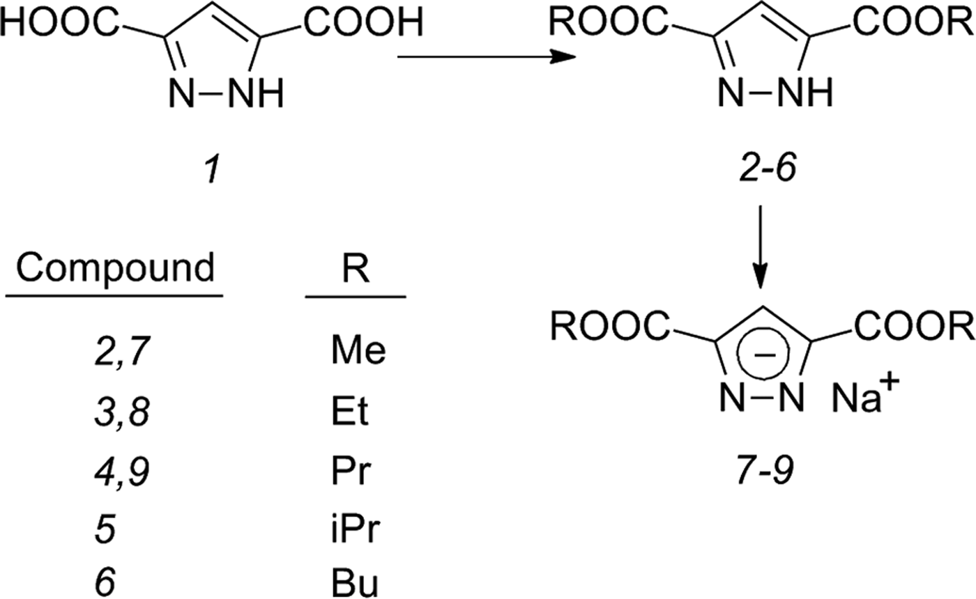
On these bases, keeping in mind the important role of azole unit in the complexation of metals by pyrazole-containing macrocycles (Sharma et al. Reference Sharma, Arán, Navarro, Ramos-Gallardo and Vegas1994; Lamarque et al. Reference Lamarque, Navarro, Miranda, Arán, Ochoa, Escartí, García-España, Latorre, Luis and Miravet2001; Escartí et al. Reference Escartí, Miranda, Lamarque, Latorre, García-España, Kumar, Arán and Navarro2002; Miranda et al. Reference Miranda, Escartí, Lamarque, García-España, Navarro, Latorre, Lloret, Jiménez and Yunta2005), we decided to study the activity against T. cruzi, Leishmania infantum and Leishmania braziliensis of some simple dialkyl pyrazole-3,5-dicarboxylates (compounds 2–6); in order to improve solubility in water, some of the corresponding sodium 3,5-bis(alkoxycarbonyl)pyrazolates (compounds 7–9) were also tested. Since initial in vitro assays showed interesting activity against different morphological forms of the mentioned protozoa, further studies on infection rates and in vivo antichagasic activity were also carried out for the most interesting pyrazole derivatives. Complementary analyses of inhibition of Fe-SOD of parasites and acute toxicity of the most active compounds were also conducted.
MATERIALS AND METHODS
Preparation of dialkyl 1H-pyrazole-3,5-dicarboxylates 2–6 and sodium 3,5-bis(alkoxycarbonyl)pyrazolates 7–9
Diesters 2–6 were prepared by acid-catalysed (HCl) esterification of the commercially available dicarboxylic acid 1 with the corresponding alcohols, following a procedure closely related to those previously reported for dimethyl (2) (Askew et al. Reference Askew, Bednar, Bednar, Claremon, Cook, McIntyre, Hunt, Gould, Lynch, Lynch, Gaul, Stranieri, Sitko, Holahan, Glass, Hamill, Gorham, Prueksaritanont, Baldwin and Hartman1997) and diethyl (3) (Schenck et al. Reference Schenck, Downes, Milne, Mackenzie, Boucher, Whelan and Bosnich1985) pyrazole-3,5-dicarboxylates (Fig. 1). Sodium pyrazolates 7–9 were prepared by treatment of esters 2–4 with sodium hydroxide, following the procedure reported for the corresponding bis(ethoxycarbonyl) derivative 8 (Reviriego et al. Reference Reviriego, Rodríguez-Franco, Navarro, García-España, Liu-González, Verdejo and Domènech2006) (Fig. 1). Analytical and spectral data of the obtained compounds are in agreement with their respective structures. Detailed experimental procedures are given as Supplementary material.
Synthesis of dialkyl pyrazole-3,5-dicarboxylates 2–6 and the sodium salts (pyrazolates) 7–9. R substituents: Me, methyl; Et, ethyl; Pr, propyl; iPr, isopropyl; Bu, butyl.

Parasites strains cultures
Epimastigotes of T. cruzi SN3 strain (IRHOD/CO/2008/SN3), initially isolated from domestic Rhodnius prolixus from Colombia (Téllez-Meneses et al. Reference Téllez-Meneses, Mejía-Jaramillo and Triana-Chávez2008) and donated to our laboratory, and promastigotes of L. infantum (MCAN/ES/2001/UCM-10) and L. braziliensis (MHOM/BR/1975/M2904) were cultivated in vitro in trypanosomes liquid medium (MTL, Gibco) with 10% inactivated fetal bovine serum (FBS, Biowest) and were kept in an air atmosphere at 28 °C in Roux flasks (Corning, USA) with a surface area of 75 cm2, as reported is previous articles (González et al. Reference González, Marín, Rodríguez-González, Hitos, Rosales, Reina, Díaz, González-Coloma and Sánchez-Moreno2005; Marín et al. Reference Marín, Clares, Ramírez-Macías, Blasco, Olmo, Soriano, Verdejo, Rosales, Gomez-Herrera, García-España and Sánchez-Moreno2013; Olmo et al. Reference Olmo, Rotger, Ramírez-Macías, Martínez, Marín, Carreras, Urbanová, Vega, Chaves-Lemaur, Sampedro, Rosales, Sánchez-Moreno and Costa2014c).
Cells culture and cytotoxicity tests
Vero cells (ECACC number 84113001) were grown in RPMI medium (Gibco), supplemented with 10% inactivated FBS in a humidified 95% air, 5% CO2 atmosphere at 37 °C for 2 days. The cytotoxicity test for Vero cells was performed according to a previously described methodology (Marín et al. Reference Marín, Ramírez-Macías, López-Céspedes, Olmo, Villegas, Díaz, Rosales, Gutiérrez-Sánchez and Sánchez-Moreno2011). In summary, Vero cells cultures were treated with stock solutions of the compounds to be tested in dimethyl sulphoxide (DMSO) to give final concentrations 100, 50, 25, 10 and 1 µ m; final concentration of DMSO in the culture medium was 0·01%. After 72 h cell viability was determined by flow cytometry (FACSVantage flow cytometer, Becton Dickinson) using propidium iodide and fluorescein diacetate to detect cells with plasma membrane damaged or intact, respectively (Marín et al. Reference Marín, Ramírez-Macías, López-Céspedes, Olmo, Villegas, Díaz, Rosales, Gutiérrez-Sánchez and Sánchez-Moreno2011). The viability percentage was calculated in comparison with the control culture containing only DMSO (0·01%). The IC50, i.e. the concentration required to give 50% of growth inhibition, was calculated using linear regression analysis from the K c values corresponding to the concentrations employed.
On the other hand, J774·2 macrophages (ECACC number 85011428) were grown in MEM (Gibco), supplemented with 20% inactivated FBS in a humidified 95% air and 5% CO2 atmosphere at 37 °C for 2 days. The cytotoxicity test for macrophages was performed according to the procedure described for Vero cells (Marín et al. Reference Marín, Ramírez-Macías, López-Céspedes, Olmo, Villegas, Díaz, Rosales, Gutiérrez-Sánchez and Sánchez-Moreno2011).
In vitro activity against extracellular forms of T. cruzi, L. infantum and L. braziliensis
The assayed compounds and the reference drugs (benznidazole or glucantime) were dissolved in DMSO as described above and added to the culture medium of epimastigotes (T. cruzi) or promastigotes (Leishmania spp.) at concentrations 100, 50, 25, 10 and 1 µ m. Cultures containing only DMSO (0·01%) were used as control. The effect of each compound was tested after 72 h using a Neubauer haemocytometric chamber according to a previously published procedure (González et al. Reference González, Marín, Rodríguez-González, Hitos, Rosales, Reina, Díaz, González-Coloma and Sánchez-Moreno2005; Ramírez-Macías et al. Reference Ramírez-Macías, Marín, Es-Samti, Fernández, Guardia, Zentar, Agil, Chahboun, Alvarez-Manzaneda and Sánchez-Moreno2012). The antiparasitic effect is expressed as the IC50, calculated by linear regression analysis from the K c values corresponding to the concentrations employed.
In vitro activity against intracellular forms of T. cruzi, L. infantum and L. braziliensis
According to previously developed experimental procedures (Ramírez-Macías et al. Reference Ramírez-Macías, Marín, Es-Samti, Fernández, Guardia, Zentar, Agil, Chahboun, Alvarez-Manzaneda and Sánchez-Moreno2012), Vero cells (for T. cruzi) or macrophages (for Leishmania spp.) were grown as described above. Cells were seeded at a density of 104 cells well−1 in 24-well microplates (Nunc) with glass round coverslips on the bottom and cultured for 2 days (near 70% cell confluence). Afterwards the cells were infected in vitro with metacyclic forms of T. cruzi or promastigotes of L. infantum or L. braziliensis (105 parasites well−1) during 24 h. The non-phagocytosed parasites were removed by washing, and then the drugs, as stock solutions in DMSO as described above, were added at final concentrations 100, 50, 25, 10 and 1 µ m. Cells with the drugs were incubated for 72 h at 37 °C in 5% CO2. Cultures containing only DMSO (0·01%) were used as control. Drug activity was determined on the basis of number of amastigotes in treated and untreated (control) cultures in methanol-fixed and Giemsa-stained preparations. The number of amastigotes was determined by analysing 200 host cells distributed in randomly chosen microscopic fields. The anti-trypanosomatid effect is expressed as the IC50 values.
Infectivity assays with T. cruzi, L. infantum and L. braziliensis
According to a previously described method (Marín et al. Reference Marín, Ramírez-Macías, López-Céspedes, Olmo, Villegas, Díaz, Rosales, Gutiérrez-Sánchez and Sánchez-Moreno2011; Ramírez-Macías et al. Reference Ramírez-Macías, Marín, Es-Samti, Fernández, Guardia, Zentar, Agil, Chahboun, Alvarez-Manzaneda and Sánchez-Moreno2012), Vero cells (for T. cruzi infection) or macrophages (for Leishmania spp. infection) were grown as described earlier. The cells were then infected in vitro with metacyclic forms of T. cruzi or promastigotes of L. infantum or L. braziliensis. The tested drugs (IC25 concentrations) were added immediately after infection and the cultures incubated for 12 h at 37 °C in 5% CO2. After removal of non-phagocytosed parasites and drugs by washing, the infected cultures were grown for 10 days in fresh medium; fresh culture medium was added every 48 h. Activity of the different compounds was determined from the percentage of infected cells and the number of amastigotes per infected cell in treated and untreated cultures, in methanol-fixed and Giemsa-stained preparations; in the case of infection with T. cruzi, number of trypomastigotes in the culture medium was also determined. The percentage of infected cells and the mean number of amastigotes per infected cell were determined by analysing 200 host cells distributed in randomly chosen microscopic fields.
In vivo trypanocidal activity
According to previous reports (Marín et al. Reference Marín, Ramírez-Macías, López-Céspedes, Olmo, Villegas, Díaz, Rosales, Gutiérrez-Sánchez and Sánchez-Moreno2011; Olmo et al. Reference Olmo, Rotger, Ramírez-Macías, Martínez, Marín, Carreras, Urbanová, Vega, Chaves-Lemaur, Sampedro, Rosales, Sánchez-Moreno and Costa2014c), BALB/c female mice (Harlan Sprague Dawley Inc.) (6–8 weeks old, 20–25 g) were infected with 105 T. cruzi metacyclic forms by the intraperitoneal route. The animals were divided into the following groups of six mice: group 1, untreated (infected with T. cruzi but not treated; study of acute and chronic phases of disease); group 2, treated with compound 3 [infected and treated for five consecutive days (5–10 days post-infection (dpi) with compound 3 (50 mg kg−1 day−1 doses; intraperitoneal route)]; and group 3, treated with compound 8 (infected and treated as indicated for compound 3). This animal experiment was performed with the approval of the Bioethical Committee of the University of Granada.
The level of parasitaemia (number of trypomastigotes per mL of blood) was recorded every 2 days from 5 to 30 dpi. A blood sample (5 µL) drawn from the mandibular vein of each treated mouse was taken and diluted 1 : 15 (50 µL of citrate buffer: 0·1 m citric acid, 0·1 m sodium citrate and 20 µL of lysis buffer at pH 7·2: 2 m Tris–HCl, MgCl2). Parasites were counted using the Neubauer chamber.
After day 60, the animals reach the chronic phase and parasitaemia decreases independently of the treatment; in fact, day 120 post-infection blood parasites are undetectable by microscopic examination.
On day 120, each mice group (1, 2 and 3) was divided into two subgroups of three animals; three subgroups (1a, 2a and 3a) were subjected to immunosuppression induced by cyclophosphamide and the other three subgroups (1b, 2b and 3b) were reserved for post-mortem study of parasites clearance in heart tissue.
The immunosuppression treatment (four intraperitoneal injections of 200 mg kg−1 of body mass of cyclophosphamide monohydrate on alternate days) was applied to mice untreated (subgroup 1a) and treated with compounds 3 (subgroup 2a) and 8 (subgroup 3a) as reported (Cencig et al. Reference Cencig, Coltel, Truyens and Carlier2011; Olmo et al. Reference Olmo, Guardia, Marín, Messouri, Rosales, Urbanová, Chayboun, Chahboun, Alvarez-Manzaneda and Sánchez-Moreno2015). Seven days after the last cyclophosphamide injection, parasitaemia reactivation was calculated as before by counting blood trypomastigotes.
Furthermore, quantitative evaluation of circulating anti-T. cruzi antibodies was performed at days 15, 60, 90 and 120 post-infection using an enzyme-linked immunoassay (ELISA); an additional evaluation of antibodies was also carried out after immunosuppressive treatment with cyclophosphamide. The sera, diluted 1 : 100 in PBS (phosphate-buffered saline), were reacted with an antigen composed of a soluble Fe-SOD of T. cruzi epimastigotes isolated in our laboratory (Longoni et al. Reference Longoni, Marín, Sauri-Arceo, López-Cespedes, Rodríguez-Vivas, Villegas, Escobedo-Ortegón, Barrera-Pérez, Bolio-Gonzalez and Sánchez-Moreno2011; Sánchez-Moreno et al. Reference Sánchez-Moreno, Gómez-Contreras, Navarro, Marín, Olmo, Yunta, Sanz, Rosales, Cano and Campayo2012a). Results are expressed as the ratio of the absorbance (Abs) of each sample at 490 nm to the cut-off value; the cutoff for each reaction was the mean of the values determined for the negative controls plus three times the standard deviation.
Non-immunosuppressed mice, untreated (subgroup 1b) and treated with compounds 3 (subgroup 2b) and 8 (subgroup 3b), were sacrificed on day 120; their hearts were removed and total DNA was extracted following the purification procedure of Wizard Genomic DNA Purification Kit (Promega). The presence of the parasite DNA was detected by PCR using two primers designed previously in our laboratory, based on the published sequence of superoxide dismutase of T. cruzi CL Brenner strain (GenBank accession No. XM_808937): iSODTc-r (5′-GTTGTCGATGGCAACCTCTT-3′) and iSODTc-d (5′-ATGAGCTTCGTCATTCCTCC-3′). These primers amplify a fragment of near 300 bp belonging to sod-b (superoxide dismutase gene b) in all strains of T. cruzi. The reaction mixture for PCR was 1 µL of DMSO, 200 nm iSODd, 200 nm iSODr, 10 mm Tris–HCl (pH 9·0), 1·5 mm MgCl2, 50 mm KCl, 0·01% gelatin, 0·1% Triton X-100, 10 mm of each dNTP, 0·5 U of Taq DNA polymerase, 1–20 ng of kDNA and HPLC water to complete a final volume of 20 µL. PCR was performed using a MyCycler thermal cycler (Bio-Rad) with the following routine temperature: 95 °C/3 min, 30 cycles of 95 °C/30 s, 55·5 °C/45 s, 72 °C/30 s and 72 °C/7 min. Finally, the amplification products were subjected to electrophoresis on 1·6% agarose gel containing GelRed nucleic acid gel stain, during 90 min at 90 V (Olmo et al. Reference Olmo, Escobedo-Ortegón, Palma, Sánchez-Moreno, Mejía-Jaramillo, Triana and Marín2014b, Reference Olmo, Rotger, Ramírez-Macías, Martínez, Marín, Carreras, Urbanová, Vega, Chaves-Lemaur, Sampedro, Rosales, Sánchez-Moreno and Costac).
Acute toxicity study
This study was carried out using female albino mice of BALB/c strain (Harlan Sprague Dawley Inc.) (6–8 weeks old, 20–25 g). A group of five mice was used as the control, and two additional groups of five mice were respectively treated with compounds 3 and 8. Five doses of 100 mg kg−1 of each compounds were initially administered intraperitoneally on alternative days, and then doses of 500 mg kg−1 of each compound were given, also on alternative days, to reach a final cumulative dose of 6 g kg−1. A week after cessation of treatment, mice sera were sent to the Biochemical Service of the Granada University where some parameters related to kidney (urea and uric acid), heart (CK-MB and LDH) and liver (AST/GOT, ALT/GTP, bilirubin and alkaline phosphatase) damage were determined with commercial kits (Cromakit) using a BS-200 Chemistry Analyzer (Shenzhen Mindray Bio-medical Electronics) as reported (Olmo et al. Reference Olmo, Rotger, Ramírez-Macías, Martínez, Marín, Carreras, Urbanová, Vega, Chaves-Lemaur, Sampedro, Rosales, Sánchez-Moreno and Costa2014c, Reference Olmo, Guardia, Marín, Messouri, Rosales, Urbanová, Chayboun, Chahboun, Alvarez-Manzaneda and Sánchez-Moreno2015).
SOD inhibition assays
Fe-SOD activity was measured in homogenates obtained from parasites cultured as described above and disrupted by sonic disintegration (Olmo et al. Reference Olmo, Clares, Marín, González, Inclán, Soriano, Urbanová, Tejero, Rosales, Krauth-Siegel, Sánchez-Moreno and García-España2014a). SOD activities were determined spectrophotometrically following a reported procedure based on the photoreduction of nitroblue tetrazolium (NBT) to formazan by superoxide ions in the presence of a sensitising dye (riboflavin) (Beyer and Fridovich, Reference Beyer and Fridovich1987). Parasite homogenate, riboflavin and the studied compounds 3 and 8 (100, 50, 25, 10, 5 and 1 µ m) were added to the assay medium containing L-methionine, NBT and Triton X-100. SOD activity was calculated from the different absorbance values at 560 nm at the beginning of assay and after UV irradiation for 10 min. Human CuZn-SOD and other reagents were obtained from Sigma Chemical Co. Differences between activities of the control homogenate and those incubated with compounds 3 and 8 were obtained according to the Newman–Keuls test. IC50 values were calculated by lineal regression analysis from the inhibition values at the concentrations employed.
Statistical analyses
Data were recorded on an Excel spreadsheet (Microsoft), and statistical analyses were performed by using SPSS software (v. 21, IBM).
RESULTS AND DISCUSSION
In vitro anti-T. cruzi evaluation
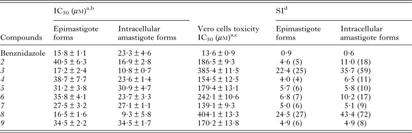
Epimastigotes of T. cruzi SN3 strain were used for the initial in vitro evaluation of compounds 2–9; this extracellular insect vector stage is easily handled in the laboratory and is very appropriate for preliminary studies of antichagasic activity (Marín et al. Reference Marín, Ramírez-Macías, López-Céspedes, Olmo, Villegas, Díaz, Rosales, Gutiérrez-Sánchez and Sánchez-Moreno2011; Fonseca-Berzal et al. Reference Fonseca-Berzal, Ibáñez-Escribano, Reviriego, Cumella, Morales, Jagerovic, Nogal-Ruiz, Escario, da Silva, Soeiro, Gómez-Barrio and Arán2016). Further studies were carried out on amastigotes, intracellular mammalian host cells forms, and consequently, more interesting from the point of view of human disease; with this aim, Vero cells were infected with metacyclic forms of the parasite, which transformed quickly into amastigotes. Vero cells were also used as a model for the study of non-specific cytotoxicity of pyrazole derivatives against mammalian cells. Compounds 2–9 as well as the standard drug benznidazole were assayed at concentrations of 1–100 µ m; DMSO used to prepare the stock solutions of the tested compounds has been shown to be non-toxic and without inhibitory effects on parasite and Vero cells growth at a final concentration of 0·01% (Marín et al. Reference Marín, Ramírez-Macías, López-Céspedes, Olmo, Villegas, Díaz, Rosales, Gutiérrez-Sánchez and Sánchez-Moreno2011). In vitro activities (IC50 values) of the studied compounds on epimastigotes and amastigotes of T. cruzi, the non-specific cytotoxicity against Vero cells (IC50 values) and the corresponding selectivity indexes (SI) are gathered in Table 1.
In vitro activity, non-specific cytotoxicity and SI found for pyrazole derivatives 2–9 and benznidazole (reference drug) on extra- and intracellular forms of T. cruzi

a IC50: concentration required to give 50% inhibition, calculated by linear regression analysis from the K c values at concentrations employed (1, 10, 25, 50 and 100 µ m); K c (culture growth constant) corresponds to the slope resulting from plotting the log of the growth measurement versus time for each drug concentration.
b Average values of four separate experiments ± standard deviations.
c Average values of three separate experiments ± standard deviations.
d SI = IC50 Vero cells/IC50 extra- and intracellular forms of parasites. In parentheses, fold increase in the SI of the compound compared with that of the reference drug.
All compounds showed good activities against the two stages of the parasite (IC50 = 9·3–40·5 µ m), of the same order or slightly smaller or larger than those of the reference drug. The most active compounds, especially against amastigotes, were diethyl ester 3 (IC50 = 10·8 µ m) and the corresponding sodium pyrazolate 8 (IC50 = 9·3 µ m). The major differences among the assayed compounds were related to non-specific cytotoxicities against Vero cells, reflected in the obtained SI values. Considering all the data included in Table 1, compounds 3 and 8, with SI values 25–72 times higher than those of benznidazole depending on the stage of the parasite, showed to be the most promising compounds and were further investigated.
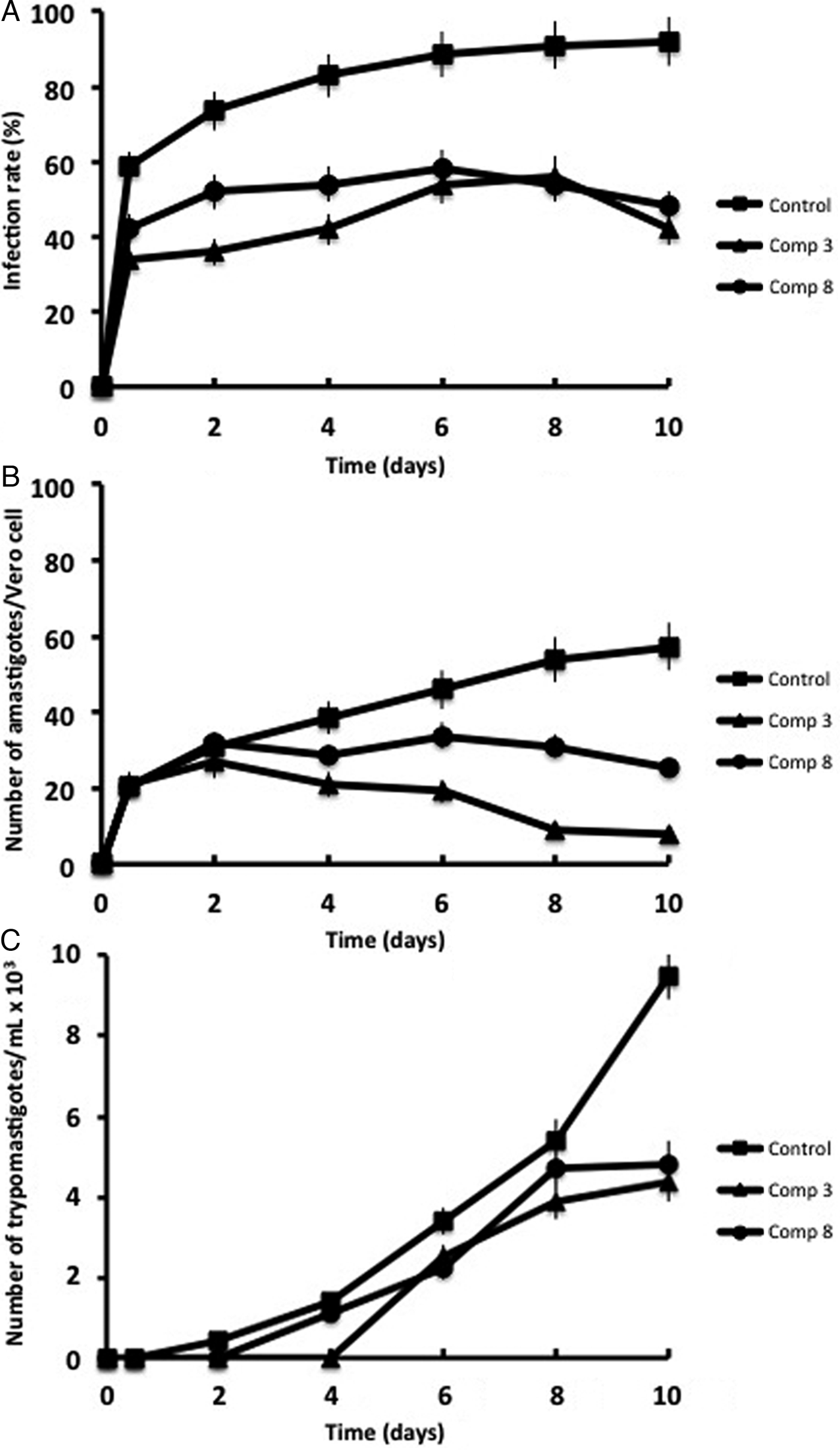
Thus, a study of T. cruzi propagation in Vero cells, with and without addition of the products showing highest activity in the previously described assays, compounds 3 and 8, was carried out (Fig. 2). During a 10-day period, different parameters such as the percentage of infection, the average number of amastigotes per infected Vero cell and the number of trypomastigotes present in the culture medium were studied. Following the infection of Vero cells with metacyclic forms, the parasites invaded the cells and transformed to amastigotes within 1 day. In the absence of drugs, the rate of host-cell infection reached its maximum, ca. 92% of invasion, on day 10 (Fig. 2A). Then the assay was carried out in the presence of compounds 3 and 8 at their IC25 concentrations in order to see their effects but minimising the number of dead parasite cells; in these cases, at the end of the experiment, the rate of infection decreased by 54% and 48% for 3 and 8, respectively, in relation to the untreated control; keeping in mind that typical values for benznidazole in this test are around 23% of reduction (Sánchez-Moreno et al. Reference Sánchez-Moreno, Marín, Navarro, Lamarque, García-España, Miranda, Huertas, Olmo, Gómez-Contreras, Pitarch and Arrebola2012c), compounds 3 and 8 were shown to be much more efficient than the reference drug.
Effect of pyrazole derivatives 3 and 8 (IC25 concentrations) on the infection rate and growth of T. cruzi in Vero cells: (A) rate of infection; (B) mean number of amastigotes per infected Vero cell; (C) number of trypomastigotes per mL in the growth medium. The represented values are the averages of four separate determinations; error bars represent standard deviations. Significant differences between the control group and groups treated with compounds 3 and 8 for α = 0·05.

On the other hand, in the absence of drugs, the average number of amastigotes per Vero cell gradually increased during the 10-day period to a value of ca. 57 (Fig. 2B). Treatment with compounds 3 and 8 remarkably reduced this number to ca. 8 and 25, respectively (86 and 56% of reduction related to the untreated control); the reference drug benznidazole is much less efficient in this assay, showing typical values of reduction in relation to the control of only 13% (Sánchez-Moreno et al. Reference Sánchez-Moreno, Marín, Navarro, Lamarque, García-España, Miranda, Huertas, Olmo, Gómez-Contreras, Pitarch and Arrebola2012c).
Finally, trypomastigotes in the culture medium are coming from the rupture of Vero cells infected by amastigotes; the number of trypomastigotes per mL found in the untreated culture medium (Fig. 2C) reached a maximum (ca. 9·5 × 103) on day 10. Once again this value was substantially reduced by compounds 3 and 8 (54 and 49%, respectively), values slightly better than those typically found for benznidazole in this assay (ca. 46% of decrease) (Sánchez-Moreno et al. Reference Sánchez-Moreno, Marín, Navarro, Lamarque, García-España, Miranda, Huertas, Olmo, Gómez-Contreras, Pitarch and Arrebola2012c).
Discrepancies sometimes observed in all these experiments between the values found for esters 2–4 and for their respective sodium salts 7–9 may be due to pharmacokinetic issues mainly related to the low solubility of esters in aqueous media.
In vivo anti-T. cruzi evaluation
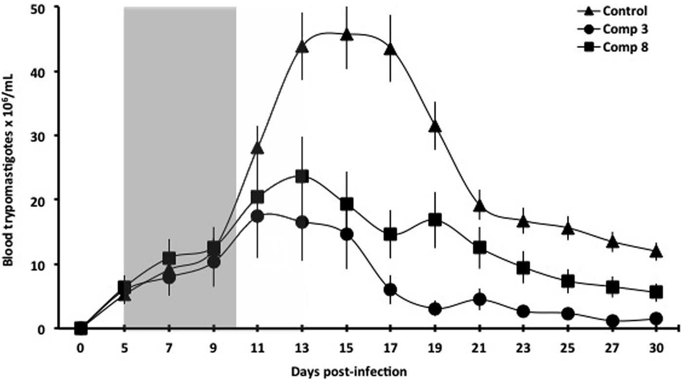
Considering the promising results obtained in the previously mentioned in vitro assays, compounds 3 and 8 were selected for the study of in vivo activity on infected female BALB/c mice. Both significant stages of Chagas disease, i.e. the acute and the chronic phases, were taken into account. For the acute phase experiments, the first 30 days after infection were considered (Fig. 3). On the days of maximum parasite load (ca. 15 days after infection) the assayed compounds decreased notably the number of trypomastigotes. At the end of the acute phase period (day 30), compounds 3 (group 2) and 8 (group 3) reduced the level of parasitaemia by 88 and 53%, respectively, in relation to the untreated control (group 1), values clearly higher than those typically found for the reference drug benznidazole (ca. 25%) (Sánchez-Moreno et al. Reference Sánchez-Moreno, Marín, Navarro, Lamarque, García-España, Miranda, Huertas, Olmo, Gómez-Contreras, Pitarch and Arrebola2012c).
T. cruzi parasitaemia in the murine model of acute Chagas disease: effect of pyrazole derivatives 3 and 8 (50 mg kg−1 day−1 doses during 5 days). Grey shading indicates the days of treatment. The represented points are the average values for each group of six mice; error bars represent standard deviations. Significant differences between the control group and groups treated with compounds 3 and 8 for α = 0·05.

None of the control animals or those treated with compounds 3 and 8 died during the treatment; however, similar assays performed in the presence of benznidazole always led to mice mortality values of about 20% (Sánchez-Moreno et al. Reference Sánchez-Moreno, Marín, Navarro, Lamarque, García-España, Miranda, Huertas, Olmo, Gómez-Contreras, Pitarch and Arrebola2012c).
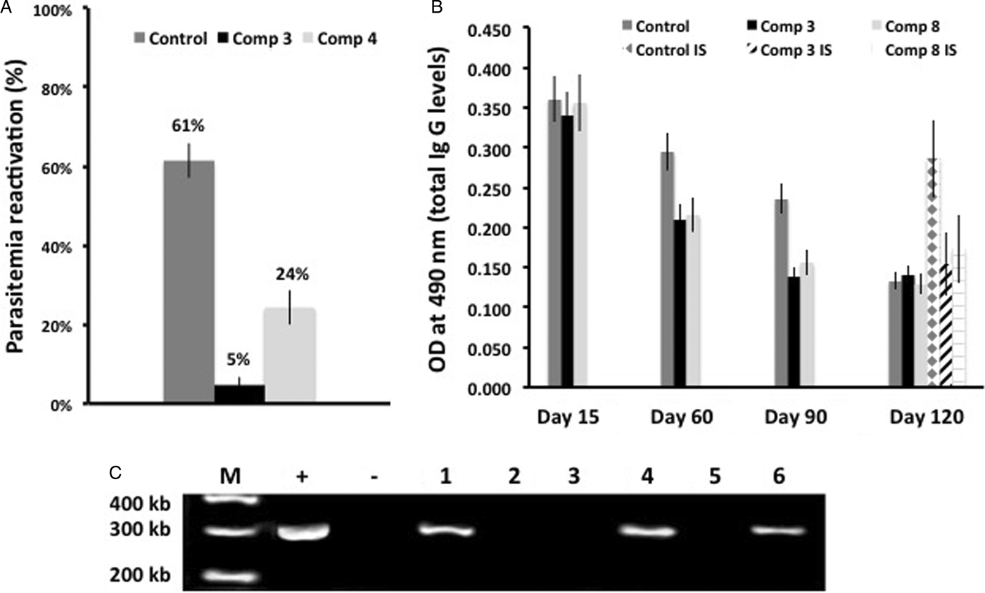
In relation to the last stage of the disease, the mice used for the study of the acute phase were kept up to day 120, after which the advanced chronic phase was reached and blood parasites could not be detected by microscopic examination. This day 120, three mice subgroups (1a, 2a and 3a) were subjected to immunosuppression induced by cyclophosphamide. This treatment allowed determining the degree of reactivation of parasitaemia and therefore the parasitological cure of animals. As shown in Fig. 4A, the percentage of parasitaemia reactivation after immunosuppression was 61% for the untreated control (subgroup 1a), while the values for compounds 3 and 8 were 5% (subgroup 2a) and 24% (subgroup 3a), respectively.
Murine model of chronic Chagas disease: (A) T. cruzi parasitaemia reactivation percentage following administration of a cyclophosphamide treatment for both the control subgroup and the mice subgroups treated with compounds 3 and 8; the indicated values correspond to averages for each subgroup of 3 mice; error bars represent standard deviations; (B) total levels of anti-T. cruzi Ig G measured by ELISA, expressed in absorbance units (OD, optical density) for the control and treated groups at different days post-infection; results are expressed as the average values for each group (six mice) or for immunosuppressed subgroups (IS; three mice); error bars represent standard deviations; (C) post-mortem PCR analysis of heart tissue of non-immunosuppressed mice at the end of experiment; lanes: (M) base pair marker; (+) PCR positive control; (−) PCR negative control; (1–3) mice treated with compound 3; (4–6) mice treated with compound 8.

Additionally, variations observed in the concentrations of antibodies against T. cruzi, showing the immunological state of the mouse, were also recorded as shown in Fig. 4B; after reaching the chronic phase (day 120), an immunological balance is achieved in all cases; this equilibrium stage is artificially broken after immunosuppression, which induces in the untreated control (subgroup 1a) a considerable increase in immunoglobulin G (IgG) levels as a result of the presence of parasite, while in animals treated with compounds 3 (subgroup 2a) and 8 (subgroup 3a), levels remain fairly constant, indicating lower parasitaemia.
Finally, results of post-mortem analysis of parasites clearance in heart tissue of immunocompetent mice (subgroups 2b and 3b) using the PCR technique are gathered in Fig. 4C. After treatment with compound 3 (subgroup 2b) the heart tissue of 66% of mice is clean of parasites (lanes 1–3), while the percentage drops to 33% in those treated with compound 8 (subgroup 3b) (lanes 4–6); all mice infected and untreated (subgroup 1b) showed positive bands in the agarose gel after PCR (data not shown).
In vitro leishmanicidal activity evaluation
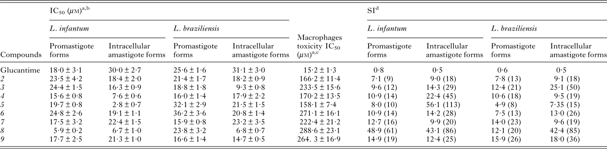
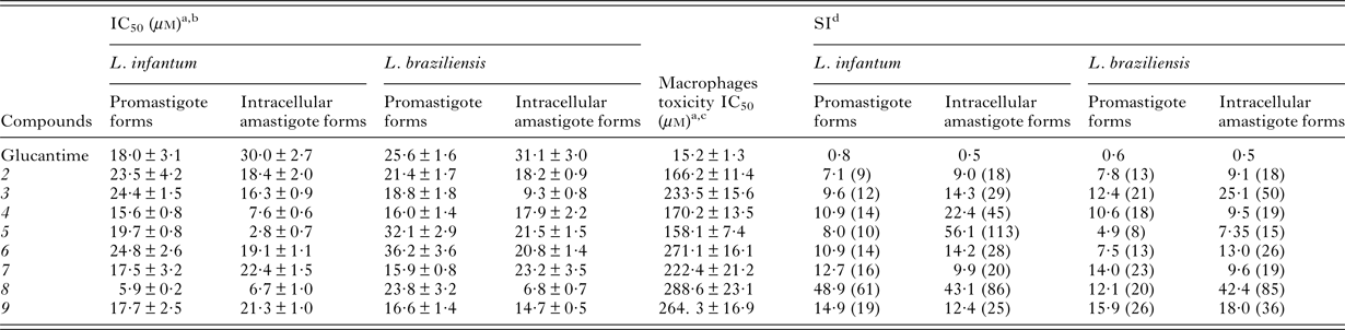
In vitro activity of compounds 2–9 on promastigote and amastigote forms of L. infantum and L. braziliensis, representative species causing respectively visceral and cutaneous leishmaniasis, the non-specific cytotoxicity against macrophages and the corresponding SI are gathered in Table 2. Early in vitro studies are usually performed on promastigotes since they are more easily handled in the laboratory (Marín et al. Reference Marín, Clares, Ramírez-Macías, Blasco, Olmo, Soriano, Verdejo, Rosales, Gomez-Herrera, García-España and Sánchez-Moreno2013). Nevertheless, results obtained for this extracellular insect stage are routinely complemented with studies conducted on amastigotes, which are the forms parasitizing the mammalian host cells and constitute the real goal of anti-leishmanial chemotherapy. Assays with amastigotes were performed by infecting macrophages with promastigotes, which were transformed into amastigotes within 1 day after infection. Pyrazoles 2–9 as well as the standard drug glucantime (meglumine antimoniate) were assayed at concentrations 1–100 µ m; the obtained data enabled the calculation of IC50 values gathered in Table 2.
In vitro activity, non-specific cytotoxicity and SI found for pyrazole derivatives 2–9 and glucantime (reference drug) on extra- and intracellular forms of L. infantum and L. braziliensis
a IC50: concentration required to give 50% inhibition, calculated by linear regression analysis from the K c values at concentrations employed (1, 10, 25, 50 and 100 µ m); K c (culture growth constant) corresponds to the slope resulting from plotting the log of the growth measurement versus time for each drug concentration.
b Average values of four separate experiments ± standard deviations.
c Average values of three separate experiments ± standard deviations.
d SI = IC50 macrophages/IC50 extra- and intracellular forms of parasites. In parentheses, fold increase in the SI of the compound compared with that of the reference drug.
Activities of these compounds against Leishmania range from ~11 times to 3/4 of those of the reference drug, depending on the concrete species and the morphological state. Once more, there are great differences in toxicities against macrophages, which are of course reflected in the corresponding SI, higher in most cases for amastigotes than for promastigotes (Table 2).
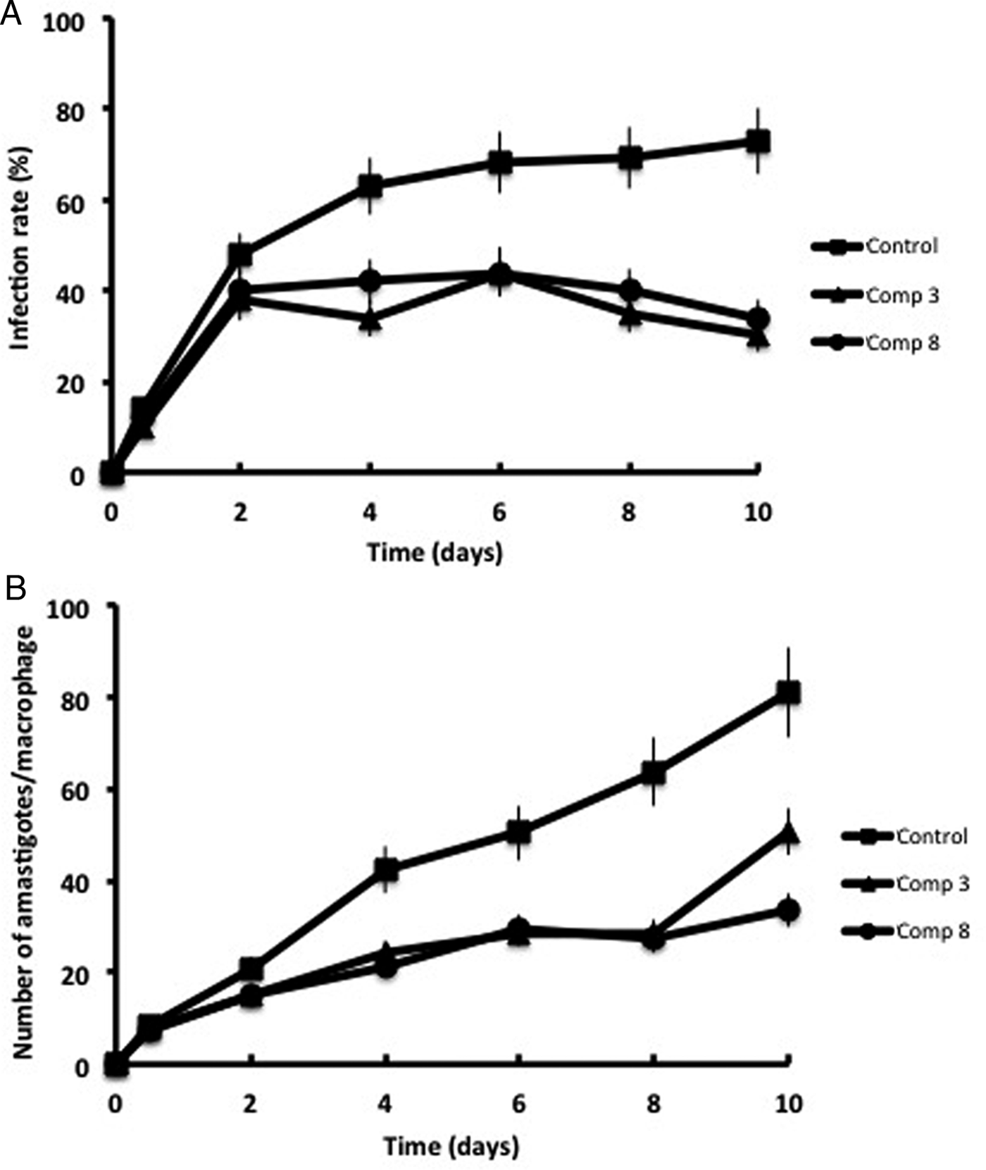
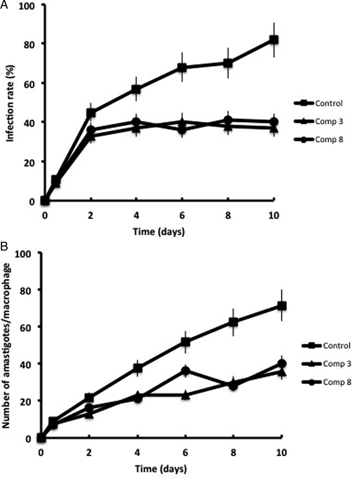
Additionally, as described above for T. cruzi, the effect of representative active compounds 3 and 8 on the propagation in macrophages of L. infantum and L. braziliensis was studied by measuring the rates of infection and the average number of amastigotes per macrophage present during a 10-days experiment. During this period the rate of infection of the host cells gradually increased reaching at the end 73% of invasion for L. infantum (Fig. 5) and 81% for L. braziliensis (Fig. 6).
Effect of pyrazole derivatives 3 and 8 (IC25 concentrations) on the infection rate and growth of L. infantum in macrophages: (A) rate of infection; (B) mean number of amastigotes per infected macrophage. The represented values are the averages of four separate determinations; error bars represent standard deviations. Significant differences between the control group and groups treated with compounds 3 and 8 for α = 0·05.

Effect of pyrazole derivatives 3 and 8 (IC25 concentrations) on the infection rate and growth of L. braziliensis in macrophages: (A) rate of infection; (B) mean number of amastigotes per infected macrophage. The represented values are the averages of four separate determinations; error bars represent standard deviations. Significant differences between the control group and groups treated with compounds 3 and 8 for α = 0·05.

For L. infantum similar experiments were conducted in the presence of the mentioned pyrazole derivatives at their IC25 concentrations (Fig. 5A). Compounds 3 and 8 were more effective than the reference drug glucantime [typical value: 24% reduction (Marín et al. Reference Marín, Clares, Ramírez-Macías, Blasco, Olmo, Soriano, Verdejo, Rosales, Gomez-Herrera, García-España and Sánchez-Moreno2013) in relation to the untreated control], displaying infection rates decreases of 59 and 53%, respectively. In relation to the number of amastigotes per macrophage (Fig. 5B), related results were obtained, showing pyrazole derivatives 3 and 8 decreases of 37 and 58%, respectively, higher than those typically obtained for the reference drug (ca. 30% reduction) (Marín et al. Reference Marín, Clares, Ramírez-Macías, Blasco, Olmo, Soriano, Verdejo, Rosales, Gomez-Herrera, García-España and Sánchez-Moreno2013).
In the case of L. braziliensis compounds 3 and 8 were subjected to a similar infection rate study (Fig. 6A), showing that they were more effective (55 and 51% decrease in relation to the control, respectively) than the reference drug for which typical values of ca. 22% decrease have been reported in this kind of assay (Marín et al. Reference Marín, Clares, Ramírez-Macías, Blasco, Olmo, Soriano, Verdejo, Rosales, Gomez-Herrera, García-España and Sánchez-Moreno2013). In connection with the average number of amastigotes per macrophage (Fig. 6B), glucantime shows typically a reduction of ca. 37% relative to the control (Marín et al. Reference Marín, Clares, Ramírez-Macías, Blasco, Olmo, Soriano, Verdejo, Rosales, Gomez-Herrera, García-España and Sánchez-Moreno2013), while compounds 3 and 8 notably decrease this value by 50 and 45%, respectively.
Acute toxicity in mice
Acute toxicity of products 3 and 8 was shown to be very low. After progressive dose increases of both compounds to reach 6 g kg−1, neither apparent toxic effects nor deaths were observed. The only finding was that in the 15–20 min immediately after administration of compounds 3 and 8 and after their transfer to the cage, treated mice displayed some hyperactivity, which ceased after this period and left no sequel or appreciable change in behaviour. Regarding the above mentioned kidney, heart and liver damage markers, no noticeable differences were observed between mice from treated and untreated groups, and the obtained values (not shown) were in the usual ranges (Olmo et al. Reference Olmo, Rotger, Ramírez-Macías, Martínez, Marín, Carreras, Urbanová, Vega, Chaves-Lemaur, Sampedro, Rosales, Sánchez-Moreno and Costa2014c, Reference Olmo, Guardia, Marín, Messouri, Rosales, Urbanová, Chayboun, Chahboun, Alvarez-Manzaneda and Sánchez-Moreno2015).
Possible deciphered of the mechanisms of action: inhibition of Fe-SOD of T. cruzi, L. infantum and L. braziliensis assays
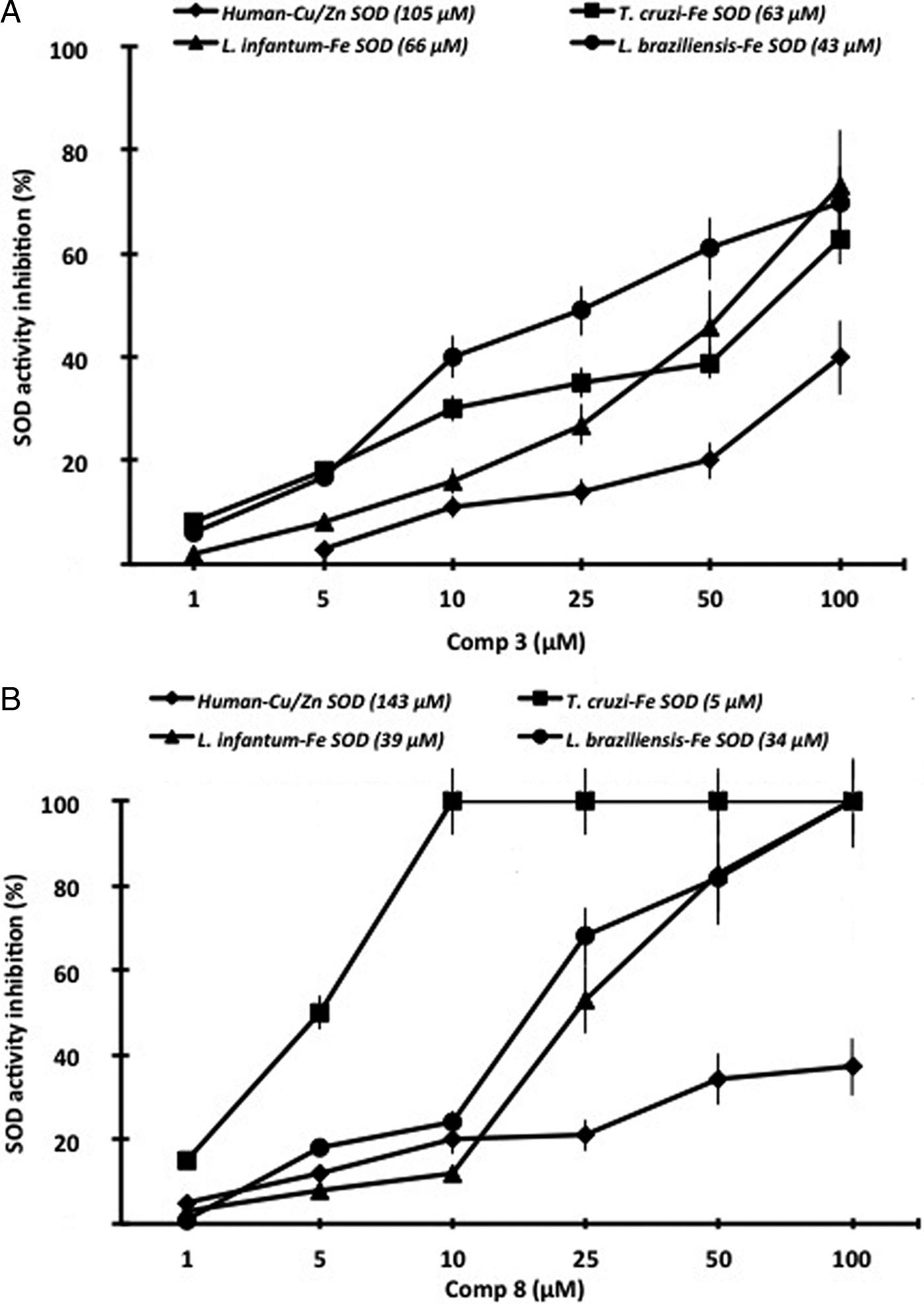
Anti-trypanosomatid activity of some pyrazole derivatives has been related to their ability to interact with Fe-SOD of parasites (Sánchez-Moreno et al. Reference Sánchez-Moreno, Sanz, Gómez-Contreras, Navarro, Marín, Ramírez-Macias, Rosales, Olmo, Garcia-Aranda, Campayo, Cano, Arrebola and Yunta2011, Reference Sánchez-Moreno, Gómez-Contreras, Navarro, Marín, Ramírez-Macías, Olmo, Sanz, Campayo, Cano and Yunta2012b, Reference Sánchez-Moreno, Marín, Navarro, Lamarque, García-España, Miranda, Huertas, Olmo, Gómez-Contreras, Pitarch and Arrebolac; Navarro et al. Reference Navarro, Sánchez-Moreno, Marín, García-España, Ramírez-Macías, Olmo, Rosales, Gómez-Contreras, Yunta and Gutiérrez-Sánchez2014). Therefore, in order to verify if this mode of action contributes to the activity of pyrazole derivatives treated in the current work, we studied the effect of compounds 3 and 8 on the parasites enzymes. Compounds were assayed at concentrations 1–100 µ m, and the inhibition data (IC50 values) obtained for CuZn-SOD from human erythrocytes, and for T. cruzi, L. infantum and L. braziliensis Fe-SOD are shown in Fig. 7. The inhibition of T. cruzi enzyme was substantially higher than that of the human origin, 1·7 and 28·7 times for compounds 3 (Fig. 7A) and 8 (Fig. 7B), respectively. Results were especially noteworthy for compound 8, which showed an IC50 value of ca. 5 µ m for T. cruzi Fe-SOD and reached 100% inhibition of this enzyme at ca. 10 µ m concentration.
In vitro inhibition (%) of CuZn-SOD of human erythrocytes, and Fe-SOD of T. cruzi epimastigotes, and L. infantum and L. braziliensis promastigotes: (A) for compound 3; (B) for compound 8. In brackets, IC50 values for the indicated SOD enzymes from different origin. Values of SOD inhibition are the averages of three separate determinations; error bars represent standard deviations. Significant differences for α = 0·05 between the inhibitory effect of compounds 3 and 8 on the CuZn-SOD of human origin and on Fe-SOD of parasites; non-significant inhibition differences between Fe-SOD of both species of Leishmania.

For L. infantum and L. braziliensis, the obtained inhibition data for compounds 3 and 8 are shown in Fig. 7A and B, respectively. In this case, inhibition of Fe-SOD from both species of Leishmania was also higher (1·6–4·3 times) than that of human erythrocytes CuZn-SOD, but values were not as remarkable as those previously found for T. cruzi enzyme. Once more, compound 8 gave the best results, but concentrations near 100 µ m were required to achieve 100% inhibition of Fe-SOD of both Leishmania species.
Concluding remarks
In the current work, we describe the high trypanocidal activity of some simple and very easily available dialkyl pyrazole-3,5-dicarboxylates against T. cruzi, L. infantum and L. braziliensis. The high efficiency of these compounds has been demonstrated by in vitro tests against various morphological forms of these parasites, and in the case of T. cruzi, by an in vivo study in a murine model of Chagas disease.
It is proposed that the activity of these compounds may be due, at least in part, to their ability to inhibit Fe-SOD enzyme, essential for parasites survival. Further studies are required to verify if the inhibition of parasites proline racemase may also be contributing to the activity shown by our pyrazole-3,5-dicarboxylic acid derivatives.
SUPPLEMENTARY MATERIAL
The supplementary material for this article can be found at https://doi.org/10.1017/S0031182017000415.
FINANCIAL SUPPORT
The authors acknowledge financial support from the former Spanish Ministry of Science and Innovation (MICINN) and now from the Ministry of Economy, Industry and Competitiveness (MINEICO) (projects Consolider Ingenio CSD2010-00065, CTQ2013-14892 and SAF2015-66690-R). F. R. acknowledges a CSIC JAE-Doc contract co-funded by EU (ESF, European Social Fund). F. O. is grateful for a FPU grant from the Ministry of Education of Spain.