Early infantile pulmonary hypertension represents a heterogeneous group of disorders, often with a very variable natural history from full recovery to persistent refractory pulmonary hypertension, right ventricular failure, and death.Reference Steinhorn 1 , Reference Hwang 2 We describe the cases of two children who both presented with severe infantile pulmonary hypertension, which was later associated with pulmonary capillary haemangiomatosis on post-mortem. Despite maximal medical therapy, both patients died from pulmonary hypertension.
Case 1
An ex 35-week-old baby girl presented to a local hospital at six weeks of age with an acute respiratory illness. She developed profound hypoxaemia in the local hospital, requiring intubation and ventilation, and she was transferred to a paediatric intensive care unit in the tertiary centre. There was no family history of cardiac or pulmonary problems in children.
Echocardiography demonstrated normal cardiac anatomy with evidence of supra-systemic right ventricular systolic pressure: estimated right ventricular systolic pressure was 78 mmHg+right atrial pressure versus systolic blood pressure 70 mmHg. There was mild right ventricular hypertrophy and a flattened interventricular septal configuration in diastole (D-shaped LV). She was treated with high-frequency oscillatory ventilation, inhaled nitric oxide 20 parts per million, and oral sildenafil. After one week, she improved clinically, was extubated and transferred to the ward. Within 24 hours, she was re-admitted to the intensive care unit, re-intubated, and ventilated for a further episode of acute respiratory distress and severe pulmonary hypertension. Echocardiography confirmed a right ventricular systolic pressure of 53 mmHg+right atrial pressure. There was mild right ventricular hypertrophy. Her pulmonary artery pressure normalised over the further two weeks and she was weaned off from ventilation. She was discharged home on sildenafil, but the cause of the pulmonary hypertension was not understood. At six months of age, there was no tricuspid regurgitation and normal left-to-right bowing of the atrial septum, and thus the sildenafil was discontinued. The interventricular septum was slightly flattened in diastole, which may have suggested elevated right ventricular pressure.
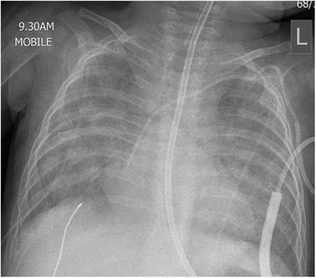
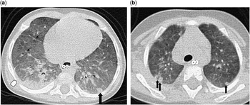
The patient presented to our tertiary-care paediatric centre in respiratory distress with hypoxaemia three days after sildenafil was discontinued. A repeat echocardiogram again showed supra-systemic pulmonary arterial pressures with the interventricular septum bowing right to left. She was admitted to the intensive care unit (ICU) and required intubation and ventilation. She had markedly reduced lung compliance and remained hypoxaemic despite high airway pressures. She achieved better oxygenation with high-frequency oscillatory ventilation and inhaled nitric oxide 20 parts per million. She was also treated with bosentan, sildenafil, and epoprostenol. The chest x-ray showed widespread diffuse alveolar opacity throughout both the lungs (Fig 1). A chest computed tomography (CT) scan demonstrated widespread symmetric ground-glass opacification, which increased in density from the apices to the lung bases (Figs 2a and b). Retrospectively, there was evidence of bilateral centrilobular nodules. There was no pleural effusion. There was minimal septal thickening.
Chest radiograph demonstrates widespread diffuse alveolar opacity throughout both the lungs.

(a) CT (image 1) scan of the thorax demonstrates widespread, bilateral ground-glass opacification. Septal thickening (thin black arrows) and a small right-sided pleural effusion (white arrow) are also present. A peripheral centrilobular density is present on the left (filled black arrow). (b) CT (image 2) multiple centrilobular densities (black arrows) together with diffuse ground-glass opacification.

Genetic testing for pulmonary surfactant protein deficiency was normal.
Cardiac catheterisation was performed, which demonstrated a structurally normal heart with moderate irreversible pulmonary arterial hypertension: pulmonary arterial pressure 52/22 mmHg (mean 36 mmHg). The pulmonary vascular resistance measured 19.9 Wood units on room air, 14.3 Wood units on 100% oxygen, and 18.5 Wood units on nitric oxide 20 ppm. Pulmonary capillary wedge pressure was 12 mmHg, and there was no evidence of pulmonary vein stenosis. A lung biopsy showed features of a non-specific interstitial pneumonitis with moderate hypertensive vasculopathy. Retrospective review of the specimen after the patient’s death also confirmed that the appearances were consistent with the above description. Pulsed intravenous methylprednisolone was administered on the basis that the clinical impression at the time was one of pulmonary hypertension secondary to interstitial lung disease. There was no clinical benefit. The patient was weaned off ventilation after four weeks and was transferred to the ward on oral sildenafil and bosentan.
Over the next four months, she had five further intensive care admissions with a similar clinical course and pronounced alveolar opacity on chest radiograph. Each episode started with a gradual increase in respiratory rate, followed by evolving chest x-ray findings, and subsequent profound hypoxaemia requiring intubation and high-frequency oscillatory ventilation. She received inhaled nitric oxide and milrinone on each occasion. Echocardiography on each occasion demonstrated supra-systemic right ventricular pressure; two further three-day pulsed courses of methylprednisolone were administered with no effect. Enteral hydroxychloroquine was used to treat interstitial lung disease, given the radiological appearances and lung biopsy findings, but had no benefit. The infant received intravenous immunoglobulin for mild hypogammaglobulinaemia.
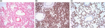
CT thorax three months later showed no change with persistant ground-glass opacity, centrilobular densities, and minimal septal thickening. Given the poor clinical course, a second lung biopsy was considered, but was deferred given the risks involved. At 12 months of age, it was decided to withdraw active medical management given the intractable nature of the disease. She died two weeks after the intensive management was withdrawn. Post-mortem examination of the lungs demonstrated marked pulmonary capillary haemangiomatosis (Figs 3a and b).
(a) Haematoxylin- and eosin-stained section shows slightly autolysed lung parenchyma from post-mortem sampling, with a striking proliferation of capillary-sized vessels indicative of haemagiomatosis (case 1). (b) Immunostaining with CD31 [platelet endothelial cell adhesion molecule-1 (PECAM-1)] decorates the endothelium of the capillaries, highlighting consolidative obliteration of the airspaces of the lung by this rich capillary vascular plexus (case 2). (c) Immunostaining with CD31 in the normal lung.

Case 2
A baby boy born at term was transferred from a district general hospital to our tertiary-care paediatric centre soon after birth for surgical input, after oesophageal atresia with tracheo-oesophageal fistula was suspected clinically and radiographically. He required intubation and ventilation for progressive hypoxaemia and hypercapnia. On his third day of life, the tracheo-oesophageal fistula was ligated, and a gastrostomy was created as his distal oesophagus could not be approximated with his proximal pouch. The infant also had a type-three laryngeal cleft and tracheomalacia that required laryngoplasty and tracheostomy, respectively. A clinical diagnosis of VACTERL association was considered, but criteria were not met. A DNA microarray study also did not identify any genetic abnormalities. There was no family history of cardiac or pulmonary problems in children.
An echocardiogram performed in the tertiary-care centre on day two of life demonstrated a small secundum atrial septal defect, a moderate peri-membranous ventricular septal defect, and a left superior caval vein-to-coronary sinus connection. A moderate patent arterial duct was present with bi-directional flow, demonstrating systemic pulmonary arterial hypertension. There was mild-to-moderate right ventricular hypertrophy. The interventricular septum was flattened (D-shaped LV) in systole and diastole.
The infant had ongoing problems with hypoxaemia, respiratory acidosis, and difficulty in ventilation. Problems were encountered with laryngeal surgical site dehiscence and occlusion of the tracheal tubes by ridges of tissue surrounding the distal tracheo-oesophageal fistula. He was managed with conventional and high-frequency oscillatory ventilation and inotropic support. Most episodes of hypercapnia were secondary to large airway obstruction, which responded to altering airway adjuncts. Repeat echocardiography demonstrated persistently elevated pulmonary arterial pressures with a right ventricular systolic pressure 60 mmHg plus right atrial pressure. The child was treated with sildenafil, bosentan, and inhaled nitric oxide 20 ppm.
Cardiac catheterisation demonstrated moderate-to-severe pulmonary arterial hypertension: 56/19 mmHg and mean 37 mmHg. The pulmonary vascular resistance measured 17 Wood units. There was no change with treatment with 100% oxygen or inhaled nitric oxide 20 ppm. There was no evidence of pulmonary vein stenosis.
High-resolution CT of the chest at 11 weeks of age demonstrated extensive dependent atelectasis with ground-glass opacity and small bilateral pleural effusions. Peripheral centrilobular nodules and septal thickening were also appreciated on retrospective review.
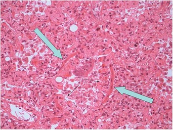
All the investigations to date failed to recognise a definite cause for his pulmonary hypertension. Despite maximal medical therapy with pulmonary vasodilators, his pulmonary arterial hypertension persisted and his clinical condition deteriorated. A trial of intravenous adenosine infusion was also administered without effect. The patient died at three months of age when treatment was withdrawn. On post-mortem examination of the lungs, pulmonary capillary haemangiomatosis was diagnosed invading the interstitium, airways, and the pulmonary vessels (Fig 4). Sanger sequencing to identify mutations in the EIF2AK4 gene was negative.
Haematoxylin and eosin (H&E) staining shows aggressive, infiltrative growth of the capillary vessels in the context of haemagiomatosis within the wall of a bronchiole. The pale blue arrows point out the smooth muscular wall of the bronchiole, whereas the thin black arrows indicate capillary-sized vessels undermining the bronchial epithelium and reducing the airway lumen (case 2).

Discussion
Pulmonary capillary haemangiomatosis is a rare condition, which was first described in 1978.Reference Waagenvort, Beetra and Spiker 3 It usually affects both the lungs, is of unknown aetiology, and is an aggressive but benign vascular neoplasm, characterised by abnormally proliferating capillaries that invade the pulmonary interstitium, airways, and blood vessels.Reference Eltorky, Headley, Winer-Muram, Garrett and Griffin 4 Most reported patients are between 20 and 40 years of age.Reference Eltorky, Headley, Winer-Muram, Garrett and Griffin 4 , Reference Ito, Ichiki and Ohi 5 There are 18 reported cases of pulmonary capillary haemangiomatosis in children and adolescents, and only two of whom were less than one-year old – a neonate and a stillborn foetus.Reference Ito, Ichiki and Ohi 5 , Reference Bartyik, Bede and Tiszlavicz 6 , Reference Oviedo, Abramson and Worthington 7 Patients typically present with signs and symptoms of pulmonary hypertension, which is progressive and causes dyspnoea, haemoptysis, and right heart failure.Reference Ito, Ichiki and Ohi 5 , Reference Takiguchi, Urama and Hiroshima 8 , Reference Frazier, Franks and Mohammed 9
The two cases we described above highlight the progressive lethal nature of the pulmonary hypertension associated with pulmonary capillary haemangiomatosis and also the diagnostic challenge it presents. Reaching the diagnosis ante-mortem, as opposed to primary pulmonary hypertension, is important as vasodilator therapy may be deleterious exacerbating pulmonary oedema.Reference Ito, Ichiki and Ohi 5 , Reference Frazier, Franks and Mohammed 9 , Reference Kothari, Jagia and Gupta 10 If diagnosed ante-mortem, adults are often listed for lung transplantation, which is currently felt to be the only curative therapy.Reference Ito, Ichiki and Ohi 5 , Reference Frazier, Franks and Mohammed 9 , Reference Kothari, Jagia and Gupta 10
Radiological investigations, particularly conventional CT and high resolution CT, may assist diagnosis. No findings are pathognomonic, but one may observe ground-glass opacity, septal thickening, and small centrilobular poorly circumscribed nodular opacities.Reference Eltorky, Headley, Winer-Muram, Garrett and Griffin 4 , Reference Ito, Ichiki and Ohi 5 , Reference Takiguchi, Urama and Hiroshima 8 , Reference Frazier, Franks and Mohammed 9 , Reference Dufour, Maitre and Humbert 11 Some authors report the findings on high-resolution CT to be specific for pulmonary capillary haemangiomatosis, which lead them to conclude that high-resolution CT scanning in particular is a very useful tool for diagnosis of this disease.Reference Takiguchi, Urama and Hiroshima 8 , Reference Frazier, Franks and Mohammed 9 , Reference Dufour, Maitre and Humbert 11
In the two patients described here, there is overlap between the CT abnormalities described in pulmonary capillary haemangiomatosis and pulmonary veno-occlusive diseaseReference Dufour, Maitre and Humbert 11 in that both patients had diffuse geographic ground-glass opacity, poorly defined centrilobular opacites, and septal thickening with small pleural effusions (Figs 2a and b). The chest x-rays did not demonstrate reticulonodular shadowing as described in adultsReference Kothari, Jagia and Gupta 10 but showed diffuse alveolar opacity (Fig 1). Both CT scans were performed without IV contrast; therefore, comment on pulmonary artery size and mediastinal lymphadenopathy is not possible.
The diagnosis is reached histologically, and thus lung biopsy is recommended to confirm the diagnosis. Early lesions will show rows of capillaries along the alveolar walls that progress to nodules and sheets of back-to-back capillaries in more advanced lesions.Reference Eltorky, Headley, Winer-Muram, Garrett and Griffin 4 , Reference Frazier, Franks and Mohammed 9 Proliferating capillaries will also compress and infiltrate the walls of pulmonary veins and venules, causing intimal fibrosis and secondary veno-occlusion.Reference Frazier, Franks and Mohammed 9 In response, there is secondary muscularisation of pulmonary arterioles and arteries inducing pulmonary hypertension. The development of pulmonary hypertension is, however, felt to be multifactorial.Reference Eltorky, Headley, Winer-Muram, Garrett and Griffin 4 Infiltration of the pulmonary artery walls, airways, and alveoli can be seen with sometimes extensive intra-alveolar haemorrhage.Reference Takiguchi, Urama and Hiroshima 8
Unfortunately, lung biopsy is not always possible in the sick patient with severe pulmonary arterial hypertension. Furthermore, a lung biopsy may miss the diagnosis by sampling an area of normal-appearing lung tissue. Pulmonary capillary haemangiomatosis has been described as having well demarcated parenchymal lesions with relatively unremarkable intervening lung tissue.Reference Frazier, Franks and Mohammed 9 Repeat lung biopsies may be warranted to detect its presence. Of note, case 1’s lung biopsy did not show pulmonary capillary haemangiomatosis, but her post-mortem lung tissue demonstrated marked disease. A recent study demonstrated pulmonary capillary haemangiomatosis in two cases among 297 lung biopsies in children with congenital heart disease over an 11-year period.Reference Aiello, Thomaz, Pozzan and Lopes 12
Lung transplantation is universally accepted as the final definitive treatment for adult pulmonary capillary haemangiomatosis. Worldwide experience with infant lung transplantation is much less than that of adults, but the evidence suggests that it is still a viable therapeutic option for infants with end-stage lung disease including pulmonary vascular disease.Reference Khan, Heinle and Samayoa 13 , Reference Conrad and Cornfield 14 A recent study reported over 80 children who underwent lung transplantation over a 26-year period.Reference Khan, Heinle and Samayoa 13 They found that overall survival for infants undergoing lung transplantation is comparable with older children and that late survival and survival in certain high-risk cohorts – for example, pre-transplant mechanical ventilation – appear to be better. Pulmonary vascular disease is recorded to be one of the most common indications for infant lung transplantation; however, the percentage of which is pulmonary capillary haemangiomatosis is not reported.Reference Khan, Heinle and Samayoa 13 , Reference Conrad and Cornfield 14 , Reference Mallory and Spray 15 Other therapies have been used with reported success in individual patients. Three paediatric cases of successful treatment in the late 1980s with recombinant alpha interferon have been reported.Reference White, Sondheimer, Crouch, Wilson and Fan 16 , Reference White 17 , Reference White, Wolf and Korones 18 Possible mechanisms of action for interferon include inhibiting proliferation of endothelial cells, smooth muscle cells, or fibroblasts and antagonising angiogenesis through its immunostimulatory actions.Reference White 17 More recently, Ginns et al reported successful remission of symptoms in a 20-year-old man after nine months of treatment with doxycycline, after failing to see improvement with alpha interferon.Reference Ginns, Roberts and Mark 19 Doxycycline was trialled on the rationale that it is a matrix metalloproteinase inhibitorReference Ishii, Iwabuchi and Kameya 20 and that it might modulate the increased matrix metalloproteinase activity associated with dysregulated angiogenesis, inducing a clinical benefit. At the time of publication, he was still symptoms-free after 18 months of therapy.
Although most cases of pulmonary capillary haemangiomatosis are sporadic, there have been reports of families with multiple affected family members, suggesting a heritable cause. Best et al identified mutations in the EIF2AK4 gene that are likely responsible for autosomal recessive pulmonary capillary haemangiomatosis in familial and some non-familial cases;Reference Best, Sumner and Austin 21 two novel mutations were found in this gene. There is likely to be additional genetic and non-genetic factors involved in the development of pulmonary capillary haemangiomatosis. This would explain why pathogenic mutations in EIF2AK4 were not found in some affected patients in the Best study,Reference Best, Sumner and Austin 21 although they did not screen the non-coding regions of the gene. It would also explain why patients present at different ages, even in the case of siblings who shared the same mutation.
Recessive EIF2AK4 mutations were also found in 13 families with pulmonary veno-occlusive disease by Eyries et al.Reference Eyries, Montani and Girerd 22 Pulmonary capillary haemangiomatosis and pulmonary veno-occlusive disease are often considered two different histological manifestations of the same disease process as they share many clinical and histopathological features. Lantuéjoul et al described capillary proliferation in 73% of lung tissue samples in cases of pulmonary veno-occlusive disease and venous and arterial changes typical of pulmonary veno-occlusive disease in the lung tissue samples of 80% of cases of pulmonary capillary haemangiomatosis.Reference Lantuéjoul, Sheppard, Corrin, Burke and Nicholson 23 This new information will allow genetic counselling for affected families, identify asymptomatic family members of affected individuals, and may allow early interventions, which could potentially delay the onset or progress of the process.Reference Best, Sumner and Austin 21 , Reference Lantuéjoul, Sheppard, Corrin, Burke and Nicholson 23
Pulmonary capillary haemangiomatosis should be considered in cases of unexplained severe infantile pulmonary hypertension and warrants lung biopsy. Early consideration of referral to a lung transplant centre should be considered in selected patients.
Acknowledgements
None.
Financial Support
The authors have no financial relationships relevant to this article to disclose.
Conflicts of Interest
The authors have no conflicts of interest relevant to this article to disclose.