Non-technical Summary
Conodonts are the feeding apparatus of the extinct conodont animals and are regarded as a significant index fossil for subdividing and correlating global marine Paleozoic and Triassic strata. It remains a challenge to differentiate the important conodont species Gnathodus pseudosemiglaber from morphologically similar species due to uncertainties in intraspecific, ontogenetic, and phylogenetic relationships between taxa. Here, in the light of population thinking, we employed quantitative morphometric methods to analyze morphological variations of G. pseudosemiglaber specimens varying from juvenile to gerontic adult, based on a conodont fauna collected from the Yudong Formation at the Yudong II section in Baoshan, southwestern China. The results provide a comprehensive taxonomic review of G. pseudosemiglaber based on its ontogenetic dynamics. Gnathodus girtyi maxwelli, a previously named species, is regarded as a junior synonym of G. pseudosemiglaber. Based on the amended systematic description of this species, we refine its biostratigraphic potential and phylogeny.
Introduction
Species is the basic biological unit used in paleontological research, thus the matter of how paleontologists define species is of utmost importance (Allmon, Reference Allmon2013). However, the concept of species is disputed constantly, and nearly 40 different species concepts have been conceived so far (Tschopp et al., Reference Tschopp, Napoli, Wencker, Delfino and Upchurch2022). Neontologists and paleontologists spent more than a century wrestling with the enigma of the species concept. Teueman (Reference Teueman1924) proposed the well-known “species problem,” which noted that it is difficult for biologists to recognize and describe a certain species in a consistent way. Allmon (Reference Allmon2013) further interpreted this problem as three interconnected questions, one of which is the species recognition problem; alternatively, to what degree can species recognized in living organisms be recognized in the fossil record? For living organisms, there is a widely adopted “biological species concept” that species are groups of interbreeding natural populations that are reproductively isolated from other such groups (Mayr, Reference Mayr, Wheeler and Meier2000). When this concept meets incomplete fossil records, the situation becomes more complicated, especially for those without living descendants. Although attempts have been made to hypothesize that fossil morphology is related to reproductive isolation in practice, it has not been clear to what extent they are related (Allmon, Reference Allmon2013). Besides, influenced by traditional typological-morphological thinking, paleontologists used to identify a species based on its morphological characteristics (Huang, Reference Huang2011).
Since the 1940s, population thinking was introduced into paleontological taxonomy (e.g., Simpson, Reference Simpson1940; Imbrie, Reference Imbrie1956). Contrary to the typological-morphological thinking that focuses on classifying species based on the presence of unique morphological characteristics, population thinking requires that the intraspecific and ontogenetic variation are incorporated into species diagnoses (Mayr, Reference Mayr1994). Under the view of population thinking, uncommon species identified in the literature based on a single, sometimes subtle, morphological feature should be re-evaluated (Huang, Reference Huang2011). From the biostratigraphical point of view, the distinguishability of fossils, especially that of index fossils, should be emphasized for the sake of more accurate subdivision and correlation of biostratigraphy. These notions confused the concept of species and phenotypes, leading to the phenomenon of excessive splitting (Fang and Rong, Reference Fang and Rong1991).
Conodonts are regarded as a group of important index fossils for the subdivision and correlation of global marine strata deposited during the Paleozoic and Triassic, due to their rapid evolutionary rate, distinctive characteristics, worldwide distribution, and abundance in marine rocks. Since conodont fossils were first recovered by Pander (Reference Pander1856), morphological features of individuals constitute the basis of conodont taxonomy, thus a large number of morphological species have been named. The discovery of conodont natural assemblages (e.g., Schmidt, Reference Schmidt1934; Rhodes, Reference Rhodes1952) revealed that conodonts are discrete elements that comprise the multielement feeding apparatuses of conodont animals (e.g., Renaud et al., Reference Renaud, Girard and Dufour2021). Specific element morphologies consistently occupy particular positions in the conodont mouth and performed different functions during feeding (Purnell et al., Reference Purnell, Donoghue and Aldridge2000; Hogancamp, Reference Hogancamp2020). The elongated S and M elements occupy the front position, forming a trapping structure, while the P1 and P2 elements are situated at the rear position, consisting of a pair of asymmetric elements respectively, which form an occlusal structure responsible for food processing (Aldridge, Reference Aldridge and Aldridge1987; Purnell and Donoghue, Reference Purnell and Donoghue1997). However, conodont fossils obtained from insoluble residues after the buffered acetic acid method from limestones are limited in their ability to provide comprehensive insights into natural assemblages. Particularly, conodonts reported from Carboniferous strata mainly are the P1 element (Merrill and Powell Reference Merrill and Powell1980; Hu et al., Reference Hu, Wang and Qi2023). Consequently, taxonomy based on natural assemblages was seen as a practically unattainable goal (Sweet, Reference Sweet1988). Although continuous studies of Carboniferous conodont apparatuses are gradually filling this shortcoming (e.g., Norby, Reference Norby1976; Lane and Ziegler, Reference Lane and Ziegler1984; Purnell, Reference Purnell1993; von Bitter and Merrill, Reference von Bitter and Merrill1998; Nestell et al., Reference Nestell, Wardlaw and Pope2016; von Bitter et al., Reference von Bitter, Norby and Stamm2022), the abundant P1 elements and their significant role in biostratigraphy further emphasizes the continued practicality of typological-morphological thinking.
Because the concept of population thinking was applied to paleontological classification, researchers began to consider the taxonomical works of conodonts from a population perspective, known as the sample population taxonomic approach (Mei et al., Reference Mei, Henderson and Cao2004; Dzik, Reference Dzik2006; Han et al., Reference Han, Orchard, Wu, Zhao, Chen, Golding, Jan, Lyu and Hashmi2022). Due to intraspecific variation, paedomorphosis (Sanz-López et al., Reference Sanz-López, Blanco-Ferrera and García López2004), and shared juvenile morphology (Hogancamp, Reference Hogancamp2020) in conodonts, it is highly possible that specimens from the same sample but at different ontogenetical stages were mistakenly identified as different morphological species, thus leading to misrecognition of conodont faunal composition, evolution, and biostratigraphical correlation.
The conodont Gnathodus pseudosemiglaber (Thompson and Fellows, Reference Thompson and Fellows1970) has a known range from the upper Tournaisian to the Visean, Mississippian (Lower Carboniferous) (Lane et al., Reference Lane, Sandberg and Ziegler1980), and plays an important role in biostratigraphical correlations in the Tournaisian strata. The first occurrence of G. pseudosemiglaber or Scaliognathus anchoralis Branson and Mehl, Reference Branson and Mehl1941, which marks the lower limit of the S. anchoralis–G. pseudosemiglaber Zone in the Baoshan Block and South China (Tian and Coen, Reference Tian and Coen2004; Hu et al., Reference Hu, Qi, Qie and Wang2020, Reference Hu, Qi, Wang, Wang and Wang2022), corresponds with the S. anchoralis–Doliognathus latus Zone by Lane et al. (Reference Lane, Sandberg and Ziegler1980) for a worldwide correlation. Lane et al. (Reference Lane, Sandberg and Ziegler1980) suggested this species range extends up to the G. bilineatus Zone where it occurs with G. bilineatus bilineatus (Roundy, Reference Roundy1926) in the middle and upper Visean. It is, however, difficult to differentiate G. pseudosemiglaber from the morphologically similar species G. bulbosus Thompson, Reference Thompson1967, and G. semiglaber Bischoff, Reference Bischoff1957, that occur with G. pseudosemiglaber in the upper part of its range (Boardman et al., Reference Boardman, Thompson, Godwin, Mazzullo, Wilhite and Morris2013). Therefore, this study employed population thinking to conduct ontogenetic research on well-preserved P1 elements in specimens from the Yudong II section in southwestern China in order to clarify the species concept of Gnathodus pseudosemiglaber.
Materials and methods
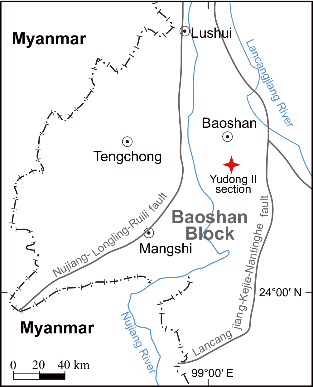
The studied Gnathodus pseudosemiglaber specimens were recovered from a 5-kg carbonate sample from a 20-cm-thick bed of microcrystalline limestone within the G. pseudosemiglaber–Scaliognathus anchoralis Zone, based on previous work conducted by Wang et al. (Reference Wang, Wang and Hu2024) in the Carboniferous Yudong Formation of the Yudong II section, western Yunnan (Fig. 1). This section yields abundant conodonts, including bispathodids, gnathodids, pseudopolygnathids, and neopolygnathids. The G. pseudosemiglaber–S. anchoralis Zone was recognized in this section, characterized by the occurrence of G. pseudosemiglaber associated with Bispathodus stabilis (Branson and Mehl, Reference Branson and Mehl1934), G. cordiformis Lane, Sandberg, and Ziegler, Reference Lane, Sandberg and Ziegler1980, and Polygnathus communis communis Branson and Mehl, Reference Branson and Mehl1934. All specimens are stored at Nanjing Institute of Geology and Palaeontology, Chinese Academy of Sciences. For more information regarding the locality and stratigraphy of this section, readers are referred to Wang et al. (Reference Wang, Wang and Hu2024).
Locality map of the Yudong II section (after Wang et al., Reference Wang, Wang and Hu2024).

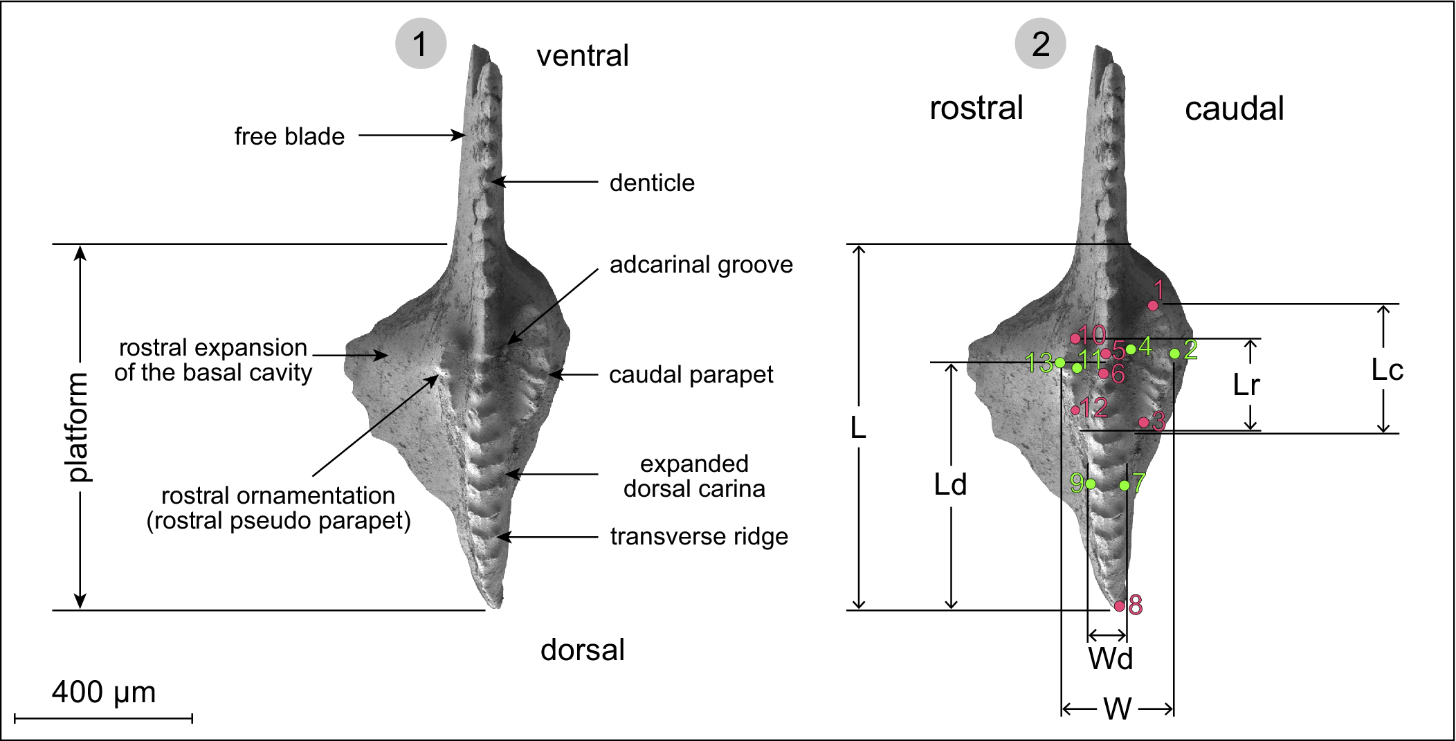
In the studied sample, YDII-C-12.0, contains 121 P1 elements, including 67 of Gnathodus pseudosemiglaber, six of Protognathodus cordiformis (Lane, Sandberg, and Ziegler, Reference Lane, Sandberg and Ziegler1980), two of Pseudopolygnathus multistriatus Mehl and Thomas, Reference Mehl and Thomas1947, and 46 of a pelagic species Bispathodus stabilis (Wang et al., Reference Wang, Wang and Hu2024). All of the specimens are amber in color, with a CAI (conodont alteration index) value of 1.5 (following Epstein et al., Reference Epstein, Epstein and Harris1977). In this sample, G. pseudosemiglaber is represented by 67 specimens of juvenile, subadult, and adult elements. Except for 15 poorly preserved specimens, the remaining 52, including 23 sinistral elements and 29 dextral elements, were measured for the following morphological parameters (Fig. 2): (1) platform length (L), (2) expanded dorsal carina length (Ld), (3) caudal parapet length (Lc), (4) rostral ornamentation length (Lr), (5) caudal parapet-rostral ornamentation width (W), and (6) expanded dorsal carina width (Wd). These parameters, which encompass the fundamental size information of a conodont, are also distinctive characteristics of G. pseudosemiglaber, namely the caudal parapet, the rostral pseudo parapet, and the expanded dorsal carina. The terminology applied in this study is according to Purnell et al. (Reference Purnell, Donoghue and Aldridge2000).
(1) Terminology used to describe Gnathodus pseudosemiglaber P1 morphology. (2) Measured morphologic parameters on a Gnathodus pseudosemiglaber P1 element: L = platform length, Ld = dorsal carina length, Lr = rostral ornamentation (or pseudo parapet) length, Lc = caudal parapet length, W = caudal parapet- rostral ornamentation width, Wd = expanded dorsal carina width. Pink points show locations of type 1 landmarks used for geometric morphometric analysis and green points show locations of type 2 landmarks. Gnathodus pseudosemiglaber sinistral P1 element from Wang et al. (Reference Wang, Wang and Hu2024, fig. 9F).

Typically, if fossil individuals extracted from a single layer came from the same population, the variations of most quantitative characters (e.g., individual size) exhibit continuity and conform to a normal distribution (Fang and Rong, Reference Fang and Rong1991; Zou and Ge, Reference Zou and Ge1995; Huang, Reference Huang2011). Therefore, the Shapiro–Wilk test (Shapiro and Wilk, Reference Shapiro and Wilk1965) was conducted to estimate whether specimens obtained from YDII-C-12.0 belong to the same population. This test examines the null hypothesis that the population is normally distributed and provides a test statistic (W) along with a probability p-value. If W is close to 1, it suggests that the sample likely came from a normal distribution. If the p-value is less than the significant level (0.05), then we can reject the null hypothesis (Huang, Reference Huang2011). Additionally, the preservation state of specimens and the consistency of their CAI serve as important indicators to judge if the studied specimens were transported.
To study the correlations among the six measured parameters of Gnathodus pseudosemiglaber, the R package corrplot (Wei and Simko, Reference Wei and Simko2017) was employed, which could provide a visual correlations heatmap among multiple variates. To reveal the dynamics during growth, the regression analysis was conducted on the six parameters for all 52 specimens.
Because the number of transverse ridges on the caudal parapet and the dorsal carina varies among specimens of different sizes, and the profiles of the rostral pseudo parapet are not regular in different specimens, it is difficult to determine whether a certain ridge in a smaller specimen corresponds to one in a larger specimen. These difficulties in identifying the homologous anatomical structures create problems for landmark-based geometric morphometrics. Therefore, we preferred methods based on measured morphological parameters rather than geometric morphometrics. Nevertheless, if the transverse ridge count changes with growth, the homologous feature is the parapet itself. Thus, the two ends of the parapet could be used regardless of ridge count.
Landmark-based geometric morphometrics was employed, which is a powerful method for quantifying biological shapes and their variation (Webster and Sheets, Reference Webster and Sheets2010). Landmarks refer to corresponding points on each specimen that match between and within populations, or biologically homologous anatomical loci recognizable on all specimens under study (Bookstein, Reference Bookstein1991; Webster and Sheets, Reference Webster and Sheets2010). Bookstein (Reference Bookstein1991) defined three types of landmarks: type 1 landmarks are those constrained to a specific point on a specific anatomical feature; type 2 landmarks are points of maximum curvature or width along certain anatomical boundaries that are anatomical features connecting type 1 landmarks; type 3 landmarks are points of true extremity that are not defined by any specific feature and rarely used. Thirteen landmarks were selected from four specimens of different sizes, which were applied to locate the positions of (1) the ventral tip of the caudal parapet (type 1), (2) the caudal tip of the caudal parapet (type 2), (3) the dorsal tip of the caudal parapet (type 1), (4) the rostral tip of the caudal parapet (type 2), (5) the denticle in front of the first expanded node on the dorsal carina (type 1), (6) the first expanded node on the dorsal carina (type 1), (7) the caudal tip of the ridge on the broadest part of the dorsal carina (type 2), (8) the dorsal tip of the dorsal carina (type 1), (9) the rostral tip of the ridge on the broadest part of the dorsal carina (type 2), (10) the ventral tip of the rostral ornamentation (type 1), (11) the caudal tip of the rostral ornamentation (type 2), (12) the dorsal tip of the rostral ornamentation (type 1), and (13) the rostral tip of the rostral ornamentation (type 2) (Fig. 2).
All landmarks were digitized with the software TpsDig2 v2.32 (Rohlf, Reference Rohlf2006). Generalized Procrustes analysis (GPA) was then undertaken on the data, which converted the points into a consensus axis but kept the size of each specimen; after that thin-plate splines (TPS) analysis was applied for comparisons among specimens of different sizes. Both methods were operated with PAST v3.25 (Hammer et al., Reference Hammer, Harper and Ryan2001).
Repository and institution abbreviation
The illustrated specimens are housed at Nanjing Institute of Geology and Palaeontology, Chinese Academy of Sciences (NIGPAS), Nanjing, China, under the catalog numbers NIGP PZ 204030–204059.
Results
Normal distribution analysis
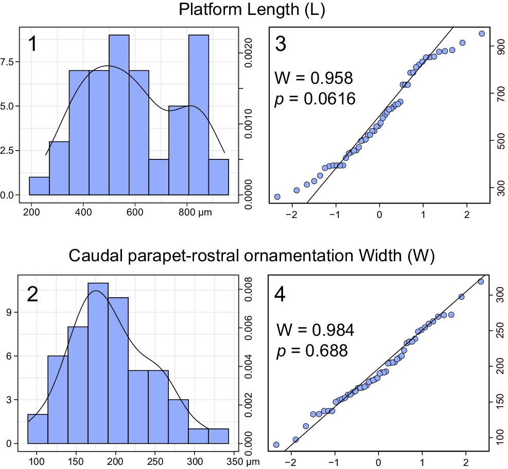
The platform length and the parapet–pseudo parapet width generally follow a normal distribution (Fig. 3.1, 3.2), although the latter parameter displays a distribution much closer to the double peak structure. According to the quantile–quantile plot (Fig. 3.3, 3.4), the distributions of our data can be described by straight lines, which means the practical distributions are close to theoretical distributions and generally follow the normal distribution. The ends of the quantile–quantile plot of the platform length show a deviation from the straight line (Fig. 3.3), which suggests the data have some extreme values. The W values of both the platform length and the parapet–pseudo parapet width are almost 1, and the p values of both the platform length and the parapet–pseudo parapet width are greater than 0.05, indicating that our data conform to a normal distribution.
Histograms and quantile–quantile plots. (1, 2) Frequency and density histogram; X-axis indicates measured value, Y-axis on the left for frequency, y-axis on the right and the black curve for density. (3, 4) Normal probability plot and results of the Shapiro–Wilk test; X-axis indicates theoretical quantiles, Y-axis indicates sample quantiles, W and p indicate test statistic and probability, respectively.

Because only 52 of 67 recovered specimens were measured, and 15 poorly preserved specimens were excluded from measurement, these results are acceptable. The preservation bias, sample bias, and the methodology by which conodonts were extracted from carbonate rocks can also result in such circumstances. Moreover, all examined specimens exhibit a consistent CAI value of approximately 1.5, indicating uniform organic metamorphism and minimal likelihood of reworked specimens. Nevertheless, there is no robust evidence denying the hypothesis that the Gnathodus pseudosemiglaber specimens of the YDII-C-12.0 came from a single population.
Correlations and regression analyses
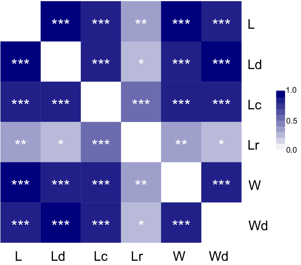
The heatmap of correlations (Fig. 4) reveals that all parameters exhibit a significant positive correlation with each other. The rostral pseudo parapet length (Lr) has a relatively weak correlation with other parameters, with which the parameter having a stronger correlation is not the platform length (L), but the caudal parapet length (Lc).
Correlations among L, Ld, Lc, Lr, W and Wd (abbreviations are given in the caption of Fig. 2). In heatmap, color represents the Spearman correlations. *, 0.01 < p value <0.05; **, 0.001 < p value < 0.01; ***, p value < 0.001.

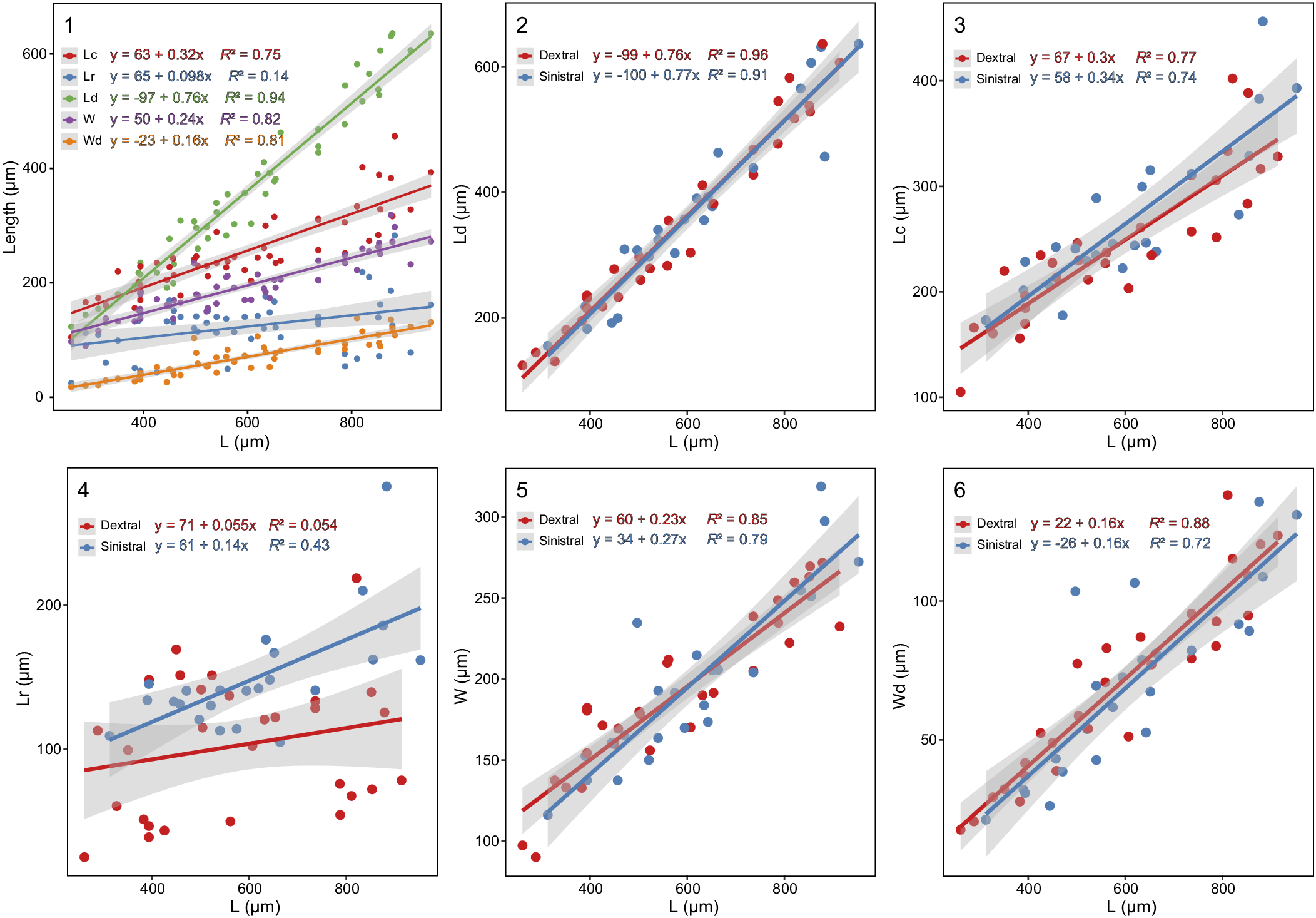
The regressions for parameters Lc, Ld, W, and Wd are relatively robust as indicated by the high coefficient values (R 2), while the result for parameter Lr is not strong (Fig. 5.1), which is consistent with the correlation heatmap. The caudal parapet length, parapet–pseudo parapet width, and dorsal carina width have a similar growth rate with each other. The growth rate of dorsal carina length to platform length is higher than for caudal parapet length, parapet–pseudo parapet width, and dorsal carina width; according to regression functions, the dorsal carina length exceeds the caudal parapet length when the platform length is about 400 μm.
(1) Linear regressions of Lc, Lr, Ld, W, and Wd versus L (abbreviations are given in the caption of Fig. 2). (2) Linear regressions of Ld versus L for both sinistral and dextral elements. (3) Linear regressions of Lc versus L for both sinistral and dextral elements. (4) Linear regressions of Lr versus L for both sinistral and dextral elements. (5) Linear regressions of W versus L for both sinistral and dextral elements. (6) Linear regressions of Wd versus L for both sinistral and dextral elements. Shadowed areas represent 95% confidence intervals.

When distinguishing the sinistral and dextral elements, regressions for parameters Lc, Ld, W, and Wd to L remain robust, which is consistent with results noted above, and exhibit similar fitting lines for both sinistral and dextral elements (Fig. 5.2, 5.3, 5.5, 5.6). However, the regression for Lr to L reveals that the fitting result of dextral elements (R 2 = 0.054) exhibits inferior performance compared to that of sinistral elements (R 2 = 0.43), and the scatter plot of dextral elements displays more dispersion (Fig. 5.4). Additionally, the rostral parapet length (Lr) of sinistral elements in Gnathodus pseudosemiglaber specimens with the same platform length (L) generally exceeds that of dextral elements (Fig. 5.4). The observed results may be attributed to variations in the ornamentation of the rostral platform in dextral elements, ranging from a single node to a pseudo parapet composed of a row of nodes. However, the ornamentation of the rostral platform commonly exhibits a pseudo parapet in sinistral elements.
Thin-plate splines
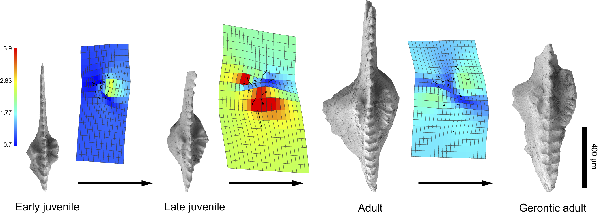
The TPS heatmap of four specimens exhibits the ontogenetic variations of the caudal parapet, rostral ornamentation, and the expanded dorsal carina (Fig. 6). The lengths of the caudal parapet and dorsal carina increase steadily during the earlier ontogenetic stage of Gnathodus pseudosemiglaber. From juvenile to adult forms, the most outstanding variation is that both the width and length of the dorsal carina grew rapidly. In addition, the length of the caudal parapet and the number of transverse ridges on it increased markedly. On the contrary, the distance between the ventral margin of the rostral pseudo parapet and the ventral margin of the dorsal carina hardly grew. From adult to gerontic forms, the most noteworthy growth occurs in both the length and width of caudal parapet and the distance between the dorsal tip and the rostral tip of the ridge on the broadest part of the dorsal carina. In addition, the scale of dorsal carina grew generally, which is evident in the specimens.
Thin-plate splines (TPS) heatmap of four representative specimens of different ontogenetic processes. Grid colors were encoded using Jacobian expansion factors to visually represent the degree of local expansion and contraction of the grids, the color spectrum ranges from yellow to red indicating expansion, and from light blue to dark blue indicating contraction.

Discussion
Taxonomy
Previously, the short caudal parapet, expanded dorsal carina, and rostral pseudo parapet on the platform were regarded as diagnostic features of Gnathodus pseudosemiglaber (Lane et al., Reference Lane, Sandberg and Ziegler1980). Based on the ontogenetic study and the revised taxonomy we have presented, the prominent morphological change of the dorsal carina through ontogeny seems to be the most useful characteristic for identifying G. pseudosemiglaber. The rostral ornamentation or pseudo parapet, however, had little change in length during the ontogenetic process.
The morphological variation of the expanded dorsal carina should be given more attention and caution in identification due to its significantly higher growth rate compared to other structures on the platform (Fig. 5). Jenkins et al. (Reference Jenkins, Crane and Mory1993) compared a newly named subspecies Gnathodus girtyi maxwelli Jenkins, Crane, and Mory, Reference Jenkins, Crane and Mory1993, with G. pseudosemiglaber and proposed that the former subspecies can be distinguished from G. pseudosemiglaber by a larger ratio of parapet length to platform length. However, only the holotype and paratype specimens of G. pseudosemiglaber were measured for the comparison, thus the result was not statistically convincing (Wang, Reference Wang2018). Our analysis demonstrates that the growth rate of the dorsal carina significantly exceeded that of both the parapet and pseudo parapet in G. pseudosemiglaber (Fig. 5), which results in a larger ratio of parapet/pseudo parapet length to platform length in juvenile specimens of G. pseudosemiglaber. Therefore, we regard G. girtyi maxwelli as a junior synonym of G. pseudosemiglaber.
The original diagnosis of Gnathodus pseudosemiglaber (Thompson and Fellows, Reference Thompson and Fellows1970) described the ornamentation on the rostral platform as “a single rounded node.” A few smaller nodes that often presented outside of the single node on the rostral platform on larger specimens were also noted by Thompson and Fellows (Reference Thompson and Fellows1970). Lane et al. (Reference Lane, Sandberg and Ziegler1980) revised the diagnosis of this species and designated the ornamentation on the rostral platform as the pseudo parapet, which consists of a fused row of small and weak nodes. However, the terminology of pseudo parapet overlooks the single rounded node.
In the population of G. pseudosemiglaber we studied here, the ornamentation on the rostral platform exhibits variations from a single node to a pseudo parapet consisting of a row of small, fused nodes. And the single node is common in dextral elements. Although it is difficult to determine if the asymmetrical rostral ornamentation between sinistral and dextral elements occurs within a pair of P1 elements from the same G. pseudosemiglaber organism, asymmetrical pairing P1 elements within one conodont species is not uncommon (Lane, Reference Lane1968), which may have enhanced occlusion efficiency, thus helping conodont animals better adapt to various food sources and predation methods (Donoghue and Purnell, Reference Donoghue and Purnell1999). Our results of the normal distribution test cannot refute the hypothesis that the studied G. pseudosemiglaber specimens were extracted from the same population. Therefore, both types of ornamentation are now considered intraspecific variations of G. pseudosemiglaber.
It is arbitrary to classify certain conodont species based on a sample that only yields juvenile or very small specimens, particularly for species with significant morphological differences between their juvenile and adult forms, such as Gnathodus pseudosemiglaber. It is preferable to consider a conodont sample in terms of populations. First, when conducting sampling, it is advisable to select samples from the same layer with consistent lithology in order to minimize the biases and errors caused by time-averaging and space-averaging. Second, try to obtain as many specimens as possible to be able to consider taxonomy in terms of population rather than individuals. Third, if there is uncertainty regarding whether the recovered specimens belong to the same population, performing the normal distribution test would be beneficial, because some morphological parameters of all individuals (e.g., individual size) in the same population should conform to a normal distribution if they exhibit continuous variation (Fang and Rong, Reference Fang and Rong1991; Zou and Ge, Reference Zou and Ge1995; Huang, Reference Huang2011).
Phylogeny
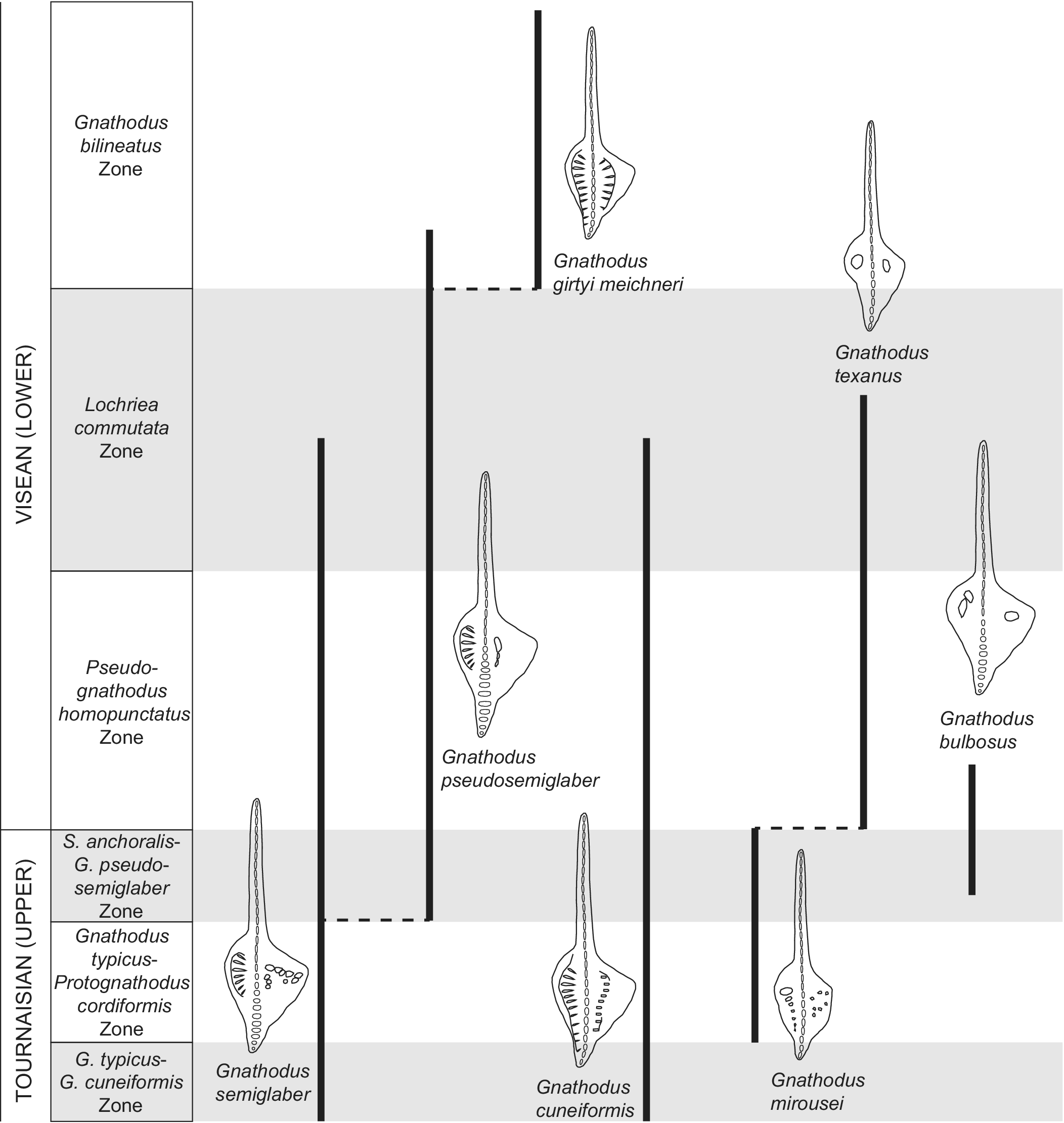
The phylogenetic relationships between the early representatives of the genus Gnathodus have been studied for many years (e.g., Thompson and Fellows, Reference Thompson and Fellows1970; Lane et al., Reference Lane, Sandberg and Ziegler1980; Belka, Reference Belka1985; Nemyrovska et al., Reference Nemyrovska, Perret-Mirouse and Weyant2006). However, it is difficult to understand the evolution of G. pseudosemiglaber until more comprehensive studies including closely related species are completed. For instance, G. delicatus Branson and Mehl, Reference Branson and Mehl1938, G. semiglaber, G. texanus (Roundy, Reference Roundy1926), and G. pseudosemiglaber are widely reported species that may refer to a variety of different, unrelated morphotypes that vary in their spatial and temporal distribution (Barrick et al., Reference Barrick, Alekseev, Blanco-Ferrera, Goreva, Hu, Lambert, Nemyrovska, Qi, Ritter and Sanz-López2022). Based on our current understanding of these Gnathodus species, the potential ancestor and descendant of G. pseudosemiglaber are discussed. Three species—G. bulbosus Thompson, Reference Thompson1967, G. semiglaber, and G. cuneiformis Mehl and Thomas, Reference Mehl and Thomas1947 (Fig. 7)—have been proposed to be the ancestor of G. pseudosemiglaber. Lane et al. (Reference Lane, Sandberg and Ziegler1980) amended the diagnosis of G. semiglaber to include G. bulbosus. However, the revision was somewhat arbitrary given the substantial morphological differences between the two species (e.g., the decorations on both the caudal and rostral platforms) (Fig. 7).
Drawings of the oral view P1 elements of Gnathodus bulbosus, G. cuneiformis, G. girtyi meischneri, G. mirousei, G. pseudosemiglaber, G. semiglaber, and G. texanus. Vertical bold lines represent conodont ranges, based on Belka (Reference Belka1985), Meischner and Nemyrovska (Reference Meischner and Nemyrovska1998), Lane and Brenckle (Reference Lane, Brenckle and Heckle2005), Sanz-López and Blanco-Ferrera (Reference Sanz-López and Blanco-Ferrera2018), and Hu et al. (Reference Hu, Qi, Qie and Wang2020), and horizontal dashed lines represent their possible phyletic relationships. Conodont zonation follows Hu et al. (Reference Hu, Qi, Qie and Wang2020). Conodont generic abbreviations: G. = Gnathodus; S. = Scaliognathus.

Here, we retain Gnathodus bulbosus as a valid species and follow the definition of Bishoff (Reference Bischoff1957) for G. semiglaber. Gnathodus bulbosus is characterized by a single circular or elongated node on the caudal platform and a single node or row of nodes perpendicular to the carina on the rostral platform (Thompson and Fellows, Reference Thompson and Fellows1970). The rostral ornamentation and carina of G. pseudosemiglaber and G. bulbosus are similar, whereas the caudal ornamentations of the two species exhibit significant differences. Besides, the G. bulbosus Zone in North America was assigned to be the latest Osagean (approximately latest Tournaisian–earliest Visean), which is higher than the first occurrence of the Scaliognathus anchoralis–Dolignathus latus Zone, whose base is correlated with the first occurrence (FO) of G. pseudosemiglaber (Tian and Coen, Reference Tian and Coen2004; Lane and Brenckle, Reference Lane, Brenckle and Heckle2005). In contrast, the other two species, G. semiglaber and G. cuneiformis, were both recorded lower than the FO of G. pseudosemiglaber (Barrick et al., Reference Barrick, Alekseev, Blanco-Ferrera, Goreva, Hu, Lambert, Nemyrovska, Qi, Ritter and Sanz-López2022). Lane et al. (Reference Lane, Sandberg and Ziegler1980) proposed that G. semiglaber gave rise to G. pseudosemiglaber within the S. anchoralis–D. latus Zone by narrowing the platform and simplifying nodose ornamentation on the rostral platform. This evolutionary process can be supported by some transitional specimens with several small nodes scattered outside the single rounded node or pseudo parapet on the rostral platform (e.g., Thompson and Fellows, Reference Thompson and Fellows1970, pl. 2, fig. 12; Sudar et al., Reference Sudar, Novak, Korn and Jovanović2018, pl. 2, figs. k, l). Some studies also proposed that G. pseudosemiglaber was derived from G. cuneiformis by reducing the length of the caudal parapet (Belka,Reference Belka1985, pl. 6, fig. 4; Belka and Groessens, Reference Belka and Groessens1986, pl. 5, figs. 4–9). The proposed transitional specimens between the two species have caudal parapets whose lengths fall within the range between that of G. cuneiformis and G. pseudosemiglaber, but display no transitional feature of dorsal carina from simplicity to expansion. Considering the stratigraphic occurrence and morphological similarity, G. semiglaber is more likely to be the ancestor of G. pseudosemiglaber. More detailed stratigraphic sampling with careful attention to all these morphological features will clarify where and when key changes occur that will strengthen phylogenetic hypotheses.
Gnathodus pseudosemiglaber was considered to potentially have given rise to G. girtyi meischneri Austin and Husri, Reference Austin, Husri, Bouckaert and Streel1974, or to G. texanus (Fig. 7), both of early Visean age (Lane and Brenckle, Reference Lane, Brenckle and Heckle2005; Nemyrovska, Reference Nemyrovska2005). Gnathodus girtyi meischneri is characterized by two parapets, while G. texanus is diagnosed by bearing one pillar-like node in both the caudal and rostral platforms (Jenkins et al., Reference Jenkins, Crane and Mory1993; Meischner and Nemyrovska, Reference Meischner and Nemyrovska1998). Gnathodus pseudosemiglaber may have evolved into G. girtyi meischneri through extending the caudal parapet and slightly narrowing the dorsal carina. Some possible transitional elements were illustrated by Nemyrovska (Reference Nemyrovska2005, pl. 6, figs. 2, 3, 5, 6, 8) and Nemyrovska et al. (Reference Nemyrovska, Perret-Mirouse and Weyant2006, pl. 3, figs. 9, 11, 18).
Subspecies of Gnathodus girtyi that possess two well-developed parapets extending nearly to the dorsal end of the carina may be confused as being homeomorphic species of G. cuneiformis (Pierce and Langenheim, Reference Pierce and Langenheim1974), but in fact these two species occur at different chronostratigraphic levels (Fig. 7). Many studies accept that G. pseudosemiglaber gave rise to G. texanus at the base of the G. texanus Zone by extremely narrowing the platform, shortening the caudal parapet, and reducing or losing the rostral ornamentation (e.g., Thompson and Fellows, Reference Thompson and Fellows1970; Lane et al., Reference Lane, Sandberg and Ziegler1980; Meischner and Nemyrovska, Reference Meischner and Nemyrovska1998). In some of the studied specimens, the rostral ornamentation is a single rounded node, suggesting a morphological similarity with the pillar-like node of G. texanus.
Several specimens that are morphologically close to Gnathodus pseudosemiglaber with shorter caudal parapet and thinner dorsal carina were also assigned to G. texanus (e.g., Thompson and Fellows, Reference Thompson and Fellows1970, pl. 2, fig. 16; Dong and Ji, Reference Dong and Ji1988, pl. 5, fig. 20; Zhang et al., Reference Zhang, Wang and Qu2006, pl. 1, figs. 15–18). However, they are more morphologically close to G. pseudosemiglaber by lacking the distinguishable single pillar-like node on the caudal platform of G. texanus and thus may represent transitional forms from G. pseudosemiglaber to an unknown descendant species rather than to G. texanus. The absence of authentic transitional forms with continuous transitional characteristics from the short parapet to the pillar-like node in ventral part of the caudal platform makes it difficult to conclude that G. pseudosemiglaber gave rise to G. texanus.
Recently, Sanz-López and Blanco-Ferrera (Reference Sanz-López and Blanco-Ferrera2018) named a new species, G. mirousei Sanz-López and Blanco-Ferrera, Reference Sanz-López and Blanco-Ferrera2018, from the Scaliognathus anchoralis Zone of the Cantabrian Mountains. This species has a pillar-like node on the ventral part of the caudal platform, and its caudal and rostral platforms are decorated with a pair of parallel rows of nodes. Because bearing only one pillar-like node in both the caudal and rostral platforms is the most diagnostic feature of Gnathodus texanus (Jenkins et al., Reference Jenkins, Crane and Mory1993; Fig. 8), G. mirousei may be the ancestor of G. texanus. Similarly, the early Visean species G. austini Belka, Reference Belka1985, which defines the G. austini Zone, preceded the G. texanus Zone in Poland and has a highly raised, simple node at the ventral end of the caudal parapet that is reminiscent of the pillar-like parapet (Belka, Reference Belka1985, pl. 4, figs. 1, 5, 6) in G. texanus, possibly supporting that G. austini is in a phylogenetic relationship with G. texanus. If the pillar-like node in the caudal platform is a synapomorphy, it is a reasonable inference that the G. mirousei gave rise to G. austini through G. texanus.
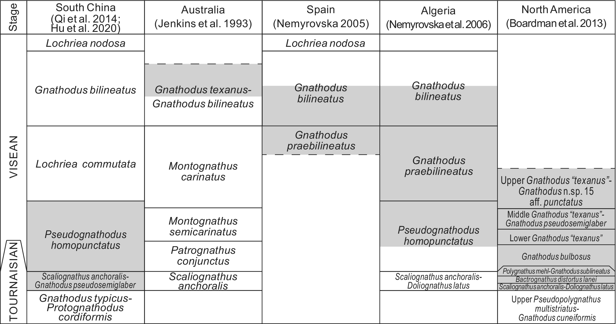
Conodont zonations and correlation of ranges of Gnathodus pseudosemiglaber among South China, Australia, Spain, Algeria, and North America. Dashed lines represent undetermined lower or upper limits of conodont zonations. Gray areas represent the distribution ranges of this species in the corresponding regions.

In summary, Gnathodus pseudosemiglaber is considered here to have most likely evolved from G. semiglaber through simplification of the rostral ornamentation. The direct descendant remains unknown, but it is likely an early subspecies of the G. girtyi group, such as G. girtyi meischneri. Although G. texanus has been considered the descendant species of G. pseudosemiglaber, it is more likely to have evolved from the more recently described G. mirousei rather than from G. pseudosemiglaber, and gave rise to G. austini.
Biostratigraphy
Gnathodus pseudosemiglaber first appears in the uppermost Tournaisian, accompanied by typical index conodonts Scaliognathus anchoralis and Doliognathus latus Branson and Mehl, Reference Branson and Mehl1941 (Barrick et al., Reference Barrick, Alekseev, Blanco-Ferrera, Goreva, Hu, Lambert, Nemyrovska, Qi, Ritter and Sanz-López2022). Tian and Coen (Reference Tian and Coen2004) established the S. anchoralis–G. pseudosemiglaber Zone in South China and correlated it with the S. anchoralis–D. latus Zone in North America, by the first appearance of S. anchoralis or G. pseudosemiglaber. However, the last appearance datum of G. pseudosemiglaber remains in debate. Lane et al. (Reference Lane, Sandberg and Ziegler1980) suggested that this species has a range overlapping with that of G. bilineatus bilineatus. Some researchers, however, have contested the possibility of G. pseudosemiglaber occurring at such a high stratigraphic level (e.g., Wang et al., Reference Wang, Qi, Hu, Sheng and Lin2014).
Here, the range of Gnathodus pseudosemiglaber is discussed based on the amended taxonomy of this species (Fig. 8). Gnathodus girtyi maxwelli, here regarded as a junior synonym of G. pseudosemiglaber, ranges within the G. texanus–G. bilineatus Zone in Queensland and New South Wales, Australia (Jenkins et al., Reference Jenkins, Crane and Mory1993). In the Cantabrian Mountains, Spain, G. pseudosemiglaber ranges from the G. praebilineatus Zone to the G. bilineatus Zone (Nemyrovska, Reference Nemyrovska2005). In Algeria, G. pseudosemiglaber ranges through the El Hariga Formation (Pseudognathodus homopunctatus Zone–G. bilineatus Zone) to the Mazzer Formation and coexists with G. girtyi, G. praebilineatus Belka, Reference Belka1985, Lochriea commutata (Branson and Mehl, Reference Branson and Mehl1941), and others (Weyant, Reference Weyant, Wagner, Winkler Prins and Granados1985; Nemyrovska et al., Reference Nemyrovska, Perret-Mirouse and Weyant2006). In North America, this species ranges from the S. anchoralis–D. latus Zone to the middle G. “texanus”–G. pseudosemiglaber Zone, and disappears at the top of the Osagean (Boardman et al., Reference Boardman, Thompson, Godwin, Mazzullo, Wilhite and Morris2013), corresponding with the lower part of Visean. It is noted that the G. bilineatus Zone has a rather long stratigraphic range spanning ca. 5 Myr (Hu et al., Reference Hu, Qi, Qie and Wang2020). In the upper part of the G. bilineatus Zone at the slope-facies Naqing section, no G. pseudosemiglaber has been recorded (Qi et al., Reference Qi, Nemyrovska, Wang, Chen, Wang, Lane, Hu and Wang2014). Therefore, G. pseudosemiglaber probably ranges from the S. anchoralis–D. latus Zone of uppermost Tournaisian to the lower part of the G. bilineatus Zone of middle Visean.
Conclusions
The classification of a sample yielding a substantial number of conodont specimens is more precisely approached through population thinking. The study of a population of Gnathodus pseudosemiglaber P1 elements offers an opportunity to present a new taxonomic description of G. pseudosemiglaber. Gnathodus pseudosemiglaber is a species of Gnathodus that is characterized by an elongated and triangular platform, a short caudal parapet, a rostral pseudo parapet, a transversely expanded dorsal carina, and a large basial cavity in the aboral view. The short parapet consists of transverse ridges on the caudal platform. The pseudo parapet comprises a row of fused small nodes or a single rounded node on the platform and exhibits a significant intraspecific variation. The transversely expanded dorsal carina occurs in large (adult) specimens and consists of isolated large nodes and ridge-like nodes. The growth rate of the dorsal carina greatly exceeded that of other structures on the platform, resulting in prominent morphological differences between juvenile and adult specimens. The previously named subspecies G. girtyi maxwelli is considered a junior synonym of G. pseudosemiglaber. Gnathodus pseudosemiglaber has a range from the Scaliognathus anchoralis–Doliognathus latus Zone of uppermost Tournaisian to the lower part of the G. bilineatus Zone of the middle-upper Visean. By combining the information of morphological similarity, transitional elements, and stratigraphic range, G. pseudosemiglaber was considered to be derived from G. semiglaber through the simplification of the rostral ornamentation, while its descendant possibly is a subspecies of the G. girtyi group, such as G. girtyi meischneri, rather than G. texanus, which is more likely to be an intermediate between G. mirousei and G. austini.
Systematic paleontology
Phylum Chordata Bateson, Reference Bateson1886
Class Conodonta Pander, Reference Pander1856
Subclass Conotonti Branson, Reference Branson1938
Order Ozarkodinina Dzik, Reference Dzik1976
Family Gnathodontidae Sweet, Reference Sweet1988
Genus Gnathodus Pander, Reference Pander1856
Type species
Polygnathus bilineatus Roundy, Reference Roundy1926, p. 13, pl. 3, figs. 10a-c; emend. Lane and Ziegler, Reference Lane and Ziegler1984.
Gnathodus pseudosemiglaber Thompson and Fellows, Reference Thompson and Fellows1970
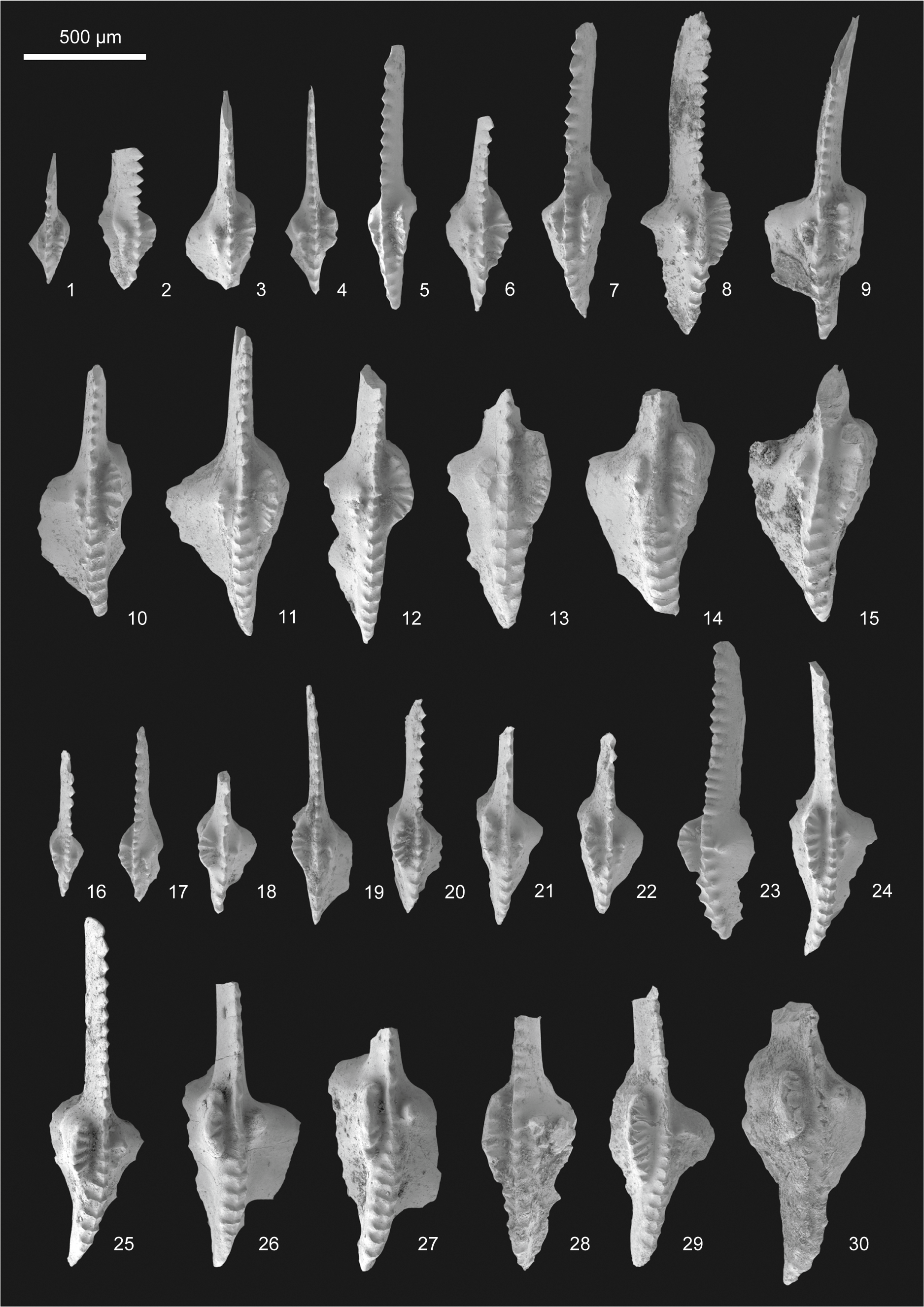
P1 elements of Gnathodus pseudosemiglaber Thompson and Fellows, Reference Thompson and Fellows1970. All oral views except for (23), which is lateral–oral view. (1–15) Sinistral elements, NIGP PZ 204030–204044; (16–30) dextral elements, NIGP PZ 204045–204059. All specimens are from sample YDII-C-12.0, Yudong II section.

Reference Rexroad and Scott1964 Gnathodus typicus; Rexroad and Scott, p. 31, pl. 2, fig. 3.
Reference Thompson and Fellows1970 Gnathodus texanus pseudosemiglaber Thompson and Fellows, p. 88, pl. 2, figs. 6, 8, 9, 11–13.
Reference Thompson and Fellows1970 Gnathodus texanus texanus, Thompson and Fellows, pl. 2, fig. 16 (only).
Reference Brenckle, Lane and Collinson1974 Gnathodus texanus; Brenckle et al., pl. 1, fig. 18.
Reference Pierce and Langenheim1974 Gnathodus typicus; Pierce and Langenheim, p. 163, pl. 3, fig. 6.
Reference Ebner1977 Gnathodus texanus; Ebner, pl. 1, figs. 6, 7.
Reference Ebner1977 Gnathodus typicus; Ebner, p. 470, pl. 1, figs. 8, 9 (only)
Reference Lane, Sandberg and Ziegler1980 Gnathodus pseudosemiglaber; Lane et al., p. 132, pl. 4, figs. 15–17, 19; pl. 5, figs. 8–15; pl. 6, fig. 14.
Reference Metcalfe1980 Gnathodus texanus pseudosemiglaber; Metcalfe, pl. 13, fig. 12.
Reference Qiu1984 Gnathodus antetexanus; Qiu, pl. 4, figs. 22, 23.
Reference Belka1985 Gnathodus pseudosemiglaber; Belka, pl. 6, figs. 5–13.
Reference Belka and Groessens1986 Gnathodus pseudosemiglaber; Belka and Groessens, pl. 5, figs. 10–15; pl. 7, figs. 1–3.
Reference Dong and Ji1988 Gnathodus pseudosemiglaber; Dong and Ji, pl. 5, figs. 23, 24.
Reference Dong and Ji1988 Gnathodus texanus; Dong and Ji, pl. 5, fig. 20 (only).
Reference Jenkins, Crane and Mory1993 Gnathodus girtyi maxwelli Jenkins, Crane, and Mory, p. 267, figs. 20A–FF.
Reference Perret, Vachard, Aguirre and Crasquin-Soleau1994 Gnathodus pseudosemiglaber; Perret et al., pl. 3, fig. 7.
Reference Perri and Spalletta1998 Gnathodus pseudosemiglaber; Perri and Spalletta, pl. 1, figs. 12–14; pl. 2, fig. 12.
Reference Tian and Coen2004 Gnathodus cuneiformis; Tian and Coen, pl. 1, fig. 3 (only).
Reference Tian and Coen2004 Gnathodus pseudosemiglaber; Tian and Coen, pl. 1, figs. 2, 12; pl. 2, figs. 15, 16.
Reference Tian and Coen2004 Gnathodus semiglaber; Tian and Coen, pl. 2, fig. 17 (only).
Reference Tian and Coen2004 Gnathodus typicus; Tian and Coen, pl. 1, figs. 5, 7, 11 (only).
Reference Nemyrovska2005 Gnathodus pseudosemiglaber; Nemyrovska, pl. 6, figs. 1, 9, 12.
Reference Nemyrovska, Perret-Mirouse and Weyant2006 Gnathodus pseudosemiglaber; Nemyrovska et al. pl. 3, figs. 2–8, 10, 14.
Reference Zhang, Wang and Qu2006 Gnathodus texanus; Zhang et al. pl. 1, figs. 15–18.
Reference Boncheva, Bahrami, Yazdi and Toraby2007 Gnathodus pseudosemiglaber; Boncheva et al., p. 341, pl. 2, figs. 3, 13; pl. 3, fig. 9.
Reference Dvořák2007 Gnathodus pseudosemiglaber; Dovrák, p. 32, pl. 2, figs. 2–6.
Reference Boardman, Thompson, Godwin, Mazzullo, Wilhite and Morris2013 Gnathodus n. sp. 12, Boardman et al., pl. 11, figs. 1–3, 7–9.
Reference Metcalfe and Aung2014 Gnathodus typicus; Metcalfe and Aung, pl. 2, figs. 1–4, 7, 8 (not 5, 6).
Reference Ahmadi, Dastanpour, Vaziri and Bahrami2016 Gnathodus pseudosemiglaber; Ahmadi et al., pl. 1, figs. 22–24.
Reference Sudar, Novak, Korn and Jovanović2018 Gnathodus pseudosemiglaber; Sudar et al., pl. 2, figs. k, l.
Reference Munkhjargal, Königshof, Waters, Carmichael, Gonchigdorj, Thassanapak, Udchachon and Davaanyam2021 Gnathodus pseudosemiglaber; Munkhjargal et al., fig. 15k.
Reference Sattari, Bahrami, Königshof and Vaziri-Moghaddam2021 Gnathodus pseudosemiglaber; Sattari et al., pl. 3, figs. 34, 35.
Reference Sattari, Bahrami, Königshof and Vaziri-Moghaddam2021 Gnathodus girtyi simplex; Sattari et al., pl. 3, fig. 17 (only).
Holotype
Department of Geology, University of Missouri (C-1003-8) from the Reeds Spring Formation, Tahlequah North, Oklahoma, USA (Thompson and Fellows, Reference Thompson and Fellows1970, pl. 2, figs. 11, 13).
Diagnosis
Carminiscaphate P1 element consisting of a free blade, and an asymmetrical, triangular platform. A short parapet parallels the carina in the ventral part of the caudal platform and bears transverse ridges. It is slightly curved away from the carina. On the rostral platform, the ornamentation, situated opposite the caudal short parapet, exhibits a pseudo parapet consisting generally 2–4 fused nodes, among which the most ventral one is elongated and much larger than others, or only a single large node. The dorsal carina transversely expands and is composed of large nodes or transverse ridges.
Occurrence
The Scaliognathus anchoralis–Doliognathus latus Zone of uppermost Tournaisian to the Gnathodus bilineatus Zone of middle Visean (see discussion for more details).
Description
The platform of the P1 element is elongated, triangular, and asymmetric. The free blade, composed of a series of fused denticles, is approximately equal in length to the platform. A short parapet on the caudal platform consists of several transverse ridges, which are not parallel but slightly radiate out from the center. The number of transverse ridges is usually equivalent to that of denticles and nodes or transverse ridges on the opposite position of the carina and is no more than five in juvenile specimens, and about eight but generally no more than ten in adult and gerontic specimens. The third most ventral transverse ridge of the parapet is usually longer than others, sometimes crosses the groove and almost reaches the dorsal tip of the ventral carina; it can be the second transverse ridge in subadults, and the first one in juveniles. The adcarinal groove between the parapet and the carina is moderately wide and deep. The ornamentation on the rostral platform varies from a single rounded node to a pseudo parapet. The latter consists of a row of small, fused nodes (commonly 2–4), among which the most ventral node is elongated. The adcarinal groove between the rostral ornamentation and the carina is slightly narrow and shallow. The carina extends to the dorsal margin of the platform, it consists of fused denticles at the ventral part but transversely expands at the dorsal part exhibiting a series of isolated large nodes or transverse ridges. The dorsal carina begins opposite the middle part of the parapet. The length and width of the expanded dorsal carina, and the ratio of that to the ventral carina significantly vary among specimens of different sizes. In juvenile specimens (Fig. 9.1–9.6, 9.16–9.20) with platform lengths ranging approximately from 250 to 500 μm, the expanded dorsal carina is absent, instead, a simple dorsal carina composed of fused denticles with wider spacing than those existing on the free blade is present, and the length of this simple dorsal carina is approximately one-half the length of the carina. In adult specimens (Fig. 9.10–9.12, 9.24, 9.25), both the length ratio and width ratio of the dorsal carina to the platform increase significantly. In some gerontic specimens (e.g., Fig. 9.13), an extremely expanded dorsal carina along with a wide caudal parapet makes the dorsal part of the caudal adcarinal groove extremely narrow, which thus makes the caudal adcarinal groove barely visible from the lateral view. The basal cavity is deep, large, and asymmetric with a broad expansion of basal cavity.
Remarks
Gnathodus pseudosemiglaber can be distinguished from G. typicus Cooper, Reference Cooper1939, by having an expanded dorsal carina. The ancestor of G. pseudosemiglaber is commonly regarded as G. semiglaber; the latter has more nodes on the rostral platform, especially the ventral part. Juvenile G. pseudosemiglaber is similar to G. texanus (Roundy, Reference Roundy1926) in size; the former is distinguished from the latter by having a parapet on the caudal platform and the ornamentation on the rostral platform that commonly is a pseudo parapet consisting of a row of small nodes. Adult G. pseudosemiglaber differs from G. texanus by the prominently expanded dorsal carina, as well as characteristics mentioned in juvenile specimens. Gnathodus pseudosemiglaber is differentiated from G. austini Belka, Reference Belka1985, by the slightly curved short parapet with the concave side toward the carina on the caudal parapet. It differs from G. cuneiformis Mehl and Thomas, Reference Mehl and Thomas1947, by having a much shorter parapet at the ventral part of the caudal platform.
Acknowledgments
We gratefully acknowledge Y. Li, Y.-F. Song, S.-R. Yang, and X.-Y. Ye for their assistance in the fieldwork. We thank A.V. Zhuravlev and an anonymous reviewer for their constructive comments and suggestions. This work was financially supported by the National Natural Science Foundation of China (grant nos. 42293281, 42172002) and the Second Tibetan Plateau Scientific Expedition and Research Program (grant No. 2019QZKK0706).
Competing interests
The authors declare none.