Introduction
Even with the advancement of assisted reproductive therapy (ART) techniques over the years, the success rate of implantation and pregnancy in in vitro fertilization and intracytoplasmic sperm injection (IVF/ICSI) programmes are still low, with nearly 25–40% of biochemical pregnancies per transfer (Coughlan et al., Reference Coughlan, Ledger, Wang, Liu, Demirol, Gurgan, Cutting, Ong, Sallam and Li2014). This low percentage is translated into unexplained infertility and recurrent implantation failure (RIF) in 10–15% of IVF/ICSI patients (Coughlan et al., Reference Coughlan, Ledger, Wang, Liu, Demirol, Gurgan, Cutting, Ong, Sallam and Li2014; Madkour et al., Reference Madkour, Bouamoud, Louanjli, Kaarouch, Copin, Benkhalifa and Sefrioui2016; EIM/ESHRE, 2017; Palermo et al., Reference Palermo, O’Neill, Chow, Cheung, Parrella, Pereira and Rosenwaks2017). RIF is considered as a clinical entity in which implantation fails to achieve a clinical pregnancy after the transfer of at least four fresh or frozen–thawed good quality embryos in a minimum of three IVF/ICSI cycles in a woman aged under 40 years old (Coughlan et al., Reference Coughlan, Ledger, Wang, Liu, Demirol, Gurgan, Cutting, Ong, Sallam and Li2014; Madkour et al., Reference Madkour, Bouamoud, Louanjli, Kaarouch, Copin, Benkhalifa and Sefrioui2016; Benkhalifa et al., Reference Benkhalifa, Zayani, Bach, Copin, Feki, Benkhalifa and Allal-Elasmi2018). To date, many options have been reported in the published literature to treat RIF, such as intrauterine insemination of cultured peripheral blood mononuclear cells (PBMC), endometrial injury, preimplantation genetic screening to eliminate the worst embryo for transfer, digital imaging during embryo culture and sequential IVF/ICSI (Bashiri et al., Reference Bashiri, Halper and Orvieto2018; Bodri et al., Reference Bodri, Milewski, Yao Serna, Sugimoto, Kato, Matsumoto and Kawachiya2018; Papanikolaou et al., Reference Papanikolaou, Chartomatsidou, Timotheou, Tatsi, Katsoula, Vlachou, Asouchidou, Zafeiratis and Najdecki2019). Recently, intrauterine insemination of autologous platelet-rich plasma (PRP; Kim et al., Reference Kim, Shin, Koo, Kwon, Choi and Kim2019) was proposed. However, the treatment choice or rescue strategy in this situation still needs earlier molecular indicator(s) or biomarker(s) that, once discovered, can prevent clinicians and patients together from falling into this kind of repeated failures by better managing embryo transfer strategy during IVF/ICSI cycles.
Successful embryo implantation requires complex molecular factors to prepare and orchestrate the cross-talk between the blastocyst and the endometrium prior to, and at the time of, implantation. Among these factors, hormones, chemokines, cytokines, growth factors and various matrix metalloproteinases (MMPs), which constitute a protease family of more than 20 members that behave as zinc-dependent proteolytic enzymes, are able to degrade specific extracellular matrix (ECM) components (Benkhalifa et al., Reference Benkhalifa, Zayani, Bach, Copin, Feki, Benkhalifa and Allal-Elasmi2018). The activity of these MMPs is essentially controlled by other molecular factors such as the tissue inhibitors of matrix metalloproteinases (TIMPs), which are considered to be essential for maintaining a balance between ECM accumulation and degradation in many physiological processes including embryo implantation (Benkhalifa et al., Reference Benkhalifa, Zayani, Bach, Copin, Feki, Benkhalifa and Allal-Elasmi2018). However, the exact role of each molecular factor in implantation has not yet been elucidated, therefore there is still an intense need for a better understanding of the molecular mechanisms of the embryo implantation process and its cross-talk with the receptive endometrium. (Rodgers et al., Reference Rodgers, Osteen, Matrisian, Navre, Giudice and Gorstein1993; Benkhalifa et al., Reference Benkhalifa, Madkour, Louanjli, Bouamoud, Saadani, Kaarouch, Chahine, Sefrioui, Merviel and Copin2015; Liang et al., Reference Liang, Diao, Huang, Lian, Chen, Li, Zhao, Li, He and Zeng2015). For this, different biomarkers were investigated from follicular fluid (FF), biopsied endometrium and embryo metabolome and secretome (Benkhalifa et al., Reference Benkhalifa, Madkour, Louanjli, Bouamoud, Saadani, Kaarouch, Chahine, Sefrioui, Merviel and Copin2015; Thompson et al., Reference Thompson, Brown and Sutton-McDowall2016; Alhilali et al., Reference Alhilali, Parham, Attaranzadeh, Amirian and Azizzadeh2019; Kaihola et al., Reference Kaihola, Yaldir, Bohlin, Samir, Hreinsson and Åkerud2019).
In IVF/ICSI, from oocyte retrieval to embryo transfer, there is a gap in the investigation of different biomarkers (from gene to single protein) to be potential predictors of implantation or RIF. Peripheral blood (PB) and/or FF, which are easily available the day of oocyte retrieval (OR) in women undergoing this treatment, could represent an optimum source of biochemical predictors of oocyte quality and embryo implantation in IVF/ICSI cycles. In addition, the FF constitutes the oocyte microenvironment and is rich in growth factors and cytokines that must play crucial roles in ovarian physiology regulation, essentially related to folliculogenesis and ovulation with MMPs, TIMPs and different other molecules, such as cell adhesion molecules. These could affect oocyte maturation and, secondly, embryo implantation. (Benkhalifa et al., Reference Benkhalifa, Madkour, Louanjli, Bouamoud, Saadani, Kaarouch, Chahine, Sefrioui, Merviel and Copin2015, Reference Benkhalifa, Zayani, Bach, Copin, Feki, Benkhalifa and Allal-Elasmi2018; Alhilali et al., Reference Alhilali, Parham, Attaranzadeh, Amirian and Azizzadeh2019).
Therefore, the present study aimed to evaluate the possibility of finding a predictive biomarker of RIF by analyzing a panel of MMPs (MMP-1, -2, -3, -7 and -9), TIMPs (TIMP1 and TIMP2), cytokines (leukaemia inhibitory factor, LIF), growth factors (vascular endothelial growth factor, VEGF) and the soluble forms of cell adhesion molecules such as intercellular adhesion molecule 1 (ICAM1) and vascular cell adhesion molecule 1 (VCAM1), in the PB (sampled at the day of OR) and in the FF of RIF and ICSI successful women undergoing ART treatment with ICSI.
Materials and methods
Study population
In total, 44 women were recruited at the Alyssa Fertility Group (AFG) Center (Alyssa Clinic, Les Berges du Lac, Tunis, Tunisia) in the period from May 2016 to July 2017. Women were aged between 25–39 years old, with no history of any disease (considered as healthy women) and with acceptable sperm quality from their partners (>1 × 10⁶/ml of spermatozoa). All these women underwent ICSI with a fresh IVF/ICSI cycle (fresh sperm, fresh oocytes and fresh embryo transfer), and were divided into two groups: RIF group (n = 22) defined as women who failed their embryo implantation after three or more ICSI treatment attempts with at least four good quality embryo transfers and the control group (n = 22), defined as women who underwent their first cycle with successful embryo implantation after ICSI treatment, and called the ICSI success group.
Controlled ovarian stimulation
An antagonist gonadotropin releasing hormone (GnRH) protocol was used for all the included women as a controlled ovarian stimulation (COS). In accordance with clinician preference, recombinant follicle-stimulating hormone (recFSH) (alone) or combined with human menopausal gonadotropin (hMG) (recFSH/hMG) was started on cycle day 2. When the leading follicle reached 14 mm, 0.25 mg of Cetrorelix (Cetrotide, Serono) was administered daily until choriogonadotropin alfa (Ovitrelle, Merck Serono) injection. All cycles received an initial gonadotropin dose of 150–225 IU/day for the first 5 days, followed by individual adjustments in the gonadotropin dose according to ovarian response. When the average diameter of leading follicles reached ≥18 mm and serum oestradiol (E2) levels were 600 pg/ml, 250 μg of choriogonadotropin alfa (Ovitrelle, Merck Serono) was administered. OR procedures were performed 36 h following Ovitrelle injection using an ultrasound-guided transvaginal oocyte aspiration.
Samples collection, biochemical measurements and analyses of investigated markers
Blood samples were collected in different tubes on the day of OR [heparin, ethylenediamine tetraacetic acid (EDTA), sodium fluoride and dry tube without anticoagulant]. Immediately after centrifugation (×3000 g for 15 min), serum and plasma samples for analyses of MMPs, TIMPs, adhesion molecules (ICAM1 and VCAM1) and cytokines were frozen and stored at −80°C until enzyme-linked immunosorbent assay (ELISA) analysis. All other analyses were carried out within 4 h and the coefficient of variation of the different parameters was 1–5%. An automatic chemistry analyzer (Architect Ci8200) performed all biochemical analyses. White blood cells and lymphocytes were obtained by determination of the complete blood count. The determination of haematological parameters was performed using a haematology automated ABX Micros 60 analyzer.
Follicular fluids were collected after oocyte pickup. Bloody or flushing FF samples were not considered for the study. The FF of each one of the included women were pooled and centrifuged immediately at 15 000 g for 15 min to eliminate cellular contents and debris, then the supernatants were aliquoted into sterile tubes and stored at −80°C until analysis.
All the MMPs from this study, along with the TIMPs, cell adhesion molecules, VEGF and LIF were analyzed in EDTA plasma, serum and FF using sandwich ELISAs (Duoset R&D Systems, Lille, France). All samples (plasma EDTA, serum and FF) were measured in duplicate with a coefficient of variation (CV) <8%.
ICSI procedure, embryo culture and transfer
Conventional ICSI procedures were carried out 3–4 h after OR using an inverted microscope (Nikon Ti2 Eclipse, Japan) equipped with micromanipulators (Narishige, Japan). All cumulus–oocyte complexes (COCs) were eliminated using hyaluronidase medium before ICSI (Hyase-10X™, Vitrolife).
Fertilization rate was calculated 16–24 h after ICSI by dividing the number of the resulting two pronuclei (2PN) zygotes by the number of matured oocytes (metaphase II) and then multiplying by 100.
Embryos were cultured in a humidified atmosphere maintained at 37°C in Trigaz incubators (6% CO2, 5% O2, q.s. N2) until day 3 or 5 post ICSI. SAGE 1-Step™ (Origio, Denmark) was the culture medium used for embryo culture from OR day until day 3 or 5 post ICSI. Embryo classification and quality until and during cleavage stage was evaluated according to BLEFCO consensus and a good quality embryo was defined as having a minimum of eight cells at day 3 post OR (Boyer and Boyer, Reference Boyer and Boyer2009). For the blastocyst, a score of 4AA to 4AB was considered as good quality based on the Gardner and Schoolcraft score system (Gardner and Schoolcraft, Reference Gardner and Schoolcraft1999). A minimum of one good quality embryo was transferred into all included women.
The serum β-human chorionic gonadotropin (β-HCG) level was measured 2 weeks after embryo transfer (ET) to detect the biochemical pregnancy. Those with positive β-HCG results (positive biochemical pregnancy) underwent ultrasonography 2 weeks later to confirm clinical pregnancy. Clinical pregnancy was defined as the presence of an intrauterine gestational sac. Luteal phase support was ongoing until 9 weeks of pregnancy using vaginal progesterone (P4) supplementation. The obstetric progress of the pregnant patients was followed up by a timely chart review.
Statistical analysis
Statistical analysis was performed using SPSS software for Windows (v.22.0). Kolmogorov–Smirnov test was used to verify the distribution normality of continuous variables. These continuous variables were compared using Mann–Whitney U-test or independent Student’s t-test as appropriate. Quantitative variables are shown as mean values ± standard deviation (SD) or median (minimum–maximum) as appropriate. Correlations between the different parameters were performed in pairs in accordance with the Spearman method. Receiver operating characteristic (ROC) curve was used to assess the predictive value of the biomarkers for the RIF group. Sensitivity, specificity and cut-off values were selected and calculated according to the Youden index. Statistical differences were considered as significant for a P-value < 0.05.
Results
In the present study, the PB and FF samples of the 44 included women were analyzed. In total, 486 oocytes were retrieved, 234 of which were metaphase 2 (MII) oocytes. The mean number of MII oocytes was almost equally divided between the two studied groups with 5 ± 2 for the ICSI success group and 5 ± 4 for the RIF group (Table 1). The rate of matured oocytes (MII) did not present a significant difference between the two groups, with 60.38 ± 22.74 and 46.06 ± 24.29 for the ICSI success and RIF groups respectively. In total, 201 oocytes were normally fertilized for the total of included women, with no significant difference in the fertilization rate between the two groups (88% ± 14% vs 80.46% ± 25.31% for ICSI success group and RIF group, respectively). Of these, 105 oocytes developed into good quality embryos at the cleavage stage (day 3 post ICSI). From the 201 fertilized oocytes, 119 embryos were transferred, 43 at the blastocyst stage and 76 at the day 3 stage, with a minimum of one good quality embryo per woman.
Clinical outcomes and characteristics of ‘RIF’ and ‘ICSI success’ women

ICSI: intracytoplasmic sperm injection, NS: non-significant difference; RIF: recurrent implantation failure.
Table 1 shows the absence of significant differences in age, oocytes and embryo characteristics between the two groups in the study. However, the number of total good quality embryos presented a difference that could be considered as at the limit of signification (P = 0.053) between the two groups, with a decrease in the RIF group compared with the ICSI success group.
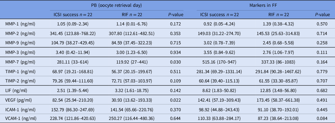
Table 2 shows that PB levels for MMP-7 and VEGF decreased significantly in the RIF group compared with the controls. However, the rest of the analyzed parameters, whether in PB or in FF, did not present any significant differences between the two groups of the study.
Comparison of biomarkers in PB and FF samples of ‘ICSI success’ and ‘RIF’ women

FF: follicular fluid; ICAM1: intercellular adhesion molecule 1; ICSI: intracytoplasmic sperm injection; LIF: leukaemia inhibitory factor; MMP: matrix metalloproteinases; PB: peripheral blood; RIF: recurrent implantation failure; TIMP: tissue inhibitor of matrix metalloproteinases; VCAM1: vascular cell adhesion molecule 1; VEGF: vascular endothelial growth factor.
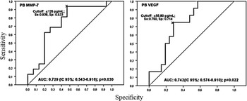
ROC curve analysis of biomarkers in PB (Fig. 1) showed that MMP-7 and VEGF levels seemed to predict RIF with an informative area under the curve (AUC) (AUC: 0.729; P < 0.030; sensitivity (Se): 0.938, specificity (Sp): 0.533; and AUC: 0.742; P < 0.022; Se: 0.750, Sp: 0.714, respectively). In addition, cut-offs at 125 pg/ml for PB MMP-7 and 55.80 pg/ml for PB VEGF were determined when the Se and the Sp reached their maximal levels in the ROC curves.
Receiver operating characteristics (ROC) curves for PB MMP-7 and PB VEGF in RIF women. AUC: Area under curve; IC: Confidence interval; MMP: Matrix metalloproteinases; PB: peripheral blood; p: P-value; Se: sensitivity; Sp: specificity; VEGF: vascular endothelial growth factor.

For correlation studies, PB MMP-7 and PB VEGF did not present correlations with any other studied parameter such as fertilization rate, embryo quality and the day of transfer (data not shown).
Discussion
According to our preliminary data, it seems that levels of MMP-7, also known as epithelial specific enzyme or matrylisin-1 (Osteen et al., Reference Osteen, Sierra-Rivera, Keller and Fox1997), were significantly decreased in the PB of RIF patients compared with controls at the day of OR. In addition, ROC curve analyses (Fig. 1) showed that the AUC value of the PB MMP-7 was informative, with a cut-off of 125 pg/ml in the circulating concentration. In women, MMP-7 is localized (Osteen et al., Reference Osteen, Sierra-Rivera, Keller and Fox1997) in the glandular and luminal epithelia of the endometrium and could initiate a ‘breakdown’ of the integrity of the luminal epithelial cells that constitute the first cell barrier encountered by the invasive trophoblast during the initial steps of implantation (Osteen et al., Reference Osteen, Sierra-Rivera, Keller and Fox1997). Its expression was revealed in the human endometrium during the proliferative phase of the menstrual cycle, but was suppressed during the early and mid-secretory phase when P4 dominates E2 levels in the natural ovulatory cycle (Rodgers et al., Reference Rodgers, Osteen, Matrisian, Navre, Giudice and Gorstein1993; Bruner et al., Reference Bruner, Rodgers, Gold, Korc, Hargrove, Matrisian and Osteen1995). P4 is crucial for the establishment of successful pregnancy and is produced by the developing follicle prior to ovulation and by the corpus luteum after ovulation in the natural cycle (Yanushpolsky, Reference Yanushpolsky2015). However, P4 has been demonstrated to inhibit and to regulate the mRNA and protein expression of MMP-7 and other MMPs in human endometrium by inducing stromal factors, such as TGF-β, in epithelial cells (Osteen et al., Reference Osteen, Rodgers, Gaire, Hargrove, Gorstein and Matrisian1994, Reference Osteen, Sierra-Rivera, Keller and Fox1997; Bruner et al., Reference Bruner, Rodgers, Gold, Korc, Hargrove, Matrisian and Osteen1995). Unfortunately, we did not measure the circulating levels of TGF-β in the participants in our study, however, and according to the published literature, this growth factor has been shown to inhibit secretion of MMP-7 by mesangial cells in vitro (Marti et al., Reference Marti, Lee, Kashgarian and Lovett1994). Therefore, this seems to affect implantation by blocking MMP-7 production in the endometrium, especially if it is present at higher concentrations at the time of implantation. However, P4 levels in our study (measured 36 h before OR) were not significantly different in RIF and ICSI success women and were all under 0.95 ng/ml (data not shown). Therefore, this finding suggests an insufficiency or a ‘hypersensitivity’ for MMP-7 against P4 inhibitory function in the RIF group of our study, whether in PB or in endometrial tissue, and this seems to disturb the probable balance between MMP-7 and P4, which is potentially ‘necessary’ to improve embryo implantation.
P4 production in COS is different from that in natural cycles, especially in the luteal phase, which is disrupted by the removal of the GCs during oocyte pick up or denudation, and which can lead to the depletion of the cell pool that is needed to produce P4 during this phase (Meinel et al., Reference Meinel, Blohberger, Berg, Berg, Dissen, Ojeda and Mayerhofer2015; Yanushpolsky, Reference Yanushpolsky2015). Therefore, after OR, MMP-7 production may also decrease, as it seems to be produced by the GCs (according to Meinel et al., Reference Meinel, Blohberger, Berg, Berg, Dissen, Ojeda and Mayerhofer2015). From that time, the corpus luteum P4 of the natural cycle is substituted by HCG or P4 supplementation during the beginning of the pregnancy (Yanushpolsky, Reference Yanushpolsky2015). However, as P4 is a potential suppressor of MMPs (Osteen et al., Reference Osteen, Rodgers, Gaire, Hargrove, Gorstein and Matrisian1994), its administration to support the luteal phase in ART cycles could negatively affect implantation by inhibiting MMP-7. In addition, Sohn et al. (Reference Sohn, Penzias, Emmi, Dubey, Layman, Reindollar and DeCherney1999) demonstrated that administration of P4 before OR affected implantation rate negatively. Moreover, vaginal P4 administration to support the luteal phase in IVF cycles, which was shown to be more tolerated by the patients, was suggested to be started within 24 to 48 h after OR to give better results in terms of embryo implantation and pregnancy rate (Yanushpolsky, Reference Yanushpolsky2015). These data supported our results that the presence of PB MMP-7 in sufficient concentrations at the time of OR was crucial for the success of embryo implantation and that PB MMP-7 could be proposed as a new biomarker of RIF to keep the circulating balance P4–MMP-7. In the FF, the levels of this small member of the MMP family were not significantly different between the two groups in our study. The findings by Bou Nemer et al. (Reference Bou Nemer, Shi, Carr, Word and Bukulmez2019), who claimed that there was no role for MMP-7 in the FF, could support our results.
Embryo implantation is a multifactorial event that includes many other molecular interactions and signalling between the blastocyst and the endometrium such as through VEGF (Benkhalifa et al., Reference Benkhalifa, Zayani, Bach, Copin, Feki, Benkhalifa and Allal-Elasmi2018). VEGF circulating levels were significantly decreased in the RIF group compared with the IVF success women, with an AUC of 0.742 in ROC curve analysis and a cut-off at 55.80 pg/ml. VEGF increases vascular permeability and is essential for the process of angiogenesis during embryo implantation, especially in trophoblast invasion into the endometrium (Valdes et al., Reference Valdes, Schutt and Simon2017). In addition, this growth factor has already been proposed as a potential marker of endometrial receptivity in a mouse model (Binder et al., Reference Binder, Evans, Gardner, Salamonsen and Hannan2014). However, data investigating the association between VEGF and RIF are limited. In a Korean study, Shim et al. (Reference Shim, Kim, Jeon, An, Lee, Kim, Ahn, Lee and Kim2018) evaluated different gene polymorphisms in the VEGF promoter region in RIF women; the authors revealed an association between the different studied polymorphisms and RIF, proposing the VEGF −2578AA genotype, −634G allele and −2578A/−1154A/−634G/936C haplotype as genetic biomarkers for susceptibility to RIF. This could be in accordance with our results, however the authors of that study did not measure the serum levels of VEGF in the participant women (Shim et al., Reference Shim, Kim, Jeon, An, Lee, Kim, Ahn, Lee and Kim2018). Our results could be supported by Vagnini et al. (Reference Vagnini, Nascimento, Canas Mdo, Renzi, Oliveira-Pelegrin, Petersen, Mauri, Oliveira, Baruffi, Cavagna and Franco2015), who investigated the relationship between RIF and different polymorphisms, including one polymorphism in the VEGF gene, the authors of this study showed an association between the VEGF −1154G/A polymorphism and RIF in Brazilian women (Vagnini et al., Reference Vagnini, Nascimento, Canas Mdo, Renzi, Oliveira-Pelegrin, Petersen, Mauri, Oliveira, Baruffi, Cavagna and Franco2015). Another study revealed an association between VEGF polymorphisms and embryo implantation in women undergoing IVF treatment with ICSI procedures and proposed VEGF +405G>C as a genetic predictive marker of embryo implantation (Boudjenah et al., Reference Boudjenah, Molina-Gomes, Torre, Boitrelle, Taieb, Dos Santos, Wainer, de Mazancourt, Selva and Vialard2014). However, Boudjenah and colleagues, in an earlier study, revealed that VEGF +405.CC, which was associated with low PB VEGF, presented higher levels of expression in women with RIF when compared with the controls (Boudjenah et al., Reference Boudjenah, Molina-Gomes, Wainer, de Mazancourt, Selva and Vialard2012). Moreover, the same laboratory confirmed that this VEGF polymorphism (VEGF +405.CC) when combined with TNF-α 308.GG, was associated with low embryo implantation rates (Boudjenah et al., Reference Boudjenah, Molina-Gomes, Torre, Boitrelle, Taieb, Dos Santos, Wainer, de Mazancourt, Selva and Vialard2014). Contrary to our results, Bansal et al. (Reference Bansal, Ford, Bhaskaran, Thum and Bansal2017) revealed high serum concentrations of VEGF in RIF women compared with controls. However, this difference could be due to blood-sampling timing [day of OR in a COS in our study vs the first half of the natural menstrual cycle in the Bansal et al. (Reference Bansal, Ford, Bhaskaran, Thum and Bansal2017) study] or to the COS used in the participants of our study (Bansal et al., Reference Bansal, Ford, Bhaskaran, Thum and Bansal2017). Therefore, these data showed that numerous VEGF polymorphisms must be responsible for the disturbance in the production rates of this protein in women with RIF and, consequently, a disturbance in angiogenesis during invasion of the blastocyst, resulting in implantation failure and RIF. However, other studies are needed to investigate the levels of VEGF in serum in women undergoing IVF/ICSI, especially at the day of OR and its association with RIF to confirm the proposal that it is a predictive biomarker for RIF.
Our results may help physicians in their choice of proposed treatments and rescue strategies in patients at risk of RIF, based on their circulating MMP-7 and VEGF levels of the day of OR of their first IVF/ICSI attempt (Coughlan et al., Reference Coughlan, Ledger, Wang, Liu, Demirol, Gurgan, Cutting, Ong, Sallam and Li2014; Bashiri et al., Reference Bashiri, Halper and Orvieto2018). Intrauterine infusion of PRP, for example, is considered as a successful treatment for infertile women with a thin endometrium, which (the latter) could be among the factors that cause RIF. Interuterine infusion had a positive effect on endometrial MMP-7 expression, which increased significantly in human endometrial stromal fibroblasts (eSF) compared with controls and had no effect on oestrogen or P4 receptor expression (Aghajanova et al., Reference Aghajanova, Houshdaran, Balayan, Manvelyan, Irwin, Huddleston and Giudice2018). The authors of this in vitro study (Aghajanova et al., Reference Aghajanova, Houshdaran, Balayan, Manvelyan, Irwin, Huddleston and Giudice2018) showed also that the use of 5% activated PRP (aPRP) had a greater effect on MMP-7 expression in eSF than other types of PRP, and this could confirm its usefulness in increasing the levels of MMP-7 in women with RIF. In addition, a freeze-all strategy and frozen–thawed ET with a modified or natural cycle are also recommended options for cases of RIF (especially if the negative influence of COS on molecular factors in the endometrium encountered in fresh IVF/ICSI cycles could be avoided), as well as the P4 supplementation effect to support the luteal phase (Mackens et al., Reference Mackens, Santos-Ribeiro, van de Vijver, Racca, Van Landuyt, Tournaye and Blockeel2017; Bashiri et al., Reference Bashiri, Halper and Orvieto2018). This strategy could be combined with autologous PRP treatment, which has had encouraging results, according to Kim et al. (Reference Kim, Shin, Koo, Kwon, Choi and Kim2019). Cytokines and growth factors such as VEGF have increased activity in PRP, and could be useful as a treatment for RIF, according to our previous results (Wu et al., Reference Wu, Diaz and Borg-Stein2016). However, a recent study showed no difference in the pregnancy results in women with history of failed implantation who used (or not) PRP treatment before frozen–thawed ET (Allahveisi, et al., Reference Allahveisi, Seyedoshohadaei, Rezaei, Bazrafshan and Rahimi2020). Intrauterine insemination of PBMC has also been recommended as a treatment for RIF (Makrigiannakis et al., Reference Makrigiannakis, BenKhalifa, Vrekoussis, Mahjub, Kalantaridou and Gurgan2015; Madkour et al., Reference Madkour, Bouamoud, Louanjli, Kaarouch, Copin, Benkhalifa and Sefrioui2016). In addition, it was shown to give better results in terms of embryo implantation when combined with a freeze-all strategy and frozen–thawed ET (Yu et al., Reference Yu, Yang, Guo, Fang, Yin, Luo, Li, Li, Zhao, Zou and Xu2014; Nobijari et al., Reference Nobijari, Arefi, Moini, Taheripanah, Fazeli, Kharazi, Hosseini, Hosseini, Valojerdi, Copin and Benkhalifa2019). This immunotherapy has been shown to increase levels of growth factors such as VEGF and to regulate the Th1/Th2 cytokine imbalance in the endometrium of women suffering from RIF, by increasing the levels of Th1 pro-inflammatory cytokines at the time of implantation (Madkour et al., Reference Madkour, Bouamoud, Louanjli, Kaarouch, Copin, Benkhalifa and Sefrioui2016; Bashiri et al., Reference Bashiri, Halper and Orvieto2018). Moreover, this treatment was also suggested to be performed with endometrium scratching (Bashiri et al., Reference Bashiri, Halper and Orvieto2018). In fact, Th1 cytokines include IL-1α, which has the ability to induce MMP-7 expression in the endometrium by opposing the P4-mediated suppression against matrylisin. Therefore, consequently, protein levels of MMP-7 should be increased by PBMC treatment in women with RIF (Osteen et al., Reference Osteen, Sierra-Rivera, Keller and Fox1997).
Our data suggested, for decreased levels of PB MMP-7 and PB VEGF on the day of OR, combining the sequential transfer strategy and PBMC treatment with a ‘soft’ preparation of the endometrium during the frozen–thawed ET cycle, with or without intrauterine insemination of the PRP prior to transfer. This strategy could increase the success rate for embryo implantation.
In conclusion, our ‘preliminary’ study showed that circulating MMP-7 and VEGF seem to play an interesting role in embryo implantation in IVF/ICSI cycles and could be proposed as circulating biomarkers for RIF. These biomarkers could help physicians, from the day of OR and from the first IVF/ICSI attempt, to use the proposed RIF treatments and rescue strategies, such as intrauterine insemination of the autologous cultured PBMC with or without frozen–thawed ET and with or without PRP treatment. However, our study is still limited and more in-depth work with a larger cohort of patients is required to confirm the usefulness of these biomarkers and to identify other circulating and FF molecular factors that could play crucial roles in modulation of embryo implantation, and by combining these to determine a final score for identifying women with RIF from their first IVF/ICSI cycle onwards.
Acknowledgements
We would like to thank Dr Oualid Triki, Professor Nizar Ben Aissia, Dr Sam Sepheur and Dr Khalil Ben Cherifa for their help and their collaboration whenever needed, as well as the technicians and all the staff in the AFG. A special thanks to all the PhD candidates and the staff of the Biochemistry Department of La Rabta University Hospital.
Financial support
The study was supported by funds of Research Laboratory LR99ES11, Ministry of Higher Education and Scientific Research of Tunisia.
Conflict of Interest
The authors declare that they have no conflict of interest.
Ethical standards
The study was approved by the Ethics Committee of Rabta Hospital. All included women were informed with details about this research study and a written informed consent was obtained from each participant. The authors assert that all procedures contributing to this work comply with the ethical standards of the relevant national and institutional committees on human experimentation and with the Helsinki Declaration of 1975, as revised in 2008.