Introduction
Childhood trauma increases risk of psychotic disorders, with an overall odds ratio of 2.78 (95% confidence interval 2.34–3.31) (Varese et al. Reference Varese, Smeets, Drukker, Lieverse, Lataster, Viechtbauer, Read, van and Bentall2012). A history of childhood trauma is also associated with affect dysregulation in bipolar disorders (Etain et al. Reference Etain, Henry, Bellivier, Mathieu and Leboyer2008; Aas et al. Reference Aas, Aminoff, Vik, Etain, Agartz, Andreassen and Melle2014a ), suicide attempts, rapid cycling, as well as lower age at disease onset (Etain et al. Reference Etain, Aas, Andreassen, Lorentzen, Dieset, Gard, Kahn, Bellivier, Leboyer, Melle and Henry2013), all markers of increased illness severity. Childhood maltreatment is a severe stressor that, in addition to behavioural and cognitive problems, is accompanied by a cascade of physiological, neurochemical and hormonal changes, which can lead to changes in both brain structure and function (Teicher et al. Reference Teicher, Andersen, Polcari, Anderson, Navalta and Kim2003, Reference Teicher, Samson, Polcari and McGreenery2006). It is well documented that childhood trauma is important for the development of stress sensitization and consequent emotional reactivity to daily stress in adult life (Myin-Germeys et al. Reference Myin-Germeys, van, Schwartz, Stone and Delespaul2001, Reference Myin-Germeys, Delespaul and van2005), including increase in negative emotion (Myin-Germeys et al. Reference Myin-Germeys, van, Schwartz, Stone and Delespaul2001; Glaser et al. Reference Glaser, van, Portegijs and Myin-Germeys2006).
Increased reactivity to stressors is an essential feature of schizophrenia, a notion dating back to Brown and Birley's seminal paper arguing that stress precipitates the onset of psychosis (Brown & Birley, Reference Brown and Birley1968). It is well known from clinical studies that patients with psychotic disorders are particularly sensitive to negative stressors, including familial high expressed emotions (Linszen et al. Reference Linszen, Dingemans, Nugter, van der Does, Scholte and Lenior1997) and other environments characterized by high levels of anger and aggression (Melle et al. Reference Melle, Friis, Hauff, Island, Lorentzen and Vaglum1996). These findings are supported by the study of Livingstone et al. (Reference Livingstone, Harper and Gillanders2009) showing that both schizophrenia and bipolar patients have higher levels of negatively valued emotions and lower levels of happiness, compared with healthy individuals.
Whereas mounting evidence has documented associations between childhood trauma and brain structure (Teicher et al. Reference Teicher, Anderson and Polcari2012; Heim et al. Reference Heim, Mayberg, Mletzko, Nemeroff and Pruessner2013), only a few studies have investigated possible functional brain correlates to maltreatment using functional magnetic resonance imaging (fMRI) (Hart & Rubia, Reference Hart and Rubia2012). Most previous fMRI studies have investigated childhood maltreatment and brain function in patients with a post-traumatic stress disorder (PTSD) (Hart & Rubia, Reference Hart and Rubia2012). An abundance of previous studies have targeted emotional processing in schizophrenia and bipolar disorders (Taylor et al. Reference Taylor, Kang, Brege, Tso, Hosanagar and Johnson2012; Corbalan et al. Reference Corbalan, Beaulieu and Armony2015), though without taking childhood trauma experiences into account.
A review including 26 studies concluded that patients with schizophrenia had reduced brain activation during emotional perception in the bilateral amygdala, visual processing areas, anterior cingulate cortex, dorsolateral prefrontal cortex, medial frontal cortex and subcortical structures compared with healthy individuals (Taylor et al. Reference Taylor, Kang, Brege, Tso, Hosanagar and Johnson2012). In addition, patients with schizophrenia had increased activation in the cuneus, parietal lobule, precentral gyrus and superior temporal gyrus (Taylor et al. Reference Taylor, Kang, Brege, Tso, Hosanagar and Johnson2012). Thus, the authors concluded that patients with schizophrenia exhibit increased brain activation during emotional processing in areas extending beyond those traditionally associated with emotion. Also, hyperactivation of the emotional regulation network, particularly the ventrolateral prefrontal cortex, has been reported in patients with bipolar disorder compared with healthy controls. This may be related to difficulties in regulating emotions in everyday life (Corbalan et al. Reference Corbalan, Beaulieu and Armony2015).
Due to the current lack of knowledge of the potential long-term effects of childhood trauma on emotional processing in psychosis, we here conducted a whole-brain fMRI analysis to investigate potential differences in brain activation in patients reporting childhood trauma compared with those not reporting childhood trauma during an emotional faces matching paradigm consisting of negative and positive faces (Hariri et al. Reference Hariri, Mattay, Tessitore, Kolachana, Fera, Goldman, Egan and Weinberger2002a , Reference Hariri, Tessitore, Mattay, Fera and Weinberger b ; Ousdal et al. Reference Ousdal, Anand, Jensen, Nakstad, Melle, Agartz, Djurovic, Bogdan, Hariri and Andreassen2012). Specifically, we tested if childhood trauma was associated with neural differentiation between negative and positive faces, which would be indicative of altered processing of emotions. In order to substantiate any imaging findings with behavioural data, we also assessed emotional ratings of the faces, in particular assessing if increased trauma was associated with stronger negativity bias reflected by less positive assessment of positive faces and more negative assessment of negative faces. Following the psychosis continuum model (Craddock & Owen, Reference Craddock and Owen2005), we pooled patients with schizophrenia and bipolar spectrum disorders, with diagnosis-specific follow-up analyses to test for specificity. The aim of the study was to investigate if high levels of childhood trauma are related to:
-
(1) Altered differentiation in brain responses to negative and positive faces.
-
(2) Rating of negative faces as more negative and positive faces as less positive.
Method
Participants
Patients were recruited consecutively from psychiatric units (out-patient and in-patient) in four hospitals in Oslo (Norway) as part of the larger Thematically Organized Psychosis (TOP) research study. A total of 101 participants with a schizophrenia spectrum diagnosis (schizophrenia: n = 26; schizophreniform disorder: n = 5; schizo-affective disorder: n = 4; other psychosis: n = 15) or a bipolar spectrum diagnosis [bipolar 1 disorder: n = 31; bipolar 2 disorder: n = 13; bipolar disorder not otherwise specified (NOS): n = 2] were included. In addition, five participants had at least one depressive episode with psychotic features. Of the participants, 76 (71.7%) were taking at least one type of antipsychotic medication, 27 (25.5%) used antidepressants, and 26 (24.5%) were taking mood stabilizers. Mean age was 31.9 (s.d. = 10.1) years and 55% were males. Mean age when first received treatment for psychosis was 26.2 (s.d. = 8.3) years. Common exclusion criteria included an unstable or uncontrolled medical condition that interferes with brain function and age outside the range of 18–65 years. The Regional Committee for Medical Research Ethics and the Norwegian Data Inspectorate approved the study. All participants gave written informed consent.
Clinical assessment
Trained physicians, psychiatrists and clinical psychologists carried out the clinical assessments. Diagnosis was based on the Structured Clinical Interview for DSM-IV Axis I disorders (SCID-I). In order to assess current symptomatology, patients underwent an abbreviated re-interview on the day of scanning, including the Inventory of Depressive Symptoms (IDS) and a short version of the Positive and Negative Symptom Scale (PANSS; Kay et al. Reference Kay, Fiszbein and Opler1987), with PANSS positive items only.
PANSS and IDS scores were available for n = 90 (89%) and n = 98 (97%), respectively. Medication at time of testing was determined through clinical interview and medical charts. Symptom severity and function were rated separately with a split version of the Global Assessment of Functioning Scale (GAF) (Pedersen et al. Reference Pedersen, Hagtvet and Karterud2007). The inter-rater reliability of the symptom assessments in the TOP NORMENT (Norwegian Centre for Mental Disorders Research) study has been shown to be good with an intra-class correlation for PANSS symptoms of 0.82 and GAF-F (functioning) of 0.85 and GAF-S (symptom) of 0.86 (Ringen et al. Reference Ringen, Lagerberg, Birkenaes, Engn, Faerden, Jonsdottir, Nesvag, Friis, Opjordsmoen, Larsen, Melle and Andreassen2008, Reference Ringen, Melle, Berg, Agartz, Spigset, Simonsen, Sundet and Andreassen2013; Berg et al. Reference Berg, Aas, Larsson, Nerhus, Hauff, Andreassen and Melle2014).
Childhood Trauma Questionnaire (CTQ)
Traumatic events in childhood were rated using a Norwegian version of the CTQ (Bernstein et al. Reference Bernstein, Stein, Newcomb, Walker, Pogge, Ahluvalia, Stokes, Handelsman, Medrano, Desmond and Zule2003). This is a 28-item self-report questionnaire (Bernstein et al. Reference Bernstein, Stein, Newcomb, Walker, Pogge, Ahluvalia, Stokes, Handelsman, Medrano, Desmond and Zule2003), yielding scores on five subscales of trauma: emotional abuse; physical abuse; sexual abuse; physical neglect; and emotional neglect. Each subscale is measured with five items rated on a five-point Likert scale from 1 (never true) to 5 (very often true). A total sum score of the CTQ was derived using all subscales and added together. No difference was observed for reports of childhood trauma between the patients with a schizophrenia spectrum disorder and patients with bipolar disorder [CTQ total score median: 42 (range 25–71) and 40 (range 25–85), respectively, Mann–Whitney test, Z = −1.11, p = 0.27]. The CTQ was measured as a continuous variable (minimum score of 5 – maximum score of 125). To test the robustness of the data, the CTQ was also measured as a dichotomized variable where patients were assigned to either a high trauma or a no/low trauma group based on the frequently used method for dichotomizing the CTQ using a median split (Roy et al. Reference Roy, Hu, Janal and Goldman2007). The median CTQ score in the total sample was 41; four individuals had this exact score. Therefore, 53 patients were assigned to the low trauma group (CTQ total score ⩽41), while 48 patients were included in the high trauma group (CTQ total score >41).
Experimental paradigm
A well-established emotional faces matching paradigm was employed (Hariri et al. Reference Hariri, Mattay, Tessitore, Kolachana, Fera, Goldman, Egan and Weinberger2002a , Reference Hariri, Tessitore, Mattay, Fera and Weinberger b ; Ousdal et al. Reference Ousdal, Anand, Jensen, Nakstad, Melle, Agartz, Djurovic, Bogdan, Hariri and Andreassen2012). Participants were instructed to select which of two stimuli (displayed at the bottom of the screen) matched a target stimulus (displayed at the top). Two conditions were included: (1) images displaying human faces expressing anger or fear (‘negative faces’) or faces expressing happiness (‘positive faces’); (2) a sensorimotor control condition in which geometrical shapes (‘forms’) were matched in the same way. The negative- and positive-face conditions were presented in two separate runs, each with four blocks of either negative or positive faces. Fearful and angry faces were intermixed within blocks of negative faces. The order of the runs was counterbalanced between subjects. Interleaved between these blocks, participants completed five shape blocks for each run. Each block consisted of six emotion-specific face trios derived from a standard set of facial affect pictures (Tottenham et al. Reference Tottenham, Tanaka, Leon, McCarry, Nurse, Hare, Marcus, Westerlund, Casey and Nelson2009). Each stimulus trio (faces or shapes) was presented for 5.4 s with no inter-stimulus interval, for a total block length of 32.6 s. The total paradigm lasted 310 s. E-prime software (version 1.0 Psychology Software Tools, Inc., USA) controlled the presentations of the stimuli. Response times and accuracy were recorded through MR-compatible ResponseGrips (NordicNeuroLab, Norway).
Behavioural data
Data on accuracy and response times from the in-scanner behavioural task were available for n = 100 (99%) of the participants. After scanning, the participants were instructed to assess the facial expressions on a laptop monitor, by rating the images from 1 to 9 based on how negative/positive they were (1 = negative, 9 = positive).
Image acquisition
MRI data were obtained with a 1.5 T Siemens Magnetom Sonata (Siemens Medical Solutions, Germany) supplied with a standard head coil at Oslo University Hospital (for detailed information, see online Supplementary material).
Image quality control and processing
fMRI datasets went through an initial quality check procedure, to detect images with poor quality due to excessive head motion, slice dropout, radiofrequency, artefacts or other noise (see, for example, Tesli et al. Reference Tesli, Kauppi, Bettella, Brandt, Kaufmann, Espeseth, Mattingsdal, Agartz, Melle, Djurovic, Westlye and Andreassen2015). T1-weighted datasets were processed using FreeSurfer (http://surfer.nmr.mgh.harvard.edu) (Fischl et al. Reference Fischl, Salat, Busa, Albert, Dieterich, Haselgrove, van der Kouwe, Killiany, Kennedy, Klaveness, Montillo, Makris, Rosen and Dale2002) providing a segmented volume used here as a mask for the T1-weigthed data for registration purposes.
fMRI data were processed using FEAT (fMRI Expert Analysis Tool) version 6.0, part of FSL (FMRIB's Software Library; www.fmrib.ox.ac.uk/fsl; Jenkinson et al. Reference Jenkinson, Beckmann, Behrens, Woolrich and Smith2012). Conventional pre-processing included motion correction (Jenkinson et al. Reference Jenkinson, Bannister, Brady and Smith2002), non-brain removal (Smith, Reference Smith2002), spatial smoothing using a Gaussian kernel of full width at half maximum (FWHM) = 6 mm (Smith & Brady, Reference Smith and Brady1997), and high-pass temporal filtering with a 90 s window. Registration from fMRI to structural space was carried out using boundary-based registration (Smith & Brady, Reference Smith and Brady1997) as implemented in FLIRT (Jenkinson et al. Reference Jenkinson, Bannister, Brady and Smith2002), and fMRI data were warped to Montreal Neurological Institute (MNI) space via the structural volume using FNIRT (http://fsl.fmrib.ox.ac.uk/fsl/fslwiki/FNIRT).
In the first-level general linear model (GLM) analysis for each run, the onset and duration of the on blocks (positive faces and negative faces, respectively) were modelled with the off blocks (geometrical shapes) as implicit baseline. The design matrix was filtered and convolved with a haemodynamic response function before the model fit. A temporal derivative was added to the model to account for regional differences in the timing of the haemodynamic response function, e.g. due to differences in acquisition time between slices. A subject-level fixed-effects analysis was performed to calculate contrast parameter estimates (COPEs) reflecting negative faces relative to positive faces.
Statistical analysis
The main analysis was performed investigating the role of the CTQ analysed as a continuous variable and activation of the negative > positive faces contrast fMRI face paradigm. As the childhood trauma data were skewed, non-parametric permutation testing (FSL's randomize tool; Winkler et al. Reference Winkler, Ridgway, Webster, Smith and Nichols2014) was applied. Threshold-free cluster enhancement was performed which does not require a predefined voxel-level cluster-forming threshold (Smith & Nichols, Reference Smith and Nichols2009). To test the robustness of the brain regions identified (and to rule out that potentially outliers were driving our findings), data were further analysed applying the CTQ as a dichotomized variable divided into high and low childhood trauma levels (CTQ divided into ⩽ or > median split). Age, sex and diagnostic group were included into the model as covariates. Spearman's correlation was performed to investigate associations between clinical features (GAF, PANSS, IDS), brain activation and the CTQ as a continuous variable. Pearson's correlation analyses were conducted to assess possible effects of antipsychotic medication use (defined daily dose) on the brain activation clusters from the negative > positive faces contrast. The Mann–Whitney test was used to investigate data not normally distributed (clinical features and behavioural data) divided into low and high trauma. Effect sizes were computed using Cohen's d (Cohen, Reference Cohen1977). Cohen's d was calculated comparing mean (M) and s.d. (s) in the high trauma group with the low trauma group (Cohen's d = M 1 – M 2/s pooled, where s pooled = √[(s 1 2 + s 2 2)/2]).
For clusters showing an association with the CTQ we further examined the activation difference for the two task conditions (positive faces v. forms and negative faces v. forms) stratified by trauma group (median split) using repeated-measures analysis of variance (ANOVA) in SPSS (IBM SPSS Statistics 21, USA). The CTQ was entered as the ‘between-subject factor’ and task condition (negative faces v. shapes og positive faces v. shapes) as ‘within-subject factors’. To assess possible diagnosis-specific associations, we performed follow-up analysis comparing brain activation between high and low trauma groups within schizophrenia and bipolar spectrum disorder groups separately.
In order to assess possible behavioural correlates we also tested for associations between average COPE values of the identified clusters from the negative > positive faces contrast and the post-scanning valence ratings of faces. We performed t tests to compare behavioural ratings of negative and positive face stimuli between trauma groups (CTQ scores divided into below or above median split), both across and within diagnostic groups.
Multiple linear regression was performed to investigate if symptom levels (GAF, PANSS and IDS) mediated the relationship between brain activation clusters and childhood trauma. Lastly, to investigate if subtypes of trauma were driving the overall CTQ score, the CTQ was also divided into subtypes of trauma (physical, sexual, emotional abuse and neglect).
Results
Demographics, clinical characteristics and behavioural performance
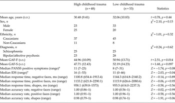
Patients with high childhood trauma (CTQ measured as a continuous variable) had lower GAF-S and GAF-F scores (Spearman's correlation: r = −0.24, p = 0.015, and r = −0.26, p = 0.01, respectively). Patients with high trauma also had more positive symptoms from the PANSS (r = 0.25, p = 0.018) and more depressive symptoms (IDS, r = 0.27, p = 0.026). There were no significant differences in accuracy or response time during scanning and the CTQ (p > 0.05). More severe symptomatology and lower function were also found in the high trauma group when dividing the CTQ into high and low trauma (see Table 1).
Sociodemographics of patients with high and low trauma

s.d., Standard deviation; GAF, Global Assessment of Functioning Scale; GAF-F, GAF-functioning; GAF-S, GAF-symptom; PANSS, Positive and Negative Syndrome Scale; IDS, Inventory of Depressive Symptomatology.
a The PANSS and IDS were not normally distributed, and a Mann–Whitney test was therefore conducted.
Main effect of task
The main effect of negative faces > forms and positive faces > forms across all participants revealed a distributed pattern of brain activation, including the prefrontal cortex, occipital cortex, hippocampus and amygdala (see online Supplementary material: Supplementary Fig. S1a, b).
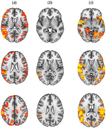
Results of analyses performed with childhood trauma as a continuous variable. Results with brain activation analysed with the Childhood Trauma Questionnaire as the continuous variable using permutation testing and different statistical thresholding. (a) t = 2, p < 0.05, uncorrected; (b) p < 0.05, threshold-free cluster enhancement (TFCE); (c) physical abuse only, p < 0.05, TFCE.

Childhood trauma and brain activation
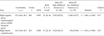
The negative > positive faces contrast revealed brain activation differences for patients with higher scores on the CTQ (measured as a continuous variable) in clusters comprising the right angular gyrus, supramarginal gyrus, middle temporal gyrus (cluster 1) and the lateral occipital cortex (cluster 2, see Fig. 1, Table 2). Dividing into high and low trauma within the same clusters, effect sizes were moderate to large (Cohen's d: 0.72–0.77). To illustrate the distribution of data we included a scatterplot of brain activation with total CTQ score (see online Supplementary Fig. S2).
Increased brain activation in patients with childhood trauma for the negative faces > positive faces contrast a

MAX, Maximum; PE, parameter estimate; s.d., standard deviation.
a Age, sex and diagnosis included in the model as covariates.
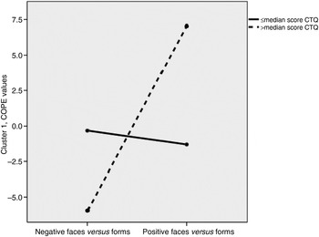
Repeated-measures ANOVA revealed that the high trauma group showed stronger brain activation for negative faces > forms than for positive faces > forms, whereas the low/no trauma group showed higher brain activation for positive faces > forms than for negative faces > forms (see Fig. 2 and online Supplementary Fig. S3).
High levels of childhood trauma moderate lower activation for positive faces and higher activation for negative faces in the right angular gyrus, supramarginal gyrus and middle temporal gyrus (cluster 1). Analysis of variance: F = 14.85, degrees of freedom = 1, p < 0.001. Contrast parameter estimates (COPE) values for > median score on the Childhood Trauma Questionnaire (CTQ): negative faces, mean 7.05 (s.d. = 15.06); positive faces, mean –5.96 (s.d. = 12.71). COPE values for ⩽ median score on the CTQ: negative faces, mean –1.30 (s.d. = 15.06); positive faces, mean –0.32 (s.d. = 12.66).

No significant associations were observed between main antipsychotic medication and brain activation in the clusters above (Pearson's correlation: r ⩽ 0.17, p ⩾ 0.16). Analyses with the sample divided into schizophrenia spectrum disorders and bipolar spectrum disorders revealed the same pattern as for the main findings (data not shown).
Associations between brain activation and clinical variables
Cluster 1 (activation of angular gyrus, supramarginal gyrus, middle temporal gyrus) and cluster 2 (the lateral occipital cortex) were associated with lower GAF-F scores (r = −0.22, p = 0.03 and r = −0.21, p = 0.03, respectively) indicating poorer functioning with relatively stronger activation of negative compared with positive faces. When symptom severity measures (PANSS, IDS and GAF scores) were added into the multivariate analysis, a history of childhood trauma was still associated with increased activation of the brain areas described above for negative faces and reduced activation for positive faces (p = 0.05).
Behavioural assessments of face stimuli
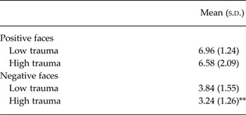
Patients with high levels of childhood trauma reported negative faces as more negative compared with patients in the no/low trauma group (t test: t = 2.41, p = 0.018, Cohen's d = 0.42; see Table 3). No significant differences were observed in the assessment of positive faces (p > 0.10). No significant associations were observed between the behavioural ratings and brain activation in the clusters reported above (p > 0.10).
Patients with high levels of childhood trauma rated negative faces as more negative, compared with patients with no/low trauma a

s.d., Standard deviation.
a Post-scanning assessment of images; after the scanning, the participants were instructed to assess the facial expression images (on a laptop in the magnetic resonance laboratory), by grading the images from 1 to 9 based on how negative/positive they were (1 = negative, 9 = positive).
** p ⩽ 0.01.
Patients with schizophrenia spectrum disorders with high trauma scores rated negative faces as more negative compared with patients in the no/low trauma group (t test: t = 3.26, p = 0.002, Cohen's d = 0.83). Positive faces were also rated as less positive (t test: t = 2.31, p = 0.025, Cohen's d = 0.20). No significant association was observed in the bipolar group (see online Supplementary Table S1). In the schizophrenia group, rating of positive faces as less positive was associated with more severe depressive symptomatology (IDS, r = −0.31, p = 0.03). Rating of negative faces as more negative was also associated with higher scores on the IDS (r = 0.42, p = 0.002).
Different subtypes of childhood trauma and COPE values
Dividing into subtypes of childhood trauma, the strongest association for the negative > positive faces contrast was found for physical abuse (see online Supplementary Table S2 and Fig. 1). A significant increase differentiation in brain response to negative v. positive faces was observed for large clusters comprising the right and left superior temporal gyrus, angular gyrus, Heschl's gyrus, insula, precentral gyrus, postcentral gyrus, and the right precentral gyrus, putamen, central opercular cortex and intracalcarine gyrus. Also neglect showed data going in the same direction with significant negative > positive faces contrast in the right middle temporal gyrus (see online Supplementary Table S3). No significant findings were observed for the other subtypes of trauma.
Discussion
In a sample comprising 101 patients diagnosed with psychosis spectrum disorders, we have demonstrated an effect of childhood trauma on altered brain activation during processing of emotional faces. Patients with high levels of childhood trauma showed stronger differentiation in brain responses between negative and positive faces in the right angular gyrus, supramarginal gyrus, middle temporal gyrus and the lateral occipital cortex compared with patients with no or low trauma scores. This effect was driven by stronger activation in response to negative faces in the high trauma group. Corroborating the imaging findings, childhood trauma was also associated with evaluating negative faces as significantly more negative, and positive faces as less positive in patients with schizophrenia. These results could suggest higher neural reactivity to negative stimuli in patients that have been exposed to high levels of childhood trauma, supporting the implication of altered attentional allocation towards negative stimuli and stronger negativity bias. The CTQ domain analyses revealed associations for physical abuse, implicating right and left superior temporal gyrus, angular gyrus, Heschl's gyrus, insula, precentral gyrus, postcentral gyrus, and the right precentral gyrus, putamen, central opercular cortex and intracalcarine gyrus. We have previously reported that physical abuse is the maltreatment most linked to cognitive impairments in larger group of patients of which the current sample is a subsample (Aas et al. Reference Aas, Steen, Agartz, Aminoff, Lorentzen, Sundet, Andreassen and Melle2012c ), indicating that physical abuse could be more strongly linked to atypical processing of emotions among the core cognitive models.
Patients with high levels of childhood trauma also had lower functioning and more severe depressive symptoms. When symptom severity were added into the multiple regression analysis, a history of childhood trauma was still associated with higher differentiation in the brain responses to negative than positive faces in the clusters identified in the fMRI analysis, thus, indicating that our findings go beyond current symptomatology.
It could be suggested that these findings arise from an attentional bias – that patients with experiences of childhood trauma give more attention to the negative faces compared with those without or with low trauma exposure. However, there were no differences in the behavioural data in task accuracy or response time in those reporting high trauma compared with all patients, indicating that other mechanisms apart from task accuracy and response time are present. Patients with childhood trauma might be hyper-alert when seeing negative faces due to their past experiences, increasing the activation in these areas in these patients. Indeed, Pollak & Sinha (Reference Pollak and Sinha2002) reported that individuals with childhood abuse history accurately identified facial displays of anger (negative emotion) on the basis of less sensory input than controls. As suggested by Pollak & Sinha (Reference Pollak and Sinha2002), this may provide a behavioural advantage for children living in threatening contexts, allowing earlier identification of a threat. It is also well documented that childhood trauma is important for the development of stress sensitization and consequent emotional reactivity to daily stress in adult life (Myin-Germeys et al. Reference Myin-Germeys, van, Schwartz, Stone and Delespaul2001, Reference Myin-Germeys, Delespaul and van2005), including increase in negative emotion (Myin-Germeys et al. Reference Myin-Germeys, van, Schwartz, Stone and Delespaul2001; Glaser et al. Reference Glaser, van, Portegijs and Myin-Germeys2006). Thus, a history of childhood trauma could influence how emotional stimuli are processed, by potentially over-focusing on negative stimuli, and under-focusing on positive stimuli.
Brain activation differences in the amygdala have previously been reported from investigations of otherwise healthy participants with trauma (van Harmelen et al. Reference van Harmelen, van Tol, Demenescu, van der Wee, Veltman, Aleman, van Buchem, Spinhoven, Penninx and Elzinga2013). Thus, the current lack of activation difference in the amygdala between trauma groups could be related to a smaller impact of trauma on amygdala functioning in patient than in healthy controls. It is generally difficult to detect activation differences in a brain region as small as the amygdala when controlling for multiple comparisons across the whole brain. However, also when we analysed childhood trauma (as a continuous variable) and the amygdala alone, no significant findings were present. It should be noted that a recent study, using a partly overlapping sample from the same dataset, did not detect activation differences between emotional processing of negative faces in bipolar v. healthy controls in the amygdala (independent of trauma) even when applying a region-of-interest analysis (Tesli et al. Reference Tesli, Kauppi, Bettella, Brandt, Kaufmann, Espeseth, Mattingsdal, Agartz, Melle, Djurovic, Westlye and Andreassen2015). Emotional processing in patients with schizophrenia and bipolar disorder has also been linked to increased brain activation in areas extending beyond those traditionally associated with emotion, possibly representing compensatory processing (Taylor et al. Reference Taylor, Kang, Brege, Tso, Hosanagar and Johnson2012) and reduced neuronal specialization.
Patients with a schizophrenia diagnosis with childhood trauma rated negative faces as more negative and positive faces as less positive than patients who did not report trauma. No significant effects were observed in the bipolar group. This could indicate that patients with schizophrenia spectrum disorders are more vulnerable for the negative effect of childhood trauma on emotion processing than patients with bipolar disorder. This is in line with previous reports of decreased self-reported arousal in schizophrenia, compared with bipolar disorder during picture viewing in a subsample of the current sample (Aminoff et al. Reference Aminoff, Jensen, Lagerberg, Andreassen and Melle2011).
Extending previous clinical investigations, our large fMRI study provides novel evidence supporting that a history of childhood trauma has long-lasting effects on brain reactivity to emotional facial stimuli in patients with a psychosis continuum diagnosis. Our findings corroborate the notion that childhood trauma is not only a phenomenon in the past but has long-term effects on brain activation and behaviours in adulthood. Our data suggest changes in brain activation patterns in psychotic patients with a history of childhood trauma, potentially serving as vulnerability towards more severe symptomatology.
As discussed by Hart & Rubia (Reference Hart and Rubia2012) in contrast to the large number of studies examining structural brain differences in individuals with and without childhood trauma, only a few have investigated functional correlates of maltreatment using imaging techniques such as fMRI. To our knowledge, our relatively large (n > 100) study is the first to conduct a whole-brain approach to investigate potential differences in brain activation between psychosis spectrum patients with childhood trauma measured as a continuous and as a dichotomized variable. Although the literature is vast in PTSD disorders (Hart & Rubia, Reference Hart and Rubia2012), much less is known about these mechanisms in schizophrenia and bipolar disorders, highlighting the descriptive value of our study.
The current study does not come without limitations. Since the fMRI assessment protocol for our healthy control group unfortunately did not include information about early trauma, we could not include a healthy control group. Thus, we were not able to investigate the role of childhood trauma on brain activation using a case–control design, but compared patients with and without high levels of childhood trauma. Without a healthy control group, it is not possible to determine whether the differences in activation between the high and low trauma groups are specific to patients with psychosis or not; the same pattern may have been observed in high and low trauma healthy volunteers if they had they been included. We also did not have neutral faces included in this paradigm, but instead positive faces, negative faces and geometrical shapes. The negative v. positive faces contrast was chosen in order to specifically capture emotional differentiation, to test if exposure to childhood trauma is associated with changes in the valence and differential brain responses to negative and positive stimuli, and not face processing per se. It should also be noted that a history of childhood trauma was assessed retrospectively. A retrospective design may be specifically challenging for this patient population given their well-established episodic memory deficits and negativity bias (Aas et al. Reference Aas, Dazzan, Mondelli, Melle, Murray and Pariante2014b ). However, the retrospective examination of childhood trauma in patients with psychosis has been found to be valid and reliable compared with other types of information, such as case notes and stable over time (Fisher et al. Reference Fisher, Craig, Fearon, Morgan, Dazzan, Lappin, Hutchinson, Doody, Jones, McGuffin, Murray, Leff and Morgan2011). As emotional abuse and neglect have also been identified as important risk factors in severe mental disorders, neglect items were included in the overall trauma score analysed (Aas et al. Reference Aas, Steen, Agartz, Aminoff, Lorentzen, Sundet, Andreassen and Melle2012c ; Larsson et al. Reference Larsson, Andreassen, Aas, Rossberg, Mork, Steen, Barrett, Lagerberg, Peleikis, Agartz, Melle and Lorentzen2013; Etain et al. Reference Etain, Lajnef, Henrion, Dargel, Stertz, Kapczinski, Mathieu, Henry, Gard, Kahn, Leboyer, Jamain and Bellivier2015). Post-hoc analysis investigating subtype of trauma revealed the largest effect of physical abuse. Further studies are warranted to test for possible differential effects of specific trauma subtypes on brain activation patterns during emotional processing.
In conclusion, along with the observed negativity bias in the assessment of emotional valence of faces, our data suggest increased brain responses to negative faces and reduced responses to positive faces in psychotic patients with childhood trauma. These findings could have implications for current symptomatology.
Supplementary material
The supplementary material for this article can be found at https://doi.org/10.1017/S0033291716002762
Acknowledgements
We thank the patients who took part in the study and the NORMENT who contributed to the data collection. We would like to specifically thank Thomas Bjella for help organizing the database. This study was funded by grants from the University of Oslo, South-Eastern Norway Health Authority (no. 2013088, no. 2006258, no. 2011096, no. 2014097), the Research Council of Norway (no. 213837, no. 223273, no. 190311/V50) and the K.G. Jebsen Foundation. This study was also funded by the NARSAD Young Investigator grant (ID: 22388) and the SCNP Young Scientist Award (to M.A.).
Declaration of Interest
None.