Myocardial dysfunction due to coronary arterial lesions represents an important complication in long-term follow-up in children suffering with Kawasaki syndrome.Reference Newburger, Takahashi and Gerber1–Reference Kato, Inoue, Kawasaki, Fujiwara, Watanabe and Toshima5 Impaired myocardial perfusion can be detected in defined regions supplied by diseased coronary arteries. Pathologic changes in coronary arteries typical in Kawasaki syndrome are ectasia, aneurysm and stenosis. At present, invasive investigations, such as cardiac catheterisation and myocardial perfusion scans, are necessary in patients with the syndrome. A non-invasive method to investigate regional myocardial function would facilitate the detection of early functional abnormalities. Thus, the number of invasive procedures could be greatly reduced.
Tissue Doppler echocardiography and strain rate imaging have recently proved to be valuable for the early detection of abnormal regional myocardial function in adults with coronary arterial disease, and in children with anomalous origin of the left coronary artery from the pulmonary trunk.Reference Voigt, Arnold and Karlsson6–Reference Mertens, Weidemann and Sutherland9 These abnormalities should also be detectable in children with Kawasaki syndrome. Exercise testing should also help discover disturbances not obvious at rest. Our study, therefore, focused on regional myocardial function at rest and under exercise as evaluated using tissue Doppler echocardiography and strain rate imaging. All patients suffered from Kawasaki syndrome during childhood, and had coronary arterial lesions, but were clinically asymptomatic. We thus aimed to evaluate the diagnostic value of tissue Doppler echocardiography in the follow-up of patients with a history of Kawasaki syndrome.
Material and methods
Population
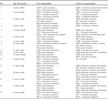
We enrolled 17 children treated for Kawasaki syndrome between 1988 and 1998 (Table 1). The population was recruited from 54 patients with the syndrome seen regularly as outpatients at the Department of Paediatric Cardiology of the University Hospital, Heidelberg. For inclusion, the patients were required to fulfill 3 criterions. The first criterion was the development of at least one coronary aneurysm at the acute stage of the disease. The second was the availability of a follow-up angiogram. The third was the ability to perform bicycle exercise testing, which is possible at an age of about 7 years.
Angiographic characteristics and development of coronary arterial lesions.

None of the patients had been treated with beta-blockers, inhibitors of angiotensin converting enzyme, or digoxin. We enrolled 17 healthy age- and gender-matched persons as a healthy control group for tissue Doppler echocardiographic studies. This control group consisted of children and adolescents investigated for a cardiac murmur, history of congenital heart disease within the family, or before an exercise test. Age and gender-matched partners were taken from a group of about 80 persons.
Parental and personal consent was obtained according to institutional ethical guidelines and the study was carried out in conformity with the Declaration of Helsinki.
Study protocol
Clinical data, and haemodynamic measurements from cardiac catheterisation procedures during follow-up, were taken from medical records. One reader blinded to tissue Doppler echocardiography findings (H.U.) reviewed digitally stored data of the initial and the most recent follow-up angiograms. Coronary arterial lesions were classified according to current guidelines as ectasia or small aneurysm, when the diameter was from 3 to 4 millimetres, a non-giant aneurysm when the diameter was from 4 to 9 millimetres, or a giant aneurysm when the diameter was greater than 10 millimetres.Reference Newburger, Takahashi and Gerber1 Coronary arterial stenosis was defined as more than a 40 percent reduction in luminal diameter.
Both the patients and their controls underwent echocardiographic examination at rest, including tissue Doppler echocardiography. On the same day, bicycle exercise testing in half-supine position was carried out at 1.5 watts per kilogram body weight. Tissue Doppler echocardiography was performed beginning with the 5th minute. At that point in time, all patients showed a stable, exercise-induced, increase in the heart rate of at least 30 percent. After acquisition of images, children were monitored for 30 minutes.
Acquisition of images
All echocardiographic studies were performed using a Vivid 5 system (General Electrics Vingmed Ultrasound, Horten, Norway) equipped with a 2.5 megaherz phased-array transducer and tissue Doppler echocardiographic technology. After completing the standard echocardiographic examination, we obtained colour-coded tissue Doppler echocardiography cine-loops of at least 3 cardiac cycles from apical 2–and 4–chamber views, and from the parasternal long and short axes. Narrow scan sectors containing usually only one ventricular wall per loop had to be used to achieve a rate of 170 to 199 colour Doppler frames per second. Nyquist limits were adjusted for each loop according to the velocity range.
Image processing and quantitative analysis
Digitally-stored cine-loops were analysed off-line using research software (System 5 Tissue Velocity Imaging version 6.0, General Electrics Vingmed) capable of extracting the strain rate from tissue velocity datasets. For each aspect of the left and right ventricular wall curved M-mode lines were drawn and manual wall-tagging was done, dividing the wall into 3 myocardial segments (basal, mid-wall, apical).Reference Voigt, Arnold and Karlsson6Timing of the opening and closing of the aortic and mitral valves was defined as described previously.Reference Voigt, Arnold and Karlsson6, Reference Jamal, Kukulski and Strotmann10 A customized application (tissue Doppler echocardiography – strain rate imaging Speqle version 3.75, Leuven, Belgium) based on the Matlab commercial software package (version 6.1.0.450, MathWorks Incorporation, Natick, USA) was used to superimpose and temporally average the heart cycles, and to process the velocity and strain rate data.
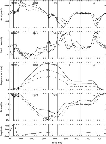
Natural strain and tissue displacement were calculated from strain rate, and velocity data as described by Urheim and colleagues.Reference Urheim, Edvardsen, Torp, Angelsen and Smiseth11 Post-systolic shortening was determined from temporal strain distribution.Reference Voigt, Lindenmeier and Exner12 Finally, the time courses of velocity, strain rate, displacement and strain were plotted as shown in Figure 1, and the corresponding numeric data imported into a custom-made Microsoft Access (version 9.0.2812, Microsoft Incorporation, USA) database.
Time course of regional velocity (top panel), strain rate (second panel from top), tissue displacement (third panel from top) and natural strain (fourth panel from top) extracted from the basal (continuous line), the mid-wall (dotted line), and the apical segment (point-dotted line) of interventricular septum at rest of a healthy subject averaged for three cardiac cycles. Vertical lines indicate valvar closure and opening, separating mechanical left ventricular cycle periods. A: late diastolic filling with atrial contraction; E: early diastolic filling (following mitral valve opening); Eject: ejection time; IVC: isovolumic contraction (between mitral valve closure and aortic valve opening); IVR: isovolumic relaxation (between aortic valve closure and mitral valve opening); PEP: pre-ejection period (from the start of QRS complex to mitral valve closure).

All examinations, image processing and quantitative analysis were done by physicians with expertise in echocardiography and tissue Doppler echocardiography (R.A., B.G., T.C.P.) who were blinded to the corresponding angiographic data.
Statistical analysis
All variables were tested for normal distribution and expressed as mean values and standard deviation. Multiple comparisons between different patient groups were made by one-way analysis of variance using Bonferroni’s post-hoc test when variables had equal variance. We also used Tamhane`s T2 test for variables with non-homogeneous variance. The values at rest and under exercise of variables without normal distribution were compared using Wilcoxon’s nonparametric test. Receiver-operator characteristic curves were constructed for variables showing significant differences between patient groups. Data analysis was performed using the statistical package SPSS release 11.0.0 (SPSS Incorporation, Chicago, USA). P values smaller than 0.05 were considered significant.
Results
Clinical characteristics
Of the 17 patients, 11 (65 percent) were male. Age at examination ranged between 5 and 23 years, with a median of 11 years. The follow-up interval, calculated between documented onset of acute Kawasaki syndrome and echocardiography, was 10.6 plus or minus 5.1 years. In 4 patients, coronary arterial lesions involved only 1 coronary artery, with 8 patients having 2 arteries affected, and 5 patients presenting with involvement of all 3 coronary arteries. All patients were asymptomatic, and reported a normal exercise tolerance at the time of the echocardiographic examination.
Angiographic follow-up
At follow-up, we found 14 coronary arteries without arterial lesions, identifying 35 arteries with persistent aneurysms. Of these, 26 were located within the left coronary artery, 9 within the right coronary artery. Segmental stenoses associated with aneurysms that were not critical, producing less than 50 percent narrowing to the adjacent normal segment, were seen in 9 arteries. These stenoses were located within the left coronary artery 6 times, and within the right coronary artery 3 times. Only one severe stenosis, producing greater than 75 percent narrowing to the adjacent normal segment, was detected in the right coronary artery of a female patient, who later underwent coronary arterial bypass surgery. In one male patient, there was functional occlusion of the left anterior descending artery directly distal of a giant aneurysm. In that patient, extensive collateral circulation originating from the left circumflex and right coronary arteries supplied the hypokinetic septum. Angiographic findings are summarised in Table 1.
Echocardiographic findings
Cross-sectional echocardiography showed no abnormalities of mural motion except for the patient discussed above with a hypokinetic area in the apical ventricular septum. M-mode- and pulsed-wave Doppler measurements showed normal findings in all cases.
For analysis of the data from tissue Doppler echocardiography, we employed only segments with good quality reproduction recordings. In examining regional longitudinal function, we used 250 left ventricular and 49 right ventricular segments both at rest and under exercise, providing 61 percent coverage of the left ventricle, and 48 percent of the right ventricle. At least one segment was obtained for each subject for left ventricular radial function. The number of segments available for analysis did not differ between patients, with 119 long-axis left ventricular segments, 7 segments per patient, and controls.
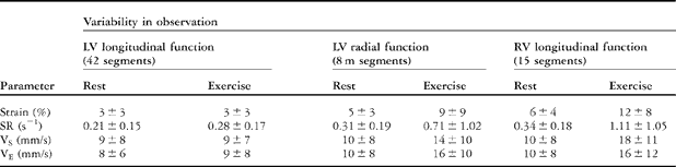
Variability of observations was assessed by examining 3 patients and 2 healthy subjects at two occasions. As shown in Table 2, variability of values under exercise was unacceptably high for parameters of left ventricular radial function and right ventricular longitudinal function. Those data were thus excluded from further analysis. On the other hand, parameters of segmental left-ventricular longitudinal function proved to be stable at rest and under exercise.
Variability in observations.
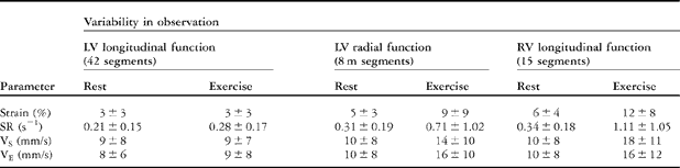
Strain: peak strain; SR: peak systolic strain rate; VE: peak early diastolic velocity; VS: peak systolic velocity.
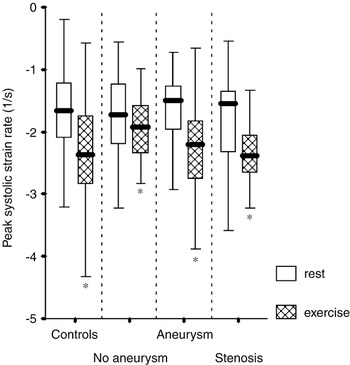
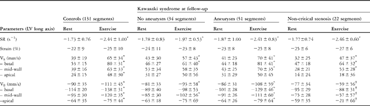
Regarding left ventricular longitudinal function, we detected no significant differences in peak systolic strain rate (Fig. 2) and peak strain in segments of healthy subjects and patients. Under submaximal exercise peak systolic strain rate increased in all investigated subgroups, whereas peak strain did not change significantly. The incidence of post-systolic shortening was under 15 percent and did not differ between subgroups. We observed no significant increase in post-systolic shortening under exercise.
Peak systolic strain rate at rest and under exercise for left ventricular segments from control subjects, and for segments supplied by arteries from patients with Kawasaki syndrome without aneurysms, with persistent aneurysms, and with residual stenosis (* p less than 0.05 versus rest).

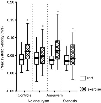
Peak systolic velocities of basal and mid-wall segments exhibited a significant increase under exercise in all groups (Fig. 3). Absolute values did not differ between patients and controls. There was also no difference within the population with Kawasaki syndrome.
Peak systolic velocity at rest and under exercise for left ventricular segments from control subjects, and for segments supplied by arteries from patients with Kawasaki syndrome without aneurysms, with persistent aneurysms, and with residual stenosis (p less than 0.05 versus rest).

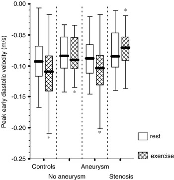
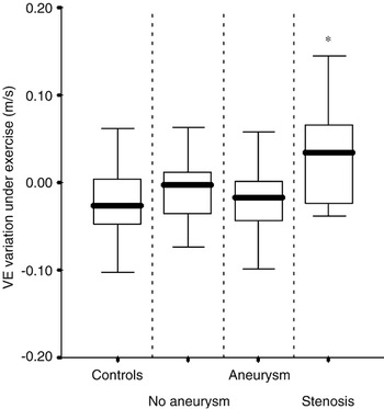
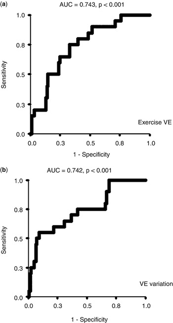
Peak early diastolic velocities exhibited a significant increase in absolute values under exercise in all segments except for those supplied by stenotic coronary arteries. Furthermore, segments with aneurysm-associated stenosis of afferent arteries showed a considerable decrease in peak diastolic velocities under exercise (Figs. 4 and 5). That finding was irrespective of segmental location in the basal, mid-wall or apical regions. This opposite trend of peak diastolic velocity enabled us to predict coronary arterial stenosis using receiver-operator characteristic analysis for peak diastolic velocities under exercise and the peak diastolic velocity variation between rest and sub-maximum exercise. Under exercise, peak diastolic velocity values under 90 millimetres per second predicted an angiographically-documented stenosis in the afferent coronary artery with 75 percent sensitivity and 64 percent specificity. This yielded an area under the receiver-operator characteristic curve of 0.743 (p less than 0.001, 95 percent confidence intervals from 0.641 to 0.845), as displayed in Figure 6a. Similarly, a decrease in the absolute value of peak diastolic velocity under exercise predicted coronary arterial stenosis with 60 percent sensitivity and 71 percent specificity (p less than 0.001, area under the curve 0.742, 95 percent confidence interval of 0.621 to 0.863 – Fig. 6b). All tissue Doppler echocardiography and strain rate imaging data for left ventricular longitudinal function are presented in Table 3.
Peak early diastolic velocities at rest and under exercise for left ventricular segments from control subjects, and for segments supplied by arteries from patients with Kawasaki syndrome without aneurysms, with persistent aneurysms, and with residual stenosis (* p less than 0.05 versus rest).

Variation in peak diastolic velocity (VE) under exercise. Note the significant opposite trend of segments supplied by arteries with aneurysm-associated stenosis (* p less than 0.05 compared to all other groups).

Receiver operating characteristic curves (a) for predicting residual coronary stenosis by peak diastolic myocardial velocity under sub maximal exercise. Simlilar curves (b) for predicting residual coronary arterial stenosis by variation in peak diastolic myocardial velocity between rest and exercise. AUC: area under receiver operating characteristic curve; VE: peak diastolic myocardial velocity.

Echocardiographic data at rest and under exercise.
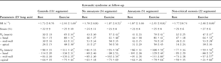
*Increase in absolute value versus rest (p less than 0.05). †Decrease in absolute value versus rest (p less than 0.05).
Strain: peak strain; SR: peak systolic strain rate; VE: eak early diastolic velocity; VS: eak systolic velocity.
Discussion
Myocardial dysfunction caused by coronary arterial pathology has become a key problem in the long term follow-up after Kawasaki syndrome.Reference Newburger, Takahashi and Gerber1–Reference Kato, Inoue, Kawasaki, Fujiwara, Watanabe and Toshima5, Reference Cheung, Yung, Tam, Ho and Chau13 To date, few studies analysed myocardial function after Kawasaki syndrome, and almost all of them investigated viability and ischaemia using different perfusion imaging techniques.Reference Dahdah, Fournier and Jaeggi14, Reference Miyagawa, Mochizuki and Murase15 Miyagawa and colleaguesReference Miyagawa, Mochizuki and Murase15 showed that pathologic redistribution of thallium during dipyridamole stress was the most sensitive marker to predict a potential cardiac event such as myocardial infarction. Dahdah and colleaguesReference Dahdah, Fournier and Jaeggi14 reported a relationship between an unfavourable response to perfusion in single-photon emission computed tomography and abnormal wall motion in echocardiography. The most severe findings were associated with persistent giant aneurysms, even in the absence of severe coronary arterial stenosis. Although nuclear perfusion has known adverse effects, and should thus not be done repetitively, attempts to perform functional myocardial imaging using echocardiographic techniques have been sporadic.Reference Pahl, Sehgal and Chrystof16, Reference Henein, Dinarevic, O’Sullivan, Gibson and Shinebourne17 Tissue Doppler echocardiography and strain rate imaging, which have proven feasible in detecting regional myocardial dysfunction non-invasively in patients with coronary arterial disease, have not yet been applied in patients with Kawasaki syndrome.Reference Voigt, Exner and Schmiedehausen7, Reference Voigt, Lindenmeier and Exner12 It was our aim to investigate whether functional changes in tissue Doppler echocardiography correlate to the degree of coronary pathology in children suffering from this syndrome.
Using tissue Doppler echocardiography, we were able to quantify left ventricular regional longitudinal function reliably at rest and under exercise. Longitudinal function represents the performance of the longitudinally oriented subendocardial myocytes, which are very sensitive to ischaemia.Reference Henein, Dinarevic, O’Sullivan, Gibson and Shinebourne17 Peak systolic strain rate, which reflects regional contractility, was normal in all groups at rest, and increased significantly under exercise. This finding demonstrates an unrestrictive systolic functional reserve in all patients studied, regardless of coronary arterial pathology. The analysis of temporal strain distribution within the cardiac cycle showed that post-systolic shortening did not occur. Post-systolic shortening is a sensitive marker for acute ischaemia.Reference Voigt, Exner and Schmiedehausen7, Reference Jamal, Strotmann and Weidemann18 Stress-induced abnormalities of wall motion, like those found in the series of Pahl and associates,Reference Pahl, Sehgal and Chrystof16 were not apparent.
As opposed to the systolic function, the severity of the coronary arterial pathology had an important effect on diastolic function. At rest, regional longitudinal diastolic function, most accurately reflected by peak diastolic velocity, was normal in all subgroups. Under conditions of exercise, however, peak diastolic velocity fell in segments supplied by coronary arteries with stenotic lesions associated with aneurysms.Reference Cerqueira, Weissman and Dilsizian19 This finding occurred independently of any basal, mid-wall, or apical segmental location.
This data suggests that the degree of reduction of flow due to the stenoses associated with the aneurysms on the one hand, and the intensity of exercise challenge on the other, were not serious enough to produce severe ischaemia or alter the properties of myocardial deformation, but were severe enough to interfere with local relaxation. That finding concurs with those of Henein and colleagues,Reference Henein, Dinarevic, O’Sullivan, Gibson and Shinebourne17 who found impaired global longitudinal function during diastole on M-Mode studies in patients with aneurysms subsequent to involvement by Kawasaki syndrome. Unlike their report, we only found pathologic myocardial relaxation in those segments associated with a stenotic coronary artery.
Exercise echocardiography with tissue Doppler echocardiography and strain rate imaging has proven feasible in our patients of providing acceptable reproducibility for left ventricular regional myocardial function. Our data suggests that diastolic function, which is more sensitive to reduced myocardial perfusion, is impaired in the setting of stenotic coronary arterial lesions. These changes occur in a state in which revascularisation is not usually recommended. Overall, exercise tissue Doppler echocardiography has the potential to identify non-invasively the presence of coronary arterial stenosis. The method can be applied in clinical follow-up of patients with a history of Kawasaki syndrome. With technical improvement, specific limitations should be overcome, and the serial use of tissue Doppler echocardiography and strain rate imaging may reduce the number of invasive investigations such as angiography and nuclear perfusion imaging.
There are certain limitations in our study. Our population did not include children with untreated severe stenotic lesions, with more than 75 percent narrowing to the adjacent normal segment, who might have developed typical ischaemic signs on exercise tissue Doppler echocardiography. Still, we were able to demonstrate that moderate coronary arterial stenosis leads to characteristic functional changes that appear early in the ischaemic cascade.Reference Nesto and Kowalchuk20
To achieve good reproducibility of measurements, only submaximal exercise was performed. Our experience from earlier studies revealed that tissue Doppler echocardiography data acquired at maximum exercise was of minor quality, and therefore not worthy of analysis.
Pharmacological stimulation during echocardiography, invasive nuclear perfusion studies, and estimation of the coronary flow reserve during catheterisation were not performed in our patients. Thus, we know neither whether there were obvious perfusion abnormalities in the segments with impaired diastolic function, nor if coronary microvascular dysfunction was present. We did not want our patients to have to undergo another invasive test in addition to angiography during cardiac catheterisation. Still, we consider the angiographic findings to be most reliable at follow-up of patients with Kawasaki syndrome. Nuclear perfusion studies are generally very problematic in children.21 In our population, any changes in perfusion might have been too minor to be visualised.
Acknowledgement
This study was supported in part by a grant from the foundation “Herzkind e.V.”, Braunschweig, Germany. Raoul Arnold and Björn Goebel contributed equally to this project.