Introduction
Numerical chromosomal abnormalities (aneuploidy) may cause an implantation failure and high rates of embryonic loss. Aneuploidy arises mainly from non-disjunction events during gametogenesis (Munné et al., Reference Munné, Alikani, Tomkin, Grifo and Cohen1995), which can also occur after fertilization (Delhanty & Handyside, Reference Delhanty and Handyside1995). Hunt & Hassold (Reference Hunt and Hassold2002) report that chromosomal abnormalities results from low efficiency of cell cycle checkpoints during the final steps of female meiosis and first mitosis of the zygote. Cell cycle errors result in unexpectedly high incidence of ploidy abnormalities in mature oocytes and apparently normally cleaving embryos (Fulka et al., Reference Fulka, First, Fulka and Moor1999).
Chromosomal analyses of cleavage stage embryos show that ploidy errors could be one of the main causes of low implantation rate of human embryos (Munné, Reference Munné2003). Wells & Delhanty (Reference Wells and Delhanty2000) observed that only about 25% of cleavage stage human embryos contained chromosomally normal cells, meaning that there is no strict correlation between morphological and ploidy abnormalities. According to Gianaroli et al. (Reference Gianaroli, Magli, Ferraretti, Fiorentino, Garrisi and Munné1997), morphological criteria alone, including blastocyst development, are not sufficient to ensure the selection of euploid and viable embryos. The identification after chromosomal analysis of euploid embryos and their selective transfer has a positive effect on the developmental outcome (Gianaroli et al., Reference Gianaroli, Magli, Ferraretti, Fiorentino, Garrisi and Munné1997). Selection of embryos based both on morphology and chromosomal status not only allows the identification of euploidy, but also reduces the risk of an abnormal implantation (Farfalli et al., Reference Farfalli, Magli, Ferraretti and Gianaroli2007). Chromosomal errors may occur in all embryo morphology grades, but are more frequently observed in arrested or fragmented embryos (Magli et al., 2000). Farfalli et al. (Reference Farfalli, Magli, Ferraretti and Gianaroli2007) demonstrated a relationship between chromosomal status and embryo developmental rate. They showed that on day 3 the incidence of monosomy and trisomy is less correlated with aneuploidy in 7- to 8-cell embryos and aneuploidy occurence is more frequent in arrested or slow cleaving embryos and in embryos with accelerated cleavage. The selection of embryos with a balanced karyotype can bring down an occurence of spontaneous abortion (Munné et al., Reference Munné, Sandalinas, Escudero, Fung, Gianaroli and Cohen2000).
According to Viuff et al. (Reference Viuff, Greve, Avery, Hyttel, Brockhoff and Thomsen2000) the procedure of in vitro production influences the chromosome complement of the embryos. Moreover, not all nuclei of the embryos could be analysed because of loss of nuclei during preparation and not all nuclei, which were observed before preparation, can be found on the slide. Gianaroli et al. (Reference Gianaroli, Magli, Ferraretti, Fiorentino, Garrisi and Munné1997) suggest that the fixation process may substantially influence the final diagnosis due to loss of cells and presence of residual cytoplasm, which can hinder an observation. Munné et al. (Reference Munné, Dailey, Finkelstein and Weier1996) characterized necessity of the correct timing for adding the fixative to obtain a good chromosome spreading.
Despite several attempts to find optimal conditions a success of chromosomal ploidy determination from embryos is lower than from somatic cells (Parkanyi et al., Reference Parkany, Chrenek, Rafay, Suvegova, Jurcik, Makarevich, Pivko, Hetenyi and Paleyanda2004, Curlej et al., Reference Curlej, Parkanyi, Bulla, Jurcik and Chrenek2007). There are at least two reasons causing this state: low success of mitotic cycle synchronization at metaphase stage in individual embryo blastomeres and a small number of blastomeres arrested at metaphase for correct chromosomal counts because of insufficient nuclei dehiscence and chromosomal overlapping (Gianaroli et al., Reference Gianaroli, Magli, Ferraretti, Fiorentino, Garrisi and Munné1997).
The goal of this paper was to determine aneuploidy rate in rabbit oocytes or embryos at different developmental stages.
Materials and Methods
Biological material
The hybrid line of New Zealand White rabbits from the stud of APRC Nitra was used. Rabbits were housed in individual flat-deck wire cages, under a constant photoperiod of 14 h of daylight. The temperature and humidity of the building were recorded continually by means of a thermograph positioned at the same level as the cages. The rabbits were fed ad libitum with a commercial diet and water was provided ad libitum with nipple drinkers. The breeding conditions were similar to intensive industrial conditions.
Retrieving of oocytes and embryos
Oocytes (18 h PC) were recovered from New Zealand White rabbit females after mating with a vasectomized male. Embryos at the 2-cell stage (24 h PC) were recovered from New Zealand White rabbit females after natural mating. The oviducts were immediately transfered from the slaughterhouse to the laboratory. Subsequently, oocytes or embryos were flushed out using prewarmed PBS (Gibco BRL) and then washed in prewarmed CO2 independent medium (Gibco BRL) supplemented with 10% FCS three times. The embryos for culture and further analysis were selected according to their morphological appearance.
Embryo cultivation and synchronization
Selected embryos were immediately transfered into k-DMEM medium (Gibco BRL) supplemented with 10% FCS and placed in a 96-well culture dish (Nunc, Denmark) under CO2 incubator conditions of 5% CO2 and 37 °C. Mitotic arrest was attained by the addition of 1 μg/ml colcemid (Gibco BRL, USA) into k-DMEM medium for either 7, 12 or 13 h incubation in the case of 2-, 4- and 8-cell embryos, respectively. In each case, several embryos were retained as a control of mitotic cycle phase, placed into unsupplemented culture medium. These embryos served as an indicator of sufficient time for the colcemid treatment.
Chromosome isolation from embryos
Mitotically arrested embryos were transfered into 39 °C-heated 0.5% pronase (Sigma) and placed into CO2 incubator for 16–20 min. Blastomeres without zona pellucida were washed in warmed CO2-independent medium (37 °C) for eliminating further ineligible effect of pronase on denuded embryo cells. For hypotonization, 1% sodium citrate (Mikrochem) solution enriched by 0.25% foetal bovine serum (Gibco BRL) and colcemid 1 μg/ml (Gibco BRL) was applied in a thermobox set at 37 °C for 15 min. Blastomere fixation was provided freshly made fixation solution, consisting of methanol (Mikrochem) and acetic acid (Lachema) adjusted to room temperature, at 3:1 ratio in a 4-well multi-dish (Nunc) under permanent microscope control until the cytoplasm was completely transparent. Fixed cells were placed onto a clean and ungreased microscopic slide into a drop of hypotonization solution. Consequently, a drop containing fixed cells was recovered by freshly made, cooled (4 °C) fixation solution, of approximately 4 cm in height. Samples were left to dry at room temperature and the chromosomes were stained by 2% Giemsa (Gibco BRL) within 8 min.
Chromosome isolation from oocytes
Cumulus cells from oocytes were removed by 5-min treatment in 0.5%. hyaluronidase solution (Sigma). Subsequently, the zona pellucida was removed by 4-min treatment in 0.5% pronase solution (Sigma). Hypotonization and chromosome isolation was performed as in case of embryos.
Chromosome observation
Stained microscopic slides were observed under a Leica light microscope (Leica Microsystems, Germany) under ×40 magnification. Found metaphase plates were converted to digital images via a camera connected to a computer and the chromosomal number was counted from each acquired picture.
Statistical analysis
Oocytes and embryos in groups were evaluated for aneuploidy rate using the chi-squared test, in which the oocyte group served as a control. Differences at p < 0.05 were considered as significant.
Results
In the present study, rabbit oocytes (n = 27) and embryos (n = 58) following in vitro culture (2-, 4- and 8-cell) were used for the detection of aneuploidy rate (Table 1).
Chromosomal analysis in rabbit oocytes and embryos, n (%).

a vs. b, p < 0.01.
The proportion of blastomeres successfully arrested at the metaphase stage was 100%, 86.1% and 92.2% for 2-, 4- and 8-cell embryos, respectively. Although the number of metaphase blastomeres after colcemid treatment was high, not all metaphase nuclei found were detectable, due to insufficient spreading and overlapping of isolated chromosomes. The proportion of analysable blastomeres attained in our study was 58.8, 83.9 and 59.8% for 2-, 4- and 8-cell embryos, respectively.
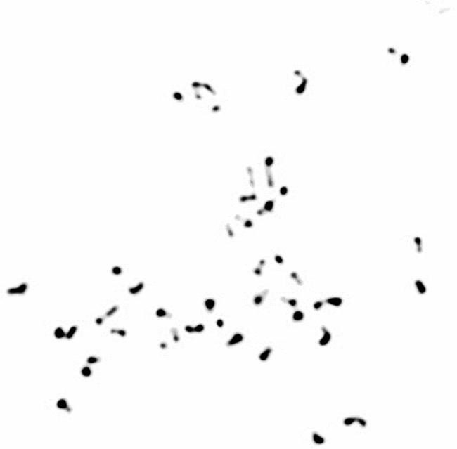
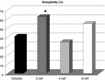
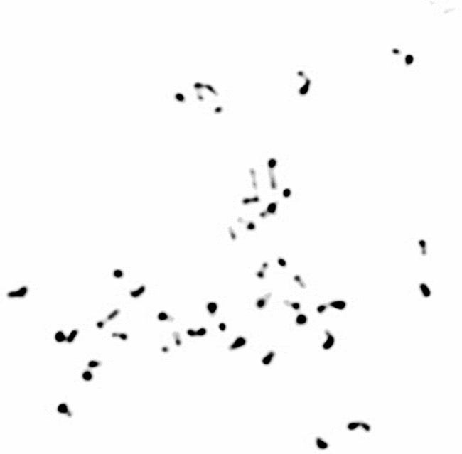
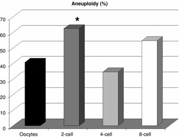
In oocytes, a 59.3% abundance of haploid sets was found. Determined percentages of blastomeres carrying the correct chromosome set was 35, 53.85 and 37.5% for 2-, 4-, 8-cell preimplantation embryo stages, respectively (Fig. 1). Of all forms of aneuploidy, hypodiploidy (hypoploidy) was the most common (29.6% for oocytes, 52.5 30.8, 54.7% for 2-, 4- and 8-cell embryos, respectively), while hyperdiploidy (hyperploidy) was observed at a lesser extent in three analysed groups (11.1% for oocytes, 10 and 3.85% for 2- and 4-cell stages, respectively). Total aneuploidy in individual groups showed 62.5% occurrence at the 2-cell stage and 54.7% at the 8-cell stage (Fig. 2). Lower aneuploidy abundance was detected in oocytes (40.7%) and in 4-cell embryos (34.65%). Blastomeres, carrying polyploid chromosomal number, were found only at low numbers in 2-cell (2.5%) and 8-cell (3.1%) stage embryos. Haploidy was detected at 11.5% and 4.7% in 4- and 8-cell embryos, respectively.
Rabbit blastomere metaphase plate, diploid number of chromosomes (n = 44), ×40 magnification.
Aneuploidy occurence in rabbit oocytes and embryos at different preimplantation stages. *, Difference is significant at p < 0.01 compared with control (oocytes).
Discussion
Chromosomal abnormalities represent one of the major causes of spontaneous abortions and implantation failures (Gianaroli et al., Reference Gianaroli, Magli, Ferraretti, Fiorentino, Garrisi and Munné1997). Although a number of various chromosomal techniques and methods exists, the question of efficient synchronization up to metaphase of embryonal cells is still not answered. Results obtained in this study show 100, 86.1 and 92.2% success of cell cycle synchronization at metaphase for 2-cell, 4-cell and 8-cell rabbit embryos, respectively. These values are higher when compared with the study by Yoshizawa et al. (Reference Yoshizawa, Miura, Narisawa, Sasaki and Zhu2001), in which 58.8% (for 2-, 4-cell embryos) and 35.0% (for 8-, 16-cell embryos) success was recorded in two groups of in vitro fertilized bovine embryos.
To summarize the number of hypo- and hyperdiploid embryos, hypodiploidy was not a result of artificial losses of chromosomes that occur due to over-spreading. Over-spread metaphases were excluded, only chromosomes in metaphase plates, which were looked complete, were counted. Furthermore, chromosome spreading occurred even when the remnants of the cytoplasm were still present. Due to this, the chromosomes were dispersed on the slide within a limited space. However, using the same culture conditions, different proportions of numerical chromosomal abnormalities at different analysed stages were found in our experiments. At the 8-cell stage, when rabbit embryonal genome is activated, 54.7% aneuploidy was found. Similarly, 56% aneuploidy was determined by Shi et al. (Reference Shi, Dirim, Wolf, Zakhartchenko and Haaf2004) in in vivo fertilized rabbit embryos at the 16-cell stage. Yoshizawa et al. (Reference Yoshizawa, Konno, Zhu, Kageyama, Kukui, Muramatsu, Kim and Araki1999) reported 80% occurrence of chromosomal abnormality in 5- to 10-cell bovine embryos, a figure higher than 54.7% at 8-cell stage in our embryos. Farfalli et al. (Reference Farfalli, Magli, Ferraretti and Gianaroli2007) in their study reported 67% occurrence of chromosomally abnormal human embryos at the 7- to 8-cell stages. Determined 62.5% aneuploidy in the 2-cell and 34.65% in 4-cell rabbit embryos differs from 13.7% attained by Iwasaki & Nakahara's (Reference Iwasaki and Nakahara1990) in 2- to 4-cell bovine embryos. Further, Roychoudhury et al. (Reference Roychoudhury, Bulla, Curlej and Chrenek2008) reported 73.33% aneuploidy occurrence in cells isolated from intact control rabbit embryos in their paper, focused on the influence of transgenesis on the aneuploidy rate. Viuff et al. (Reference Viuff, Greve, Avery, Hyttel, Brockhoff and Thomsen2000) published that cytogenetic investigations may result in an unreliable estimation of the number of polyploid embryos because only a few cells from the embryo can be accurately analysed. The cause for polyploidy can be induced by in vitro culture or originate from gametogenesis (Viuff et al., Reference Viuff, Greve, Avery, Hyttel, Brockhoff and Thomsen2000). In our study, polyploid cells were found just in a small percentage of 2-cell and 8-cell embryos, 2.5% and 3.1% respectively. Also, Viuff et al. (Reference Viuff, Greve, Avery, Hyttel, Brockhoff and Thomsen2000) found 9% polyploidy only in 8-cell bovine embryos. According to the criterion of Shi et al. (Reference Shi, Dirim, Wolf, Zakhartchenko and Haaf2004), morula and blastocyst stages were excluded in our study from the analysis to avoid the problem that single embryos with higher than average number of euploid or aneuploid cells may strongly influence the statistics. Rabbit matured oocytes used in our experiment exhibited 40.7% aneuploidy; this finding is almost in agreement with the statement that about 30% of human oocytes carry a chromosomal imbalance (Farfalli et al., Reference Farfalli, Magli, Ferraretti and Gianaroli2007). This condition has several clinical consequences as approximately one-third of spontaneous abortions are aneuploid. The most obvious link to the increase of aneuploidy in oocytes is maternal age (Farfalli et al., Reference Farfalli, Magli, Ferraretti and Gianaroli2007).
In conclusion, the convenient timing of colcemid effect at a desirable concentration gave us a higher efficiency in the synchronization of embryo cell nuclei up to metaphase stage than had been published before. These two aspects in our study were found to be critical for achieving a good number of potentially analysable blastomeres for chromosome screening.
Acknowledgements
This work was supported by the Slovak Research and Development Agency under the contract No. LPP-0126–06. We are grateful to Dr Alexander V. Makarevich from the Animal Production Research Centre for critical reading of the manuscript.