INTRODUCTION
Sympathetic ganglia are essential elements for the transmission of control signals from the central nervous system (CNS) to numerous target organs (Klimaschewski et al., Reference Klimaschewski, Kummer and Heym1996; McLachlan, Reference McLachlan2003; Szurszewski and Miller, Reference Szurszewski, Miller and Johnson2006; Fasano and Niel, Reference Fasano and Niel2009). Neurons in these ganglia receive inputs from preganglionic neurons in the spinal cord, dorsal root ganglia neurons and visceral organs such as the gastrointestinal tract and genitourinary systems. Thus, sympathetic neurons can participate in a variety of reflexes that control essential physiological functions of visceral organs. The physiology and pharmacology of these neurons have been studied in detail, and the nature of their synaptic connections is well established (Klimaschewski et al., Reference Klimaschewski, Kummer and Heym1996; Li and Horn, Reference Li and Horn2006; Szurszewski and Miller, Reference Szurszewski, Miller and Johnson2006).
Neurons in sympathetic ganglia are individually surrounded by a tight envelope of satellite glial cells (SGCs), which are distinct from Schwann cells, and share several similarities with astrocytes. In contrast to the large amount of available information on the neurons, relatively little is known about SGCs in these ganglia, and in particular regarding the function of these cells. Pathological changes in sympathetic ganglia following nerve injury have been investigated extensively. In numerous studies, axotomy of the postganglionic fibers was found to cause striking morphological and physiological changes in the neurons; for example retraction of synaptic contacts associated with decline in synaptic transmission (Matthews and Nelson, Reference Matthews and Nelson1975; Shadiack et al., Reference Shadiack, Sun and Zigmond2001; De Stefano et al., Reference De Stefano, Leone, Moriconi, Del Signore, Petrucci and Paggi2007). It appears that the neuronal changes are accompanied by biochemical and morphological changes in SGCs, and it is likely that SGCs participate in the neuronal reactions to injury (Matthews and Nelson, Reference Matthews and Nelson1975; De Stefano et al., Reference De Stefano, Leone, Moriconi, Del Signore, Petrucci and Paggi2007). SGCs are found also in sensory ganglia, and have been recently a focus of intensive study (for reviews see Hanani, Reference Hanani2005; Ohara et al., Reference Ohara, Vit, Bhargava, Romero, Sundberg and Charles2009 and also articles in this issue). These cells are endowed with a variety of receptors and appear to contribute to pathological conditions, such as chronic pain. It can be proposed that SGCs in sympathetic ganglia are similar in these aspects to their counterparts in sensory ganglia, but this needs to be verified experimentally.
One of the hallmarks of most types of glial cells is that they are interconnected by gap junctions (Rouach et al., Reference Rouach, Avignone, Même, Koulakoff, Venance and Blomstrand2002), which enables them to function as an electrical syncytium. This arrangement allows not only the passage of ions, but also a variety of metabolites and signaling molecules with molecular weight of up to 1000. It is well established that coupling among astrocytes in the brain and retina is important for regulating the extracellular K+. There is electron microscopic evidence for the presence of gap junctions in SGCs in sympathetic ganglia (Elfvin and Forsman, Reference Elfvin and Forsman1978), but their functional significance has not been explored. It should be noted that the identification of gap junctions is not a sufficient evidence that the cells are connected by gap junctions because the observed gap junctions could be by one cell on itself (reflexive gap junctions, see Herr, Reference Herr1976). There is ample evidence that peripheral nerve injury leads to greatly augmented gap-junction-mediated SGC coupling in sensory ganglia (Hanani et al., Reference Hanani, Huang, Cherkas, Ledda and Pannese2002; Cherkas et al., Reference Cherkas, Huang, Pannicke, Tal, Reichenbach and Hanani2004; Ohara et al., Reference Ohara, Vit, Bhargava and Jasmin2008), but currently there is no information on whether similar changes occur in sympathetic ganglia under pathological conditions.
OBJECTIVE
The objective of this work was to answer the following questions: (1) Are SGCs in sympathetic ganglia functionally coupled? (2) What is the extent of this coupling? (3) Is the coupling altered following nerve injury? The method employed in this work was intracellular injection of a fluorescent dye that crosses gap junctions (dye coupling).
METHODS
Animals and preparations
Experiments were done on mice of either sex, 3–5 months old. Superior cervical ganglia were obtained from Balb/c mice (n = 20), and superior mesenteric ganglia from C57BL mice (n = 11). C57BL mice were used because the dissection of the ganglia was easier in them than in other strains. The experimental protocol was approved by the Institutional Animal Care and Use Committee. The animals were killed by CO2 inhalation and ganglia were removed and placed in cold (4°C) Krebs solution (pH 7.4) containing (mM): 120.9 NaCl, 5.9 KCl, 14.4 NaHCO3, 2.5 MgSO4, 2.5 CaCl2, 1.2 NaH2PO4 and 11.5 glucose. The ganglia were pinned onto the Sylgard bottom of a chamber superfused with Krebs solution bubbled with 95% O2 and 5% CO2 at 23–24°C.
Inflammation models
Submandibular inflammation was induced by injecting 20 µl of 1:1 complete Freund's adjuvant (CFA) solution (Sigma, St Louis, MO, USA) in saline under the submandibular skin of mice anesthetized with isoflurane. Seven days after the injection the animals were euthanized with CO2 and the superior cervical ganglia on both sides were removed and studied in vitro.
Colonic inflammation was induced by instilling dinitrobenzoate sulfonate (DNBS, 50 µl, 100 mg/ml in 50% ethanol) into the distal colon via anal tube (see Huang et al., Reference Huang, Belzer and Hanani2010); the tip of the tube was located 1.2–1.5 cm from the anal verge. Ten to 12 days later the animals were killed with CO2 and the superior mesenteric ganglion was removed. For both models, untreated animals were used as controls.
Intracellular labeling
Dishes with the attached ganglia were placed on the stage of an upright microscope (Axioskop FS2, Zeiss, Jena, Germany) equipped with fluorescent illumination and a digital camera (Pixera, Los Gatos, CA, USA), connected to a personal computer. SGCs were injected singly with the fluorescent dye Lucifer yellow (LY, Sigma) 3% in 0.5 M LiCl solution from a glass microelectrode, connected to a preamplifier (WPI, wpiinc.com). The tip resistance of the microelectrodes was 80–120 MΩ. Cell coupling was assayed by intracellular injection of LY and observation of dye passage to other cells (dye coupling). LY was injected by hyperpolarizing current pulses with 100 ms duration and 1–1.5 nA amplitude at 5 Hz, for 3–5 min; for further details see Huang et al. (Reference Huang, Cherkas, Rosenthal and Hanani2005). Living labeled cells were observed and photographed during the experiments, which allowed cell identification. The gap junction blocker carbenoxolone (Sigma) was added to the bathing medium at 50 µM.
Statistical analysis
Fisher's exact test was used for comparison; p < 0.05 is considered as statistically significant.
RESULTS
Morphological features of SGCs in superior cervical ganglia
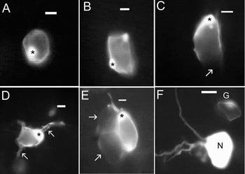
Intracellular injection of LY into SGCs in superior cervical ganglia revealed a number of morphological features of these cells. The sheath formed by SGCs around the neurons cells was very thin, except in the nuclear region, where it was much thicker. The flat processes of a given SGC were seen to ensheathe all (Fig. 1A,B), or part the neuronal surface (Fig. 1C). In some cases, glial extensions having a tube-like shape covered neuronal processes (Fig. 1D). Neurons were frequently labeled; they were much larger than SGCs and had several long processes (Fig. 1F). Neurons were not found to be coupled among themselves or to SGCs.
Intracellular injections of Lucifer yellow (LY) into cells in mouse superior cervical ganglia. The injected cell in each case is marked with an asterisk. (A) An SGC was injected with LY, and no dye passage to other cells is seen. This cell forms a complete envelope around the neuron. (B) The dye passed from the injected SGC to another SGC around the same neuron. (C) An example of an SGC that was not coupled to other cells, and did not make a complete envelope around the neuron. The arrow indicates a region that apparently was not covered by the SGCs processes, but may have been covered by another SGC that was not labeled with dye. (D) The injected SGC made a complete envelope around the neuron and also formed tube-like covers around the proximal part of two neuronal processes (arrows). (E) An example of inter-envelope coupling in a ganglion from an animal that was injected 7 days previously with CFA in the submandibular region. The injected SGC is coupled to glial envelopes around two neighboring neurons (arrows). (F) An example of a LY-injected neuron (N). Neurons were considerably larger than SGCs and had several long processes. Neurons were not coupled to other neurons or SGCs. An SGC (G), which was injected separately is seen. Scale bars, 10 µm (A–E), and 20 µm (F).

Glial coupling in superior cervical ganglia is increased by submandibular inflammation
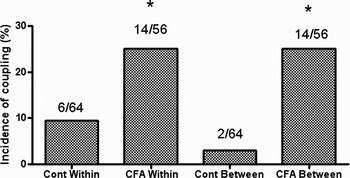
We defined two types of SGC coupling – between SGCs forming the envelope around a given neuron (Fig. 1B), and between SGCs forming the envelopes of different neurons (Fig. 1E). Injecting LY into SGCs in control ganglia resulted in most cases in the labeling of a single cell (Fig. 1A,C,D), that is, the injected cell was not coupled to any other cell (see Fig. 2 for summary of data). In other cases, the dye spread to other SGCs surrounding the same neuron (Fig. 1B). In a very small number of cases, the injected cell was coupled to a SGC forming the envelope of an adjacent neuron. The passage of the dye from the injected cell to the neighboring ones could be followed over time, and was largely complete within 3–5 min.
Summary of dye coupling experiments in mouse superior cervical ganglion. The histograms show how submandibular CFA injection increased the incidence of SGC coupling. ‘Cont Within’ indicates the incidence of coupling between SGCs around a given neuron, and ‘CFA Within’ indicates the same parameter 7 days after CFA injection. Coupling between SGC envelopes is indicated by ‘Cont Between’ and ‘CFA Between’. Both types of coupling were significantly augmented by CFA. The numbers above the bars indicate the proportion between coupled cells and the total number of injected cells. The asterisks indicate p < 0.05.

It has been reported that nerve injury or inflammation lead to an increase in SGC coupling in sensory ganglia (Hanani et al., Reference Hanani, Huang, Cherkas, Ledda and Pannese2002; Huang et al., Reference Huang, Belzer and Hanani2010). To find out how peripheral inflammation alters SGC coupling in superior cervical ganglia, we performed dye coupling experiments in ganglia from mice 7 days after the injection of CFA under the submandibular skin. We found that in these animals there was a 2.6-fold increase in the number of injected cells that were coupled to other SGCs ensheathing the same neuron. An even greater effect (over 8-fold) was found in the incidence of SGCs coupled to SGCs around different neurons (Fig. 1E). Neurons (n = 50) were not found to be coupled to neurons or SGCs in either control or treated animals.
To test whether the glial coupling was via gap junctions, we did the LY injection in ganglia from CFA-treated mice in the presence of the gap junction blocker carbenoxolone (50 µM). This completely blocked the inter-envelope coupling induced by CFA (in 0/30).
DNBS-induced colonic inflammation augments glial coupling in the superior mesenteric ganglion
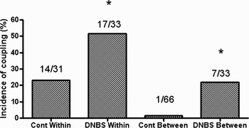
To examine whether the augmentation in glial coupling is a general feature of sympathetic ganglia, we injected LY into SGCs in mouse superior mesenteric ganglia. We found that the general pattern of SGC structure and coupling was quite similar to that described above for the superior cervical ganglia. In control ganglia, coupling incidence of SGCs within the envelope was 23%, and between different envelopes it was 1.5%. Qualitatively, colonic inflammation induced by DNBS led to similar changes in the superior mesenteric ganglion as found in the superior cervical ganglia after submandibular inflammation. Ten days after the induction of colonic inflammation, the incidence of coupling between SGCs forming the envelope around a given neuron from control animals increased by 2.2-fold. Coupling between SGCs forming the envelope of different neurons was 14.6-fold greater than that in control (see Fig. 3 for numerical data). Neurons did not display coupling (0/31).
Summary of dye coupling experiments in mouse superior mesenteric ganglion. The histograms show how colonic injection of DNBS increased the incidence of SGC coupling. ‘Cont Within’ indicates the incidence of coupling between SGCs around a given neuron, and ‘DNBS Within’ indicates the same parameter 10 days after DNBS injection. Coupling between SGC envelopes is indicated by ‘Cont Within’ and ‘DNBS Within’. Both types of coupling were significantly augmented by DNBS. The numbers above the bars indicate the proportion between coupled cells and the total number of injected cells. The asterisks indicate p < 0.05.

Is the increase in cell coupling due to systemic or local effect?
The observed changes in glial coupling could be due to local or systemic effects (or to a combination of the two). Although most investigators believe that nerve injury affects only the involved ganglia, there is evidence for some spread of changes from ipsilateral to contralateral dorsal root ganglia following sciatic nerve ligation (Brázda et al., Reference Brázda, Klusáková, Svízenská, Veselková and Dubový2009). To address this question with respect to sympathetic ganglia, we examined dye coupling in superior mesenteric ganglia from mice in which CFA had been injected 7 days earlier into the submandibular region. The results were clear-cut: in ganglia from the treated mice (n = 3) the coupling within the envelope was 3/30 (10%), compared with 5/35 (14.3%) in controls (n = 5). The incidence of coupling between envelopes was 1/30 (3.3%) in treated mice and 1/35 (2.9%) in controls; in both cases p > 0.05. It can be concluded that the effects induced by submandibular CFA injection were not systemic, but localized to ganglia projecting to the region exposed to CFA.
CONCLUSIONS
• Under control conditions dye coupling among SGCs in mouse sympathetic ganglia is not extensive, and is largely limited to cells around a given neuron.
• Following peripheral inflammation, dye coupling among SGCs is markedly increased, particularly between SGCs forming envelopes around different neurons. This coupling was blocked by a gap junction blocker. The effect appeared to be confined to ganglia projecting to the region affected by the inflammation, rather than systemic.
DISCUSSION
The results described above provide novel information on gap-junction-mediated coupling between SGCs in murine sympathetic ganglia. In ganglia from control animals coupling among SGCs was quite limited, but was greatly augmented following peripheral inflammation.
We studied two different sympathetic ganglia, the superior cervical ganglion, which is paravertebral, and the superior mesenteric ganglion, which is prevertebral. The results obtained for these two ganglia were very similar, and therefore can be probably generalized for other sympathetic ganglia. Under control conditions the extent of coupling among SGCs in both ganglia was small, which holds for both coupling around the same neuron, and for coupling between different envelopes, which was negligible. We have obtained similar results for SGCs in the intrinsic ganglia of the guinea-pig urinary bladder (Hanani et al., Reference Hanani, Maudlej and Härtig1999), which are parasympathetic. This similarity suggests that coupling among SGCs in autonomic ganglia is not extensive, unlike that found among astrocytes. It should be added that the effects were apparently limited to the ganglia projecting to the affected region and were not systemic, as evidenced by the absence of cross-influence.
It is interesting to note that glial cells in the enteric nervous system, which is also a part of the autonomic nervous system, is very extensive (Hanani et al., Reference Hanani, Zamir and Baluk1989). Enteric glia are very different from SGCs, and morphologically resemble astrocytes (Gabella, Reference Gabella1972; Komuro et al., Reference Komuro, Baluk and Burnstock1982).
In both inflammation models, the area affected by the inflammation was not extensive and the proportion of ganglionic neurons projecting into them was likely to be very small. However, our dye coupling results indicate that SGCs that surround a large proportion of the neurons in the ganglia were affected by the inflammation. This implies that the inflammation triggered changes that spread over a large portion of the ganglion. Such spread has been well documented for sensory ganglia following localized peripheral injuries (Stephenson and Byers, Reference Stephenson and Byers1995; Thalakoti et al., Reference Thalakoti, Patil, Damodaram, Vause, Langford and Freeman2007; Huang et al., Reference Huang, Belzer and Hanani2010). The most likely mechanism that can mediate such spread is the release of diffusible signaling molecules from the affected neurons. The nature of these molecules is still obscure, but they could be inflammatory mediators such as cytokines (Miller et al., Reference Miller, Jung, Bhangoo and White2009).
The functional implications of our findings remain to be explored, but a reasonable approach is to view them in comparison with the better studied CNS glial cells. Glial activation (reactive gliosis) is the most prominent pathologic response of glial cells, and has been studied extensively in astrocytes and microglia (for reviews see Raivich et al., Reference Raivich, Bohatsche, Kloss, Werner, Jones and Kreutzberg1999; Pekny and Nilsson, Reference Pekny and Nilsson2005). The expression of intermediate filaments, in particular glial fibrillary acidic protein (GFAP) and vimentin, is greatly increased in reactive astrocytes. Nerve damage causes a large upregulation of GFAP in SGCs in sympathetic ganglia, and it was suggested that the damage led to SGC gliosis (Elfvin et al., Reference Elfvin, Björklund, Dahl and Seiger1987). We have proposed that augmentation in gap junctions could be part of the SGC activation in sensory ganglia (Huang et al., Reference Huang, Belzer and Hanani2010), and it is quite conceivable that the same idea holds for sympathetic ganglia. Glial abnormalities can have wide ranging implications for ganglionic function. For example, they could adversely affect the neuronal microenvironment. It has been shown that augmented SGC coupling in sensory ganglia leads to hyperexcitability of neurons, and to abnormal firing and pain (Ohara et al., Reference Ohara, Vit, Bhargava and Jasmin2008; Huang et al., Reference Huang, Belzer and Hanani2010). It can be hypothesized that the increase in SGC coupling reported here can similarly lead to abnormal neuronal activity in sympathetic ganglia. Obviously, more research on glia–neuron interaction in sympathetic ganglia is required, and it is likely that such research would be most rewarding.
ACKNOWLEDGEMENTS
Supported by the Israel Science Foundation (Grant No. 212/08), by the US-Israel Binational Science Foundation (BSF, Grant No. 2007311).
Statement of interest
None.