Introduction
Petrous internal carotid artery (ICA) aneurysm is rare; however, rupture of the aneurysm can be life-threatening. Aneurysm may be misdiagnosed as a glomus jugular tumour, and unintentional intervention may result in massive bleeding.Reference Oyama, Hattori, Tanahashi, Kito, Maki and Tanahashi1 The pathogenesis of petrous ICA aneurysms has been reported to be related to trauma, congenital disease, arterial dissection, tumour invasion, radiation therapy, fibromuscular disease, iatrogenic injury and chronic otitis media.Reference Auyeung, Lui, Chow and Chan2–Reference Baker, Rizk, Carroll and Lambert7
Herein, we report a case of petrous ICA aneurysm rupture induced by Eustachian tube catheterisation, and describe successful intervention by endovascular coiling compaction.
Case report
An 84-year-old woman presented at a local otolaryngology clinic with progressive otalgia due to acute exacerbation of chronic otitis media. She was treated with peri-oral antibiotics, ear irrigation and Eustachian tube catheterisations for approximately two months. In addition to otolaryngological symptoms, she had myelodysplastic syndrome, and was treated by repeated blood transfusion to improve her symptomatic anaemia by the haematologist at our hospital. She was referred to us with left-sided massive otorrhoea following Eustachian tube catheterisation.
At the initial visit, a perforation was observed at the patient's left tympanic membrane, but no active bleeding could be identified. Her left hearing was scaled out. As there was no obvious otorrhoea, she was advised to carefully monitor her symptoms. One month later, she had left otorrhoea again and visited our hospital by ambulance. Her otorrhoea stopped upon arrival at the hospital. She was hospitalised for a blood transfusion as progressive anaemia was found. We could not identify any cause related to the otorrhoea, despite examinations that included contrast-enhanced computed tomography (CT).
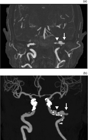
The patient was discharged, and did not experience any otorrhoea until two months later when she suffered another massive otorrhoea episode with epistaxis during left-sided ear cleaning at a third clinic visit. Gauze was packed into her nasal cavity and left ear canal, and she was hospitalised. She received blood transfusions for progressive anaemia. Contrast-enhanced CT and CT angiography revealed an aneurysm and adjacent stenosis at the left petrous internal carotid artery (ICA) (Figure 1). The aneurysm protruded into the left middle-ear cavity, which was devoid of normal bony barriers.
(a) Coronal contrast-enhanced computed tomography (CT) scan and (b) CT angiography image, showing a left-sided petrous internal carotid artery (ICA) aneurysm (arrows), with ICA stenosis at the distal portion of the aneurysm (arrowhead).

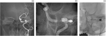
Coil embolisation to the petrous ICA aneurysm and percutaneous transluminal angioplasty were performed under general anaesthesia. Conventional angiography to the right proximal ICA via the right femoral artery was carried out. Figure 2a shows the aneurysm, which measured 15.0 mm × 9.0 mm × 8.0 mm, located at the right proximal ICA next to left middle ear. Embolisations to the aneurysm were performed using: the Target 360 Ultra Coil (4 mm × 15 cm; Stryker, Kalamazoo, Michigan, USA), Target 360 Nano Coil (3 mm × 6 cm, 2.5 mm × 4 mm, and 1.5 mm × 3 cm; Stryker) and Target Helical Nano Coil (2 mm × 6 cm; Stryker) (Figure 2b). Subsequently, we dilated the stenosis at the distal portion of the aneurysm with a Gateway percutaneous transluminal angioplasty balloon catheter (via a 2.5 mm × 9 cm monorail system (Stryker), at 6 atm, for 3 seconds) using Chikai 14 soft tip, 200 cm micro guidewire (Asahi Intecc J-Sales, Tokyo, Japan) and a Marksman micro catheter (eV3; Covidien, Irvine, California, USA) (Figure 2c). Stenting was not performed because she had anaemia associated with myelodysplastic syndrome, and she was not receiving dual antiplatelet therapy.
(a) Frontal view of left-sided petrous internal carotid artery (ICA) aneurysm and ICA stenosis treated by percutaneous transluminal angioplasty. (b) Coil embolisation of the aneurysm was performed. (c) Stenosis of the ICA was dilated at the distal portion of the aneurysm.

The CT scans revealed no obliterations and extravasations, and no recoils. No further rebleeding occurred and the patient had no symptoms of brain infarction. The left tympanic membrane closed naturally and there was no discharge accumulation in her left ear approximately one month later.
Discussion
We present a rare case of a left-sided petrous internal carotid artery (ICA) aneurysm accompanied by stenosis of the ICA at the distal portion of the aneurysm. Because the patient was susceptible to bleeding due to myelodysplastic syndrome, even air pressure from Eustachian tube catheterisation may have caused the aneurysm to rupture. This is the first report of an ICA aneurysm ruptured by Eustachian tube catheterisation.
In the present case, repeated Eustachian tube catheterisations may have inflicted traumatic pressure to the site of the ICA aneurysm, where pressure could have increased because of stenosis at the distal portion. As the patient suffered left-sided chronic otitis media, we suspected that the pseudoaneurysm was induced by chronic inflammation, which resulted in ICA stenosis, and the aneurysm ruptured following repeated Eustachian tube catheterisations. The reduced coagulation associated with myelodysplastic syndrome may have also contributed to the bleeding.
Rupture of ICA aneurysms may present with various symptoms, such as headache and tinnitus, and may include IInd to XIIth cranial nerve deficits.Reference Murai, Shirokane, Kitamura, Tateyama, Matano and Mizunari8 Moreover, the patient may experience bleeding as epistaxis and otorrhoea, which can be life-threatening, as in the present case. When the patient reported herein visited our hospital, CT and CT angiography were necessary to detect any abnormality, including ruptured aneurysms, even though the bleeding had stopped. At the second visit, we performed CT examination; however, as the bleeding symptoms were not active and we did not observe the massive bleeding on-site, we chose to monitor the patient with careful observation. We reached the proper diagnosis upon the patient's third visit to our clinic with massive bleeding.
Regarding iatrogenic bleeding of aneurysms, Oyama et al. reported a patient who developed repeated haemorrhages after undergoing an earpick procedure.Reference Oyama, Hattori, Tanahashi, Kito, Maki and Tanahashi1 Surgery such as mastoidectomy, paracentesis and middle-ear procedures may also result in excessive bleeding.Reference Oyama, Hattori, Tanahashi, Kito, Maki and Tanahashi1,Reference Schmerber, Vasdev, Chahine, Tournaire and Bing9,Reference Barrett and Lawrence10 To the best of our knowledge, there are no case reports of an ICA aneurysm rupture caused by Eustachian tube catheterisation. Aneurysm rupture may not be the result of mild stimulation only, such as the air pressure by Eustachian tube catheterisation; however, in the present case, it may have happened because of a myelodysplastic syndrome episode that weakened the repair mechanism of the vascular wall, resulting in bony deficiencies between the petrous ICA and middle-ear cavity.
Endovascular procedures, surgical ligation and conservative treatment have been used to treat petrous ICA aneurysms. Murai et al. reported that less than 10 per cent of cases had complications, such as ischaemia and rebleeding, among 88 cases.Reference Murai, Shirokane, Kitamura, Tateyama, Matano and Mizunari8 As our patient had a high risk of aneurysm in open surgical ligation procedures because of myelodysplastic syndrome, with bleeding and anaemia tendencies, we chose to perform an endovascular coiling embolisation technique, which was successful.
• Petrous internal carotid artery (ICA) aneurysms are very rare vascular lesions, which may present with otalgia and life-threatening massive otorrhoea
• Surgery, earpick procedures and Eustachian tube catheterisation may result in massive otorrhoea
• Computed tomography (CT) angiography or angiography should be performed prior to surgery in patients with the ICA next to the middle ear
• A CT angiography should be performed immediately when a patient reports massive otorrhoea
• Endovascular occlusion is an option for treatment, to avoid open surgical ligation complications
Competing interests
None declared