INTRODUCTION
Throughout their lifetime, organisms face a dilemma when challenged by severe environmental conditions that threaten survival or breeding. They either avoid the constraints, by migrating or hibernating, adjust their life style to the challenges, by altering physiological properties, or manipulate their habitat for example, by releasing compounds that mitigate or divert the negative effects. A well-characterized example of avoidance strategy in parasitic nematodes is the climate-driven migration of infective larvae into the gastrointestinal mucosa of their hosts and the arrestment of development for a period of weeks or months (Dunsmore, Reference Dunsmore1961; Connan, Reference Connan1969; Michel, Reference Michel1974; Herd and McNaught, Reference Herd and McNaught1975; Smith et al. Reference Smith, Jackson, Jackson and Williams1983; Armour and Duncan, Reference Armour and Duncan1987; Borgsteede and Eysker, Reference Borgsteede and Eysker1987; Ashworth and Kennedy, Reference Ashworth and Kennedy1999; Sommerville and Davey, Reference Sommerville and Davey2002). By delaying maturation, removing the impact on host mortality and avoiding the shedding of eggs when probability to fail development is high, nematodes improve their chances of surviving and their expected life-time reproductive output.
Tissue migration during larval development most frequently occurs without arrestment and can include distal movements across multiple organs or be limited to the gastrointestinal tissue (Anderson, Reference Anderson2000). This strategy has been widely documented among nematode species and often explained as reminiscent of a primordial behaviour associated with maturation and completion of individual's life cycle (Anderson, Reference Anderson2000; Audebert et al. Reference Audebert, Vuong and Durette-Desset2003; Chubb et al. Reference Chubb, Ball and Parker2010). An alternative hypothesis is that there is a reproductive advantage for the transient movements of larvae into host's tissues (Read and Skorping, Reference Read and Skorping1995). This was supported by the finding that migratory nematodes grew bigger and faster than their close relatives settled in the gastrointestinal lumen – this group also included larvae with a tissue phase in the gastrointestinal wall (Read and Skorping, Reference Read and Skorping1995). More recently, theoretical work by Parker et al. (Reference Parker, Ball and Chubb2009) proposed that the selective advantage for larval migration critically depends on the survival – mortality ratio encountered in the tissues compared to the gastrointestinal lumen and how this changes over time. Simulations show that the survival rate of incoming larvae is initially higher in the tissue than in the lumen but decreases with time, or parasite development, as the host responds to control the infection (e.g. acquired immunity). By migrating back into the lumen mortality decreases, parasites complete maturation, and shedding of eggs into the environment is facilitated (Parker et al. Reference Parker, Ball and Chubb2009). Importantly, the contrasting mortality risk of the two habitats could explain the evolution of both distal larval migrations and the movements limited to the gastrointestinal mucosa.
Tissues and gastrointestinal lumen are clearly different in terms of their immunological, biochemical and physiological properties. Crucially, these conditions can vary, sometimes in a predictable way during the course of the infection and can alter the habitat for incoming larvae (Mulcahy et al. Reference Mulcahy, O'Neill, Fanning, McCarthy and Sekiya2005). Seasonality in the intensity of infection can amplify these changes and host characteristics, namely age, breeding status and co-infections can create additional sources of variability in host-larval interactions that can affect larval migration. Thus, how do changes in host properties and parasite intensities in the lumen influence the number of larvae in the tissue? And, across a host population, what is the pattern of larval abundance in the tissue? In this study, we examine the hypothesis that between-host variability in number of tissue larvae is influenced by the conditions encountered at the site of infection and by the environmental cues priming infective larvae during their free-living stage. Larvae of the gastrointestinal nematodes Graphidium strigosum and Trichostrongylus retortaeformis were extracted from the gastrointestinal tissue of free-living European rabbits (Oryctolagus cuniculus) and intensity examined in relation to host immunity, parasite intensity in the lumen and seasonal weather changes. The modulatory effects of host age, sex and breeding status, as well as co-infection with the second parasite, were also considered as they play an important role in the variability of the host's response to these infections (Cattadori et al. Reference Cattadori, Boag, Bjornstad, Cornell and Hudson2005, Reference Cattadori, Boag and Hudson2008). Three, not mutually exclusive predictions were examined. First, if weather is the driving factor of larval variability in the tissue then infective larvae become programmed during the free-living stage on the herbage and intensities in the tissue will be associated with seasonal thermal changes, regardless of the host immune conditions or intensity of infection in the lumen. Second, if host-acquired immunity plays an important role, then the number of larvae in the tissue will proportionally vary with the strength of the immune response. Specifically, assuming that the immune response in the tissue is lower than in the lumen, the number of larvae is expected to increase in the gastrointestinal wall of adult individuals, while fewer larvae are predicted in the tissue of young hosts. Third, if intensity-dependent parasite processes (immunity excluded) affect variability of larvae in the gastrointestinal wall, higher levels of infection in the lumen will be associated with more tissue larvae, while immunity or weather conditions will not contribute to this pattern. Previously, we showed that G. strigosum and T. retortaeformis exhibit contrasting dynamics of infection and rabbit immune responses (Cattadori et al. Reference Cattadori, Boag, Bjornstad, Cornell and Hudson2005, Reference Cattadori, Boag and Hudson2008; Murphy et al. Reference Murphy, Nalpas, Stear and Cattadori2011). As such, we predicted contrasting patterns between the infective larvae of the two species: density-dependent mechanisms were expected to cause most of the variation in G. strigosum larvae in the stomach tissue while immunity was expected to influence T. retortaeformis in the small intestine walls.
MATERIALS AND METHODS
The parasite-host system
Graphidium strigosum and Trichostrongylus retortaeformis are widespread gastrointestinal helminths of the European rabbit (Oryctolagus cuniculus) (Audebert and Durette-Desset, Reference Audebert and Durette-Desset2007). Both parasites have a direct life cycle with free-living stages; rabbit infection occurs by ingestion of third-stage infective larvae with contaminated herbage. Graphidium strigosum inhabits the stomach while T. retortaeformis colonizes the small intestine; the pre-patent period is about 12 days for T. retortaeformis and 42 days for G. strigosum (Audebert et al. Reference Audebert, Hoste and Durette-Desset2002; Massoni et al. Reference Massoni, Cassone, Durette-Dusset and Audebert2011). Long-term monitoring of 2 populations of rabbits from Scotland showed that both infections occur with high intensities and a strong seasonal pattern, with the majority of transmission occurring during the spring-summer months (Cattadori et al. Reference Cattadori, Boag, Bjornstad, Cornell and Hudson2005, Reference Cattadori, Boag and Hudson2008). G. strigosum intensity increases exponentially with host age and with no evidence of parasite mortality or strong immune regulation (Cattadori et al. Reference Cattadori, Boag and Hudson2008; Murphy et al. Reference Murphy, Nalpas, Stear and Cattadori2011). In contrast, T. retortaeformis intensity peaks in juveniles and decreases in adults as a consequence of a robust immune response that reduces or clears the infection (Cattadori et al. Reference Cattadori, Boag, Bjornstad, Cornell and Hudson2005; Murphy et al. Reference Murphy, Nalpas, Stear and Cattadori2011; Thakar et al. 2012). Challenges of rabbits with 20 000 T. retortaeformis third-stage larvae lead to a recovery rate from the small intestine mucosa of between 2% and 16% in the first 10 days post-infection, no larvae were retrieved after this period (Audebert et al. Reference Audebert, Vuong and Durette-Desset2003). Rabbits infected with this nematode in autumn shed few eggs during the winter but higher numbers in the following spring, indicative of larval arrestment (Michel, Reference Michel1952; Eysker, Reference Eysker1978; Michel, Reference Michel, Borgsteede, Armour and Jansen1978). No information is available on the tissue phase of G. strigosum larvae although Martin et al. (Reference Martin, Thomas and Urquhart1957) reported a clear delay in larval development following a high infection dose.
Larval extraction
A population of rabbits was sampled randomly from an agricultural ecosystem in Perthshire, Scotland (Latitude 5°29′40″N, Longitude 3°9′55″W) every month, from January 2008 to January 2011 (Cattadori et al., unpublished data). Animals were processed within a few hours of sampling, the gastrointestinal tract was removed from each rabbit and the stomach and small intestine independently processed. The organs were gently washed to remove the parasites from the lumen (see details below), individually frozen at −20 °C and shipped to The Pennsylvania State University (USA) for larval extraction. Previous work has shown that the recovery of larvae from gastrointestinal tissues is not affected by long-term storage at −20 °C (Ciordia et al. Reference Ciordia, Vegors and Bizzell1957). Records of host sex, age and breeding status were also collected for each individual (Cattadori et al. Reference Cattadori, Boag, Bjornstad, Cornell and Hudson2005, Reference Cattadori, Boag and Hudson2008). Animal field procedures were executed in accordance with UK regulations and pre-approved by the Institutional Animal Care and Use Committee of The Pennsylvania State University.
In the laboratory, samples were slowly thawed, weighed after removing the accumulated water and cut into small pieces to facilitate tissue digestion. Samples were placed into lid protected glass jars and filled up with a solution of 4·5% HCl, 1·9% pepsin powder and 93·6% deionised H2O (adapted from Herlich, Reference Herlich1956). The jars were stored in an incubator at 37 °C, after 90 min the contents were poured through 2 nested sieves (top to bottom sieves: 212 μm and 100 μm), carefully washed and the digested tissue with the larvae collected into a cylinder. The undigested tissue was placed back into the jar with fresh digesting solution and incubated for an additional 30 min. This procedure was repeated until the tissue was completely digested and all the larvae collected. The digested material was set aside at room temperature for 24 h, the supernatant was then gently removed and the larvae stored in a 1:1 solution of 10% formalin and water in 14 ml vol. tubes. Finally, the full content of each tube was carefully inspected under a stereomicroscope, G. strigosum and T. retortaeformis were counted and stages identified following the protocols of Audebert et al. (Reference Audebert, Cassone, Hoste and Durette-Desset2000) and Massoni et al. (Reference Massoni, Cassone, Durette-Dusset and Audebert2011). Larvae appeared as typical ex-sheathed L3 or L4 stages under the microscope, however, the extraction technique prevented identification of their status (active or arrested) or their exact location in the gastrointestinal tissue.
Parasite intensity in the lumen
The number of G. strigosum and T. retortaeformis in the lumen (throughout the text defined as intensity of infection, sensu Margolis et al. Reference Margolis, Esch, Holmes, Kuris and Schad1982) was quantified by opening longitudinally the stomach or the small intestine and washing the organs onto a 100 μm sieve by carefully passing the tissue through the fingers to remove the food content, the mucus and the worms in the mucus and lumen (Boag, Reference Boag1985). Between 4% and 50% of the total washed content was inspected in 10 ml aliquot counts and the total helminth intensity was estimated; no stage classification was performed for these counts.
Host immune response
Antibody data have been previously quantified from the blood serum of each sampled rabbit (Cattadori et al. unpublished data). This information was used as a measurement of the host immune response to the 2 parasites. Briefly, species-specific antibodies IgA and IgG were measured against G. strigosum and T. retortaeformis using their excretory/secretory (ES) by-products as a source of antigen and an enzyme-linked immunosorbent assay (ELISA). Antibodies against ES products can be used as a reliable alternative to somatic compounds (Hewitson et al. 2009). In our case, they showed negligible cross-reactivity between the two nematodes (Cattadori et al., unpublished data). Data were expressed as standardized antibody optical density index (OD index) (Murphy et al. Reference Murphy, Nalpas, Stear and Cattadori2011). We have previously suggested that IgA and IgG are involved in the removal or death of L3 and adult stages of T. retortaeformis in the lumen but no clear effects have been observed for G. strigosum (Murphy et al. Reference Murphy, Nalpas, Stear and Cattadori2011; Pathak et al. Reference Pathak, Pelenski, Boag and Cattadori2012; Thakar et al. Reference Thakar, Pathak, Murphy, Albert and Cattadori2012). Yet, antibodies may stimulate larval migration for both parasites species.
Weather variables
Daily weather data were available from the meteorological station at The James Hutton Institute (Invergowrie, Scotland) located 12 miles from our field site. The temperature variables (air, grass and soil temperature) were highly correlated (Pearson's product moment correlation r, range: 0·94–0·87, for all P < 0·0001) and exhibited a strong seasonal pattern, the average coldest month was December and the hottest was July (Supplementary Fig. 1S, online version only). Total rainfall fluctuated among months, the driest months ranged from late winter to late spring, and weak correlations were observed with temperature variables (Pearson's correlation r, range: 0·06–0·11, for all P < 0·05). Photoperiod was also weakly associated with temperature (Pearson's correlation r, range: 0·16–0·33, for all P < 0·0001) and rainfall (Pearson's correlation r: −0·21, P < 0·001) (Suppl. Fig. 1S, online version only).
For every weather variable, the arithmetic mean was calculated for 15-day periods over the 3 years of study, so that every month was represented by 2 periods, period 1: from days 1 to 15 and period 2: from days 16 to 31. By knowing on which day of the month a rabbit was sampled (i.e. period 1 or period 2) and the age of the individual (in months), every rabbit was related to the correct weather period. We assumed that infective larvae were conditioned by weather stimuli shortly before infecting a host; hence, the weather variables in the last month and an half of the rabbit lifespan were used.
Statistical analysis
Differences in the number of larvae retrieved from the tissue of subgroups of rabbits for example, by host sex, age (young and adults), breeding and non-breeding condition as well as month of sampling, were examined using linear mixed effect models (LME-REML, www.r-project.org), where the number of tissue larvae was used as a response variable while host condition, or seasonality, were used as the fixed explanatory term. To identify how immunity, parasites in the lumen and weather variables could explain between-host variability in the intensity of G. strigosum or T. retortaeformis larvae in the gastrointestinal tissue, linear mixed effect models were also applied for which antibodies, intensity of infection in the lumen and weather data were used as fixed terms. To take into account the confounding effect that high quantities of ingested food could result in large numbers of larvae in the tissue, the linear relationship between log-transformed larval numbers in the tissue and log-transformed host body mass was estimated and the residuals of the larval component used as a response variable. Larvae in the tissue were not scaled over nematodes in the lumen to avoid the confounding effect of co-variation between the 2 variables and to allow us to examine how intensity of infection in the lumen was associated with tissue larvae (i.e. no proportion data were used). Fixed variables were log-transformed where necessary. The month of sampling, host sex and age were included as a random variables (described as standard deviation of the intercept), with age nested into sex and month, where month is the highest nested variable; different nested combination were used based on the combination of fixed effects. Random variables were represented as factorial components. Data exploration started from a full additive model and the appropriate combination of nested random components, the contribution of two-way interactions between fixed effects and the role of quadratic terms were considered. Analyses were repeated for each parasite independently; however, to examine the effect of co-infection with the second parasite, the abundance and species-specific antibody responses against the second nematode were also investigated. In this case, the rabbit identity code, ID, was included as a random effect to take into account the 2 parasite species in the same host. The minimum parsimonious models were selected and presented as final results.
RESULTS
In total, 791 rabbits infected with either one of both nematode species were sampled between January 2008 and January 2011, of these 431 were males (54%) and 360 females (46%); 141 individuals were classified as kittens (age classes: 1 to 3 months old), 220 were juveniles (age classes: 4 and 5 months old) and 430 were adults (age classes: 6 and more than 8 months old).
Graphidium strigosum larvae in tissue
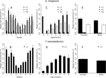
Three helminth stages were recovered from the stomach tissue: third- and fourth-stage larvae (L3 and L4) and adults, L3 was the most abundant stage and L4 the least (coeff±s.e.: L4 vs L3 −1·37 ± 0·06 and adult vs L3 −0·52 ± 0·06, N = 1747, for both P< 0·001–note that the large sample size is a consequence of having the same individual with different parasite stages in the tissue; to take this into account, the individual ID is included as a random effect in the LME) (Fig. 1). The number of parasites recovered differed among months (coeff±s.e.: −0·05 ± 0·01 N = 581, P < 0·001); across these months L3 was consistently higher than L4 (two-way interaction month-L4 stage vs L3: P < 0·01) but not adults (Fig. 1A). The number of G. strigosum extracted from the tissue increased with host age (coeff±s.e.: 0·24 ± 0·04, N = 570, P < 0·0001) and was mainly driven by L3 (two-way interaction age-L4 or age-adult vs L3, for both: P < 0·0001) (Fig. 1C). There was no significant difference in the number of G. strigosum retrieved from the stomach tissue in each host sex (Fig. 1E). The effect of host breeding status was also examined by focusing on 2 phases of the female reproductive cycle, pregnancy (i.e. individuals carrying fetuses) and nursing (i.e. individuals with exposed nipples). There was a tendency for more G. strigosum to be extracted from non-nursing than nursing females (coeff±s.e.: −0·36 ± 0·18, N = 216, P = 0·051) and this was driven mainly by adult worms (P < 0·04). No differences were found between pregnant and non-pregnant females.
Graphidium strigosum and Trichostrongylus retortaeformis recovered from the stomach and small intestine tissue, respectively; mean (±s.e.) over 3 years by month of sampling (A and B), host age (C and D) and sex (E and F).

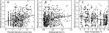
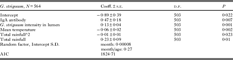
Adult helminths in the stomach wall were unexpected and the visual examination of a few tissues under the stereomicroscope revealed that adults were settled just under the epithelial lining in an elongated shape, suggesting active behaviour. A positive relationship was found between adults retrieved from the stomach tissue and total nematodes in the lumen (coeff±s.e.: 0·23 ± 0·03, N = 576, P < 0·0001) but no significant association was observed with anti-G. strigosum antibodies. Since we were interested in variability in larval abundance in the tissue, analyses were based on the combined L3 and L4 data and their relationship with host antibody responses, intensity of infection in the lumen and weather. The minimum parsimonious model showed that larval number from the stomach tissue (residual variable used, details in Statistical analysis section) were associated positively with antibody IgA, intensity of G. strigosum infection in the lumen and negatively with mean air temperature, a linear and quadratic relationship was also found with total rainfall (Fig. 2 and Table 1). Analysis repeated using adult rabbits only, showed results consistent with the general patterns based on the whole host population, suggesting that most of the observed trends are driven by the adult cohort (Supplementary Table 1S, online version only). In the younger group (rabbits from 2 to 5 months old) arrested larvae were positively related to IgA and negatively associated with mean temperature, no correlation was found with the intensity of G. strigosum in the lumen (Suppl. Table 1S, online version only).
Relationships between Graphidium strigosum tissue larvae and (A) intensity of infection in the lumen, (B) serum antibody IgA and (C) mean air temperature. Pair-wise linear relationships (LME) between the significant variables are reported. Tissue larvae (y-axis) are the residual of the linear relationship between log-transformed larvae in the tissue and log-transformed host body mass.

Linear mixed model between Graphidium strigosum tissue larvae (residual variable) as a response and host antibodies, parasite intensity in the lumen and weather variables as fixed explanatory effects

The possible roles of T. retortaeformis co-infection and anti-T. retortaeformis antibodies were explored and G. strigosum larvae in the stomach tissue were positively associated with T. retortaeformis intensity in the small intestine lumen (P < 0·05; the minimum model also included variables as reported in Table 1), no association was found with antibodies.
Trychostrongylus retortaeformis larvae in tissue
Only third-stage T. retortaeformis larvae were recovered from the digestion of the small intestine (Fig. 1). The number of larvae differed among months (coeff±s.e.: 0·04 ± 0·02, N = 704, P < 0·05), a few larvae were found in the tissue in summer and a larger amount in winter (Fig. 1B). Larvae in the tissue increased with rabbit age (coeff±s.e.: 0·10 ± 0·04, N = 704, P < 0·0001) and more larvae were extracted from female than male rabbits (coeff±s.e.: −0·20 ± 0·09, N = 704, P < 0·05, the negative coefficient is because females were coded 1 and males 2) (Fig. 1D and F). Fewer larvae were collected from pregnant than non-pregnant females (coeff±s.e.: −1·01 ± 0·40, N = 76, P < 0·05) while no significant differences were observed between nursing females and females not nursing.
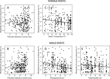
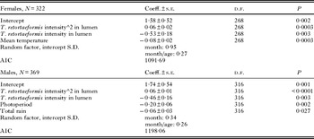
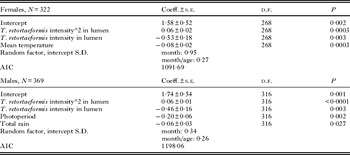
Given that we found a significant host sex-biased parasitism in the L3 from the small intestine tissue, analyses were then performed on each host sex, independently. The number of larvae from female hosts (residual variable used) was significantly related to the intensity of infection in the lumen (both linear and quadratic term) and negatively associated with mean temperature (Fig. 3 and Table 2). For male rabbits, the minimum model showed that larvae were associated with T. retortaeformis intensity in the lumen (both linear and quadratic term) and negatively correlated with total rainfall and photoperiod duration (Fig. 3 and Table 2). For both host sexes, antibodies did not contribute to the pattern observed. Analyses were repeated using adult and juvenile rabbits. For adult females, larvae in tissue were associated with parasite intensity in the lumen (both linear and quadratic term) and negatively associated with temperature (Supplementary Table 2S, online version only)). No significant minimum model was found for juvenile females (Suppl. Table 2S, online version only). For both adult and juvenile males, larvae were positively associated with intensity of infection in the lumen and negatively with temperature (Suppl. Table 2S, online version only).
Relationships between Trichostrongylus retortaeformis tissue larvae in female and male rabbits and (A and B) intensity of infection in the lumen, (C and D) mean temperature and mean photoperiod hours, and (E) total rainfall. Pair-wise linear relationships (LME) are reported from (C) to (E), no non-linear relationship was fitted for (A) and (B). Tissue larvae (y-axis) are the residual of the linear relationship between log-transformed larvae in the tissue and log-transformed host body mass.

Linear mixed model between Trychostrongylus retortaeformis tissue larvae (residual variable) as a response and parasite intensity in lumen and weather variables as fixed explanatory effects, by host sex
The contribution of G. strigosum co-infection on T. retortaeformis tissue larvae was explored. For female hosts, larvae were positively associated with G. strigosum intensity (P < 0·001; the minimum model also included the significant effect of T. retortaeformis intensity) while for males a positive association was found with anti-G. strigosum IgA (P < 0·05; the minimum model also included variables as in Table 2).
DISCUSSION
We used a natural nematode-herbivore system and examined the pattern of between-host variability in number of larvae from the gastrointestinal tissue. In addition to investigating the role of host immune responses, parasite intensity-dependent constraints and seasonal weather fluctuations in affecting the trends observed, we also explored how changes in host properties (i.e. age, sex and breeding status) and co-infection with the second nematode species contributed to this variability. Both nematode larvae exhibited a strong seasonal pattern marked by a fewer larvae recovered in the summer, during host reproduction and emergence of newborns. Tissue larvae were found across the entire host age range, from 2-month-old kittens to 8+ month-old adults, and the number increased with host age. These trends were affected by host sex and breeding conditions although their contribution was not consistent between the 2 parasite species. For G. strigosum, larvae in the stomach tissue were positively associated with IgA and intensity of infection in the lumen but negatively related to mean air temperature. For T. retortaeformis, larvae in the small intestine wall were related to intensity of infection in the lumen and mean air temperature. The general pattern that emerges from these findings suggests that intensity of infection and/or host immunity could be important sources of heterogeneity in the number of tissue larvae, this was more apparent as hosts became older. The seasonal weather conditioning of free-living larvae probably contributes to affect the duration of larvae in the tissue and some arrestment during the autumn-winter period cannot be excluded. These observations support theoretical predictions that the incoming larvae move into the gastrointestinal wall to escape potential sources of mortality in the lumen (Parker et al. Reference Parker, Ball and Chubb2009). We suggest that these causes can be host and parasite driven while weather cues may be more critical in tuning the duration of the tissue phase.
We have recently shown that laboratory rabbits consistently reduce or clear T. retortaeformis in the lumen via the activation of antibodies and eosinophils, and also neutrophils, as a response to larval establishment (Murphy et al. Reference Murphy, Nalpas, Stear and Cattadori2011; Thakar et al. Reference Thakar, Pathak, Murphy, Albert and Cattadori2012). We have also shown that individuals develop a strong IFNγ response in the small intestine mucosa following a primary infection, suggesting an inflammatory reaction to microflora and bacterial infiltration during larval tissue migration and parasite colonization (Murphy et al. Reference Murphy, Nalpas, Stear and Cattadori2011; Thakar et al. Reference Thakar, Pathak, Murphy, Albert and Cattadori2012). It is possible that by damaging the mucosa, creating chronic inflammation and diverting the tissue immune responses toward other tasks, persistent nematode infections can facilitate larval movements in the gastrointestinal tissue as hosts become older. The lack of a significant relationship between antibodies and T. retortaeformis tissue larvae was probably caused by an immune system that is constantly challenged by a parasite with rapid development. In other words, while the antibody response remains relatively high because of the exposure to previous and current infective stages, larvae move in-and-out of the small intestine wall rapidly. Moreover, variability in host age, gender and breeding status may have prevented the detection of significant patterns in the immune-larval migration relationship. For G. strigosum larvae we found a strong relationship with serum IgA indicating that antibodies can be important in larval variability between hosts, however, this should be interpreted with caution. We previously showed that rabbits developed a classical Th2 response against this parasite and worms did damage the stomach tissue, yet infection persisted in the stomach lumen, where we found relatively low levels of IgA and IgG in the mucus compared to the blood serum (Murphy et al. Reference Murphy, Nalpas, Stear and Cattadori2011, Pathak et al. Reference Pathak, Pelenski, Boag and Cattadori2012). Therefore, while we do not excluded that antibodies can contribute to larval migration the role of immunity in driving this pattern is still obscure.
For both nematode species, numbers of larvae in the tissues were positively related to the intensity of infection in the gastrointestinal lumen, suggesting that intra-specific competition for resources (food and space) or compounds produced by adult stages may be key factors in larval migration and between-hosts heterogeneity in accumulation of tissue larvae. Indeed, intensity-dependent parasite constraints appear to become more critical in adult rabbits compared to younger individuals, where infection is still relatively low, especially for G. strigosum. However, while the level of infection in the lumen appears to be a crucial cause of larval variability in the gastrointestinal tissue, differences in host age and breeding status can alter host physiology and tissue properties, in addition to the intensity of infection in the lumen, and contribute to amplify individual variability in the amount of tissue larvae. Surprisingly, we found adult G. strigosum burrowing under the epithelial lining and increasing in number with host age. We suggest that this is a strategy either to reduce species-specific competition or to evade the host immune effectors circulating in the stomach mucus. As rabbits become older and intensity of infection increases, more adult worms adopt this behaviour although changes in female breeding status can modify this trend, as well as parasite intensity in the lumen (Cattadori et al., unpubl. data). This observation has important implications for the ability of this parasite to persist without apparent mortality and accumulate with host age, despite a robust immune response (Cattadori et al. Reference Cattadori, Boag and Hudson2008; Murphy et al. Reference Murphy, Nalpas, Stear and Cattadori2011).
A negative relationship was found between tissue larvae and mean air temperature. One possible explanation is that environmental cues stimulate the timing of larvae in the tissue. The lower the thermal energy accrued on the pasture before infection, the higher the probability for longer residency in the tissue and, probably, larval arrestment. This agrees with previous studies that found an inverse relationship between temperature and propensity of larvae to arrest (Hutchinson et al. Reference Hutchinson, Lee and Fernando1972; McKenna Reference McKenna1973). Seasonality can play a strategic role in delaying larval maturation in the tissue but also in influencing parasite intensity in the lumen and thus, contributes to the number of larvae in the gastrointestinal tissues. Rainfall and photoperiod played a part with temperature to the climatic stimulus, although their effect was not consistent over time or among host groups. We were unable to distinguish arrested from migrating larvae, however, we do not exclude that arrestment affected both nematode species, especially during the cold months. Indeed a high number of larvae were collected from the tissues in the autumn-winter period, when the risk of infection is a relatively low and the majority of hosts are adult individuals (Cattadori et al. Reference Cattadori, Boag, Bjornstad, Cornell and Hudson2005; Reference Cattadori, Boag and Hudson2008).
Free living rabbits are commonly co-infected with both G. strigosum and T. retortaeformis (Cattadori et al. Reference Cattadori, Boag and Hudson2008) and thus we investigated the role of the second parasite on tissue larvae. For both nematodes, the number of larvae extracted from the gastrointestinal tissue was positively correlated with the intensity of the second parasite in the lumen or the antibody response against this second parasite. The two nematode species inhabit separate parts of the gastrointestinal tract and interactions are expected to occur via excretory/secretory parasite compounds flowing from G. strigosum down towards T. retortaeformis or alternatively, mediated by the host immune response. At the current time it is unclear which mechanism drives the positive co-variation between larval intensity in the tissue and co-infection in the lumen; more work is needed to confirm that this pattern is biologically meaningful.
Despite the data on the parasite and the host, no details were available on the intensities of parasite stages in the gastrointestinal lumen, larval location in the tissue or timing of the last infection. This prevented us from quantifying the rate of larval migration and the duration of the larval phase. Yet, using two closely related gastrointestinal nematodes we showed that variability in host properties (including immunity), intensity of infection in the lumen and seasonality can influence the number of larvae in the gastrointestinal tissue. The intrinsic mechanisms underlying the patterns observed are still obscure; the next challenge is to reveal the processes driving larval movements and how they change with host status and intensity of infection in a seasonal environment.
ACKNOWLEDGMENTS
Authors would like to thank Carolyn Mahan, Jake Warner and Alexander Hernandez (PSU) for useful comments on the preliminary manuscript. We are also grateful to Douglas Allan for allowing the sampling of rabbits around his farm and to Kathleen Creppage for technical assistance. This study was supported by NSF (DEB-LTREB 0716885).