Introduction
Auditory neuropathy is defined as any degree of hearing loss, with preservation of cochlear function but aberrant cochlear nerve conduction on electrophysiological testing.Reference Starr, Picton, Sininger, Hood and Berlin 1 Many phenotypes of auditory neuropathy exist. The pathophysiological abnormality has been ascribed, variably, to outer hair cell function, the synaptic interface between inner hair cells and the auditory nerve, and axonal and myelin integrity within the auditory nerve itself.Reference Starr, Sininger and Pratt 2 Differences between pre- and post-synaptic derangements can be determined based on electrophysiological patterns.Reference McMahon, Patuzzi, Gibson and Sanli 3 Recent work suggests that auditory neuropathy patients with a pre-synaptic pathology may have better cochlear implantation outcomes as compared to auditory neuropathy patients with post-synaptic pathology.Reference Tavartkiladze 4
Methods of hearing rehabilitation in auditory neuropathy patients include frequency modulation systems, hearing amplification and cochlear implantation.Reference Madden, Rutter, Hilbert, Greinwald and Choo 5 In particular, cochlear implantation outcomes in auditory neuropathy patients are highly variable,Reference Madden, Hilbert, Rutter, Greinwald and Choo 6 and likely depend largely on the specific physiological derangement – pre-or post-synaptic – leading to auditory nerve function impairment.
Imaging characteristics of the cochlear modiolus are infrequently studied. In certain conditions, such as in enlarged vestibular aqueduct syndrome, the modiolus may be deficient or calcified.Reference Naganawa, Ito, Iwayama, Fukatsu, Ishigaki and Nakashima 7 However, modiolar characteristics in patients with auditory neuropathy have not previously been reported. This study sought to characterise the radiological appearance of the modiolus in auditory neuropathy patients using computed tomography (CT), specifically by investigating the degree of ossification present, as represented by radiological attenuation, and the potential diagnostic implications for auditory neuropathy.
Materials and methods
The Institutional Review Board of the Beijing Friendship Hospital, Capital Medical University, China, approved this study. A retrospective review of all patients who underwent cochlear implantation from April 2007 to March 2014 was performed.
Seven implanted ears from seven paediatric patients who had been diagnosed with auditory neuropathy were identified. All subjects had worn binaural hearing aids for at least six months without benefit. Pre-operative high-resolution temporal bone CT was performed in a standard fashion, and the mid-modiolar cut was utilised for image analysis. Inclusion criteria were as follows: profound hearing loss on age-appropriate testing, type A tympanometry, absent stapedial reflex, intact otoacoustic emissions, and no evidence of inner-ear abnormalities or cochlear nerve deficiency on imaging.
Fifteen paediatric subjects were selected from the remaining cochlear implant recipients to serve as the comparison group. All subjects had worn binaural hearing aids for at least six months without benefit. Temporal bone CT and magnetic resonance imaging (MRI) were performed in a standard fashion in all cases. All patients demonstrated profound hearing loss on age-appropriate testing, type A tympanometry, absent otoacoustic emissions, and showed no evidence of inner-ear abnormalities or cochlear nerve deficiency on imaging.
All subjects in both the auditory neuropathy group and the comparison group underwent cochlear implant surgery via standard mastoidectomy with a facial recess approach. Cochlear (Sydney, Australia) and Med-El (Innsbruck, Austria) devices were used.
Pre-operative imaging was reviewed in a blinded, retrospective fashion. Quantitative measurement of the attenuation at the midpoint of the modiolus, at the middle turn of the cochlea, was performed for the implanted ear in all subjects. Because of their widespread use in diagnostic radiology and standardised nature, Hounsfield units (HU) were chosen for attenuation measurement. Data were analysed with SPSS 19.0 statistical software (IBM, Armonk, New York).
Results
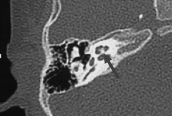
Seven paediatric subjects, 5 males and 2 females, were included in the auditory neuropathy group (mean age, 2.5 years; range, 1.3–6 years). In all subjects, pre-operative CT revealed no inner-ear abnormalities, and pre-operative MRI confirmed the presence of a cochleovestibular nerve bundle. Ossification of the scala tympani and vestibuli was not observed. Three patients were implanted with Cochlear CI24R(CA) devices and the other four received Med-El Pulsar devices. Temporal bone CT imaging of one auditory neuropathy subject is presented in Figure 1.
Computed tomography image of patient with auditory neuropathy. Arrow denotes midpoint of modiolus at the middle turn, where attenuation was measured.

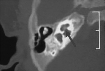
The comparison group comprised 15 paediatric subjects, 12 males and 3 females (mean age, 2.4 years; range, 1–6.3 years). There was no statistically significant difference between the mean ages of the auditory neuropathy group and the comparison group (p = 0.926, student's t-test). In all subjects, pre-operative CT revealed no inner-ear abnormalities, and pre-operative MRI confirmed the presence of a cochleovestibular nerve bundle. Ossification of the scala tympani and vestibuli was not observed. Twelve patients were implanted with Cochlear CI24R(CA) devices and three received Med-El Pulsar devices. Temporal bone CT imaging of one comparison subject is presented in Figure 2.
Computed tomography image of comparison patient with profound sensorineural hearing loss. Arrow denotes midpoint of modiolus at the middle turn, where attenuation was measured.

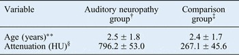
Values for attenuation, measured in Hounsfield units, at the midpoint of the modiolus at the middle turn of the cochlea are reported in Table I. In the auditory neuropathy group, the mean (± standard deviation (SD)) attenuation was 796.2 ± 53.0 HU (range, 756.1–879.1 HU). In the comparison group, the mean (± SD) attenuation was 267.1 ± 45.6 HU (range, 206.1–375.1 HU). There was statistically significantly less ossification in the modiolus in the comparison subjects as compared to the auditory neuropathy patients (p < 0.05, student's t-test).
Attenuation at midpoint of modiolus, at middle turn of cochlea*

Data represent means ± standard deviations.
* As measured on computed tomography of the temporal bone. † n = 7; ‡ n = 15. **p = 0.92, student's t-test; § p < 0.05, student's t-test. HU = Hounsfield units
Discussion
Auditory neuropathy, a form of hearing loss usually diagnosed in childhood, is characterised by any degree of hearing loss with intact cochlear function and derangement of auditory neural conduction on electrophysiological studies. Although proposed aetiologies vary, it is undisputed that the neonatal auditory pathway is highly susceptible to noxious insult. Severe neonatal hyperbilirubinemia has been associated with sensorineural hearing loss (SNHL), including auditory neuropathy.Reference Madden, Rutter, Hilbert, Greinwald and Choo 5 , Reference Shapiro and Nakamura 8 , Reference Saluja, Agarwal, Kler and Amin 9 For example, audiological studies in children with serum bilirubin levels exceeding 20 mg/dl have reported auditory dysfunction in 26–87 per cent of cases involving either cochlear or retrocochlear function.Reference Baradaranfar, Atighechi, Dadgarnia, Jafari, Karimi and Mollasadeghi 10 – Reference Nickisch, Massinger, Ertl-Wagner and von Voss 12 In addition, bilirubin encephalopathy has been reported to be associated with auditory neuropathy in 53 per cent of affected patients.Reference Nickisch, Massinger, Ertl-Wagner and von Voss 12 Despite these associations, the pathophysiology of auditory neuropathy remains unclear.
In this study, we sought to characterise the appearance of the modiolus on CT imaging in patients with auditory neuropathy. We hypothesised that auditory neuropathy patients would display different degrees of attenuation at the modiolus, as compared to a comparison group with profound SNHL without auditory neuropathy. It is plausible that increased ossification of the modiolus may either impair neuronal transmission or reflect a morphological change inhibiting neuronal transmission. In order to maximise homogeneity across subjects, all patients in the auditory neuropathy group and comparison group had bilateral profound hearing loss; all met criteria for and underwent cochlear implantation after deriving no benefit from an appropriate trial of amplification.
We found that the auditory neuropathy patients had statistically significantly higher attenuation values than a similar control group with SNHL. Calcification, which is defined as abnormal calcium deposition in soft tissues, is distinct from ossification, which represents increased mineral deposition in cartilaginous or bony structures. The higher attenuation values in the auditory neuropathy group indicate a greater degree of ossification of modiolar bone in this group as compared to the control group with SNHL.
The reason for the increased ossification at the modiolus remains unknown, but may be of clinical significance. Increased modiolar bone ossification may represent a lower density of spiral ganglion neurons, or may be present as a sequela of prior inflammation and subsequent healing. Furthermore, the observed imaging characteristics in auditory neuropathy patients may be congenital or acquired; longitudinal imaging was not available to identify any temporal trend. It is possible that a neonatal insult, such as hyperbilirubinemia, led to pathophysiological changes in the otic capsule, including the modiolus, which critically houses the spiral ganglion. It must be emphasised, however, that a causal relationship cannot be inferred by these results.
A comparison can be drawn to post-meningitic deafness. Labyrinthitis ossificans, which is the gravest otological sequela of meningitis, results in diffuse cochlear ossification, leading to hearing loss and posing a significant problem for future cochlear implantation.Reference Suga and Lindsay 13 , Reference Green, Marion and Hinojosa 14 However, a notable difference between labyrinthitis ossificans and our auditory neuropathy population is that the latter displayed no ossification of the scala tympani, which is the most common focus of involvement in labyrinthitis ossificans. This may suggest a similar pathophysiological process of inflammation and ossification, but a different target location.
Finally, it is important to emphasise the heterogeneity of auditory neuropathy. In our study, only auditory neuropathy patients with bilateral profound hearing loss, who met cochlear implantation criteria, and who had no evidence of cochlear nerve deficiency or aplasia, were analysed. Cochlear implantation outcomes in patients with auditory neuropathy are highly variable, with some subjects performing at the level of peers with SNHL but with others falling short.Reference Trautwein, Sininger and Nelson 15 – Reference Miyamoto, Kirk, Renshaw and Hussain 17 Furthermore, when auditory neuropathy is associated with anatomical cochlear nerve deficiency or aplasia, performance is expectedly poorer.Reference Walton, Gibson, Sanli and Prelog 18 In the auditory neuropathy population in this study, therefore, differing aetiologies may be at play, thus introducing heterogeneity into the study population.
-
• Diagnostic criteria for auditory neuropathy include sensorineural hearing loss in the setting of intact cochlear function
-
• In addition, there is aberrant neural conduction on audiological and electrophysiological testing
-
• Hearing loss in auditory neuropathy patients is variable
-
• Cochlear implants provide auditory benefit to select patients
-
• Increased modiolar ossification is often noted on computed tomography, but aetiology and clinical relevance remain unclear
The limitations of the present study include its retrospective design. Furthermore, long-term auditory performance data following cochlear implantation were not available, although those data are being gathered. Future investigations will attempt to correlate long-term auditory and speech performance with quantitative radiological findings.
Acknowledgement
Line Wang was partially supported by a special project of the Ministry of Health, People's Republic of China (grant number: 201202001).