Introduction
The Taenia saginata human taeniasis–bovine cysticercosis complex is a cosmopolitan zoonosis of medical, veterinary and economic importance, with humans and cattle as hosts of the adult and larval (metacestodes) stages, respectively. Taeniasis results from the ingestion of raw or undercooked beef contaminated with the larval stage. In humans, the adult intestinal worm causes minimal symptoms such as anal pruritus and occasional abdominal pain, general weakness, sickness, diarrhoea, weight loss and constipation. Control of the parasite demands reliable information of the prevalence. Unfortunately, there are few data on T. saginata infections in Venezuela. The only official figures from the relevant authorities report 1962 taeniasis cases in the states of Lara and Portuguesa during the period 2004–2013, but without identifying either their origin (rural vs. urban) or the species (T. saginata or T. solium) of the adult Taenia infections (MPPP de la salud 2004–2013). In this study we report four T. saginata cases occurring in residents of distinct urban populations of the Aragua state in Venezuela.
Materials and methods
Ethical aspects
All four patients were informed of the goals of the study and were willing to participate in the evaluation questionnaire and subsequent clinical treatment, and signed an informed consent form.
Evaluation and treatment of the patients
Four female patients with suspected Taenia spp. adult infections were referred to us by local doctors between November 2013 and August 2016. Each patient was interviewed and provided a stool sample for parasitological and molecular investigations. One week later, following the finding of eggs and proglottids in their stools, the patients were asked to return for treatment.
All four patients were treated under medical surveillance, according to the WHO/FAO/OIE protocol (2005). After fasting for 10 h, a single oral dose of praziquantel (Cisticide®, Merck) (10 mg/kg), was given to each patient. One hour later, the patients received a saline purge (oral Fleet Phospho-soda) and were then kept on a light diet (juices and soups) for 12 h. Faeces were collected for 6 h and examined for eggs and adult Taenia proglottids and scolices. Further faecal samples were examined at monthly intervals for four months in order to confirm successful treatment. In addition, we requested faecal samples from the patients’ family members for similar parasitological investigation.
Parasitological and molecular diagnosis
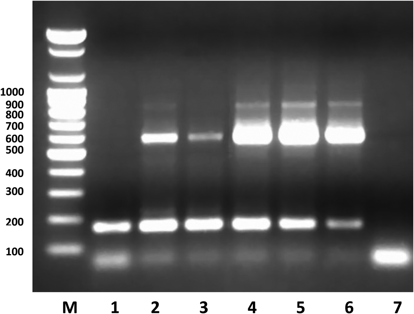
Direct (Lugol and saline solution) and concentration methods (Kato and Willis-Molloy) were used for the isolation of Taenia eggs in fresh faecal samples (Botero & Restrepo, Reference Botero and Restrepo2012). Adult parasites were identified by morphological examination of expelled proglottids. Discrimination between T. saginata and T. solium was made through uterine branch quantification in gravid proglottids and through scolex morphology. For molecular diagnosis, DNA, prepared by extraction from proglottids using Chelex 100 resin, was submitted to a PCR multiplex protocol designed to discriminate between T. saginata and T. solium, (González et al., Reference González, Montero, Harrison, Parkhouse and Gárate2000, Ferrer et al., Reference Ferrer, Useche and Rangel2010). Briefly, DNA extracted was characterized using specific primers: PTs7S35F1 direct primer 5′-CAGTGGCATAGCAGAGGAGGAA-3′, PTs7S35F2 direct primer 5′-CTTCTCAATTCTAGTCGCTGTGGT-3′ and PTs7S35R1 reverse primer 5′- GGACGAAGAATGGAGTTGAAGGT-3′. As negative control, distilled water replaced DNA. Samples of bona fide DNA from T. solium and T. saginata were used as positive controls. The amplification products were evaluated by 2% agarose gel electrophoresis, stained with 0.5 µg/ml ethidium bromide. Identification of T. saginata was based on the identification of the 600 and 170 nucleotide bands. Only the 170 nucleotide component is amplified from DNA of T. solium.
Results and discussion
In this study, we report four confirmed cases of T. saginata taeniasis from Aragua state, Venezuela. The four patients were female and from four different, geographically separated urban municipalities; specifically, patient 1 was from Francisco Linares Alcántara, patient 2 was from Sucre, patient 3 was from Libertador and patient 4 was from Girardot. Although patients 1, 2 and 3 had never been outside their municipalities, patient 4 had been in Colombia for one month a year prior to her treatment.
All the patients had bought their meat at local butchers and had sought medical consultation for the following reasons: (1) expulsion of faecal proglottids (all four patients); (2) abdominal pain, diarrhoea, reflux/dyspepsia and epigastric pain (patients 1, 2 and 4); (3) weight loss (patient 4). Significantly, patients 1, 2 and 3 admitted consuming raw meat during the process of preparing their meals; the other said that she had not knowingly consumed raw meat. All the patients were successfully treated with praziquantel without recurrence up to four months post treatment.
Eggs and gravid proglottids of Taenia spp. were detected in the stools of all four patients. After subsequent treatment, adult worms were expelled by all the patients and measured 1.4–6.0 metres long. Scolices were only recovered from patients 1 and 4. The quantification of uterine branches of the gravid proglottids confirmed the parasite to be T. saginata in all four cases, in agreement with a similar conclusion from the examination of the scolices recovered from patients 1 and 4. Finally, the PCR analysis provided unambiguous identification of T. saginata (fig. 1). Thus, amplification of the DNA of all four patients yielded the 170 and 600 nucleotide components characteristic of T. saginata. (fig. 1).
Amplification products obtained by multiplex PCR from proglottid DNA. Molecular weight marker (M), T. solium DNA positive control (1); T. saginata DNA positive control (2); DNA sample patient 1 (3); DNA sample patient 2 (4); DNA sample patient 3 (5); DNA sample patient 4 (6); negative control (7). The number of nucleotides is given in the ordinal. Nucleotide sequences of 170 and 600 bases are amplified from the DNA of T. saginata, whereas only the 170 base band is amplified from T. solium DNA. Details in Materials and methods.

Post treatment parasitological examination of the stools of patients 1, 2 and 3, detected the presence of Taenia spp. eggs at day 3. Thereafter, all the samples (until four months) were negative for Taenia eggs. Similarly, coprological examination of the patients’ families failed to detect Taenia eggs, although family members of patients 1 and 3 were infected with Trichuris trichura and Blastocystis hominis respectively. The family members of patient 4 did not provide a faecal sample.
In conclusion, we have reported four confirmed cases of T. saginata taeniasis in urban zones of the Aragua state. The fact that each infected individual lives in geographically distinct urbanizations makes it unlikely that infections had a common origin, and suggests a problem in the efficiency of meat inspection that demands urgent attention, as indeed has recently confirmed by Rodrigo Agudo, the food assessor for FEDENAGA (Federación Nacional de Ganaderos, Venezuela) (Notitarde, 31 October 2018).
Author ORCIDs
G. Rojas, 0000-0002-3256-4770; C. Medina, 0000-0001-7613-7327; E. Ferrer, 0000-0002-4173-6642; R.M.E. Parkhouse, 0000-0001-5967-3291; M. Cortez, 0000-0002-2071-9413
Acknowledgements
We thanks to Florencio Mendoza, Dirección General de Salud Ambiental, Ministerio del PPP la Salud, Maracay, Aragua, Departamento de Parasitologia, UC-Sede Aragua, Laboratorio Clinico Ladymar CA, and to Everling Salas and Jesus Perez for their valuable collaboration.
Financial support
This work was supported by Fondo Nacional de Ciencia, Tecnologia e Innovacion, Venezuela, FONACIT (Grants 2012000398 and 2012000395) and Fundación Centro de Investigaciones Biomédicas de la Universidad de Carabobo (FUNDABIOMED-UC).
Conflict of interest
None.
Ethical standards
All procedures performed in this study, involving human participants were in accordance with ethical standards of the institutional and/or national research committee and with the 1964 Helsinki declaration and its later amendments or comparable ethical standards. All the four patients were informed of the goals of the study, and were willing to participate in the evaluation questionnaire and subsequent clinical treatment, and signed an informed consent form.