Introduction
The formation of more than one oocyte within one ovarian follicle is a relatively rare event but known since the 1900s (Hartman, Reference Hartman1926). This phenomenon is known as polyovularity or binovularity in the literature and confirmed by histological investigation of the ovarian tissues (Papadaki, Reference Papadaki1978; Gougeon, Reference Gougeon1981) and laparoscopic egg collection procedures during assisted reproductive treatments (Ron-El et al., Reference Ron-El, Nachum, Golan, Herman, Yigal and Caspi1990; Safran et al., Reference Safran, Reubinoff, Porat-Katz, Werner, Friedler and Lewin1998; Vicdan et al., Reference Vicdan, Isik, Dagli, Kaba and Kisnisci1999; Rosenbusch and Hancke, Reference Rosenbusch and Hancke2012; Cummins et al., Reference Cummins, Koch and Kilani2016). Today the most accepted definition of polyovular follicle is the inclusion of two oocytes within a common zona pellucida or their fusion in the zonal region (Ron-El et al., Reference Ron-El, Nachum, Golan, Herman, Yigal and Caspi1990).
The data regarding the incidence of polyovularity and significance of gametes originated from polyovular follicles are limited in the literature and the low number of cases complicate the ability to make a decision about the fertilization and implantation potential of these oocytes. In a recent case report, it was shown that a genetically normal embryo can result from a conjoined oocyte and subsequent pregnancy and live birth can be achieved (Cummins et al., Reference Cummins, Koch and Kilani2016). However, it has been suggested that polyovular follicles may lead to dizygotic twins, mosaicism and tetraploidy in the embryo (Papadaki, Reference Papadaki1978; Zeilmaker et al., Reference Zeilmaker, Alberda and van Gent1983), hence use of these embryos is not recommended unless no other embryos are available (Cummins et al., Reference Cummins, Koch and Kilani2016).
In the present study, we report one case of a conjoined oocyte and another case with recovery of two separate oocytes in a cumulus cell complex and details of their early embryonic development. We reviewed the literature to evaluate information regarding the origin, incidence and significance of polyovularity in reproductive health.
Case 1
A 27-year-old nulligravida patient with regular menstrual cycles and normal somatic karyotypes attended our sperm donation programme due to their partner’s azoospermia diagnosis. Controlled ovarian stimulation was performed with a gonadotropin-releasing hormone (GnRH) antagonist protocol. Recombinant follicle-stimulating hormone (FSH) (225 IU, Gonal-F; Serono) was administered on day 2 of the menstrual cycle. Ovarian response was monitored with transvaginal ultrasound (TV-USG) and serum oestradiol level. When the leading follicle exceeded 13 mm in diameter, 0.25 mg of GnRH antagonist (Cetrotide; Serono) was started daily until the day of the last trigger. For ovulation induction, 250 μg human chorionic gonadotrophin (hCG; Ovitrelle, Serono) was administered before 36 h of oocyte retrieval. In total, 13 oocytes were retrieved by TV-USG guided aspiration. After denudation, 11 oocytes were at the metaphase-II stage with extruded polar body (MII), one was immature with a germinal vesicle (GV) and one was a binovular zona pellucida containing a mature (MII) and a small-sized immature oocyte (GV, not included in total oocyte count) with connected zona pellucida divided by a thin layer (Fig. 1 A). Intracytoplasmic sperm injection (ICSI) was performed to all mature oocytes including one in binovular zona pellucida. The following day, 10 MII oocytes showed normal fertilization [two pronuclei (2PN) with two polar bodies] including MII of the conjoined oocyte (Fig. 1 B). The embryos were cultured in single-step medium, Continuous Single Culture Complete (CSCM-C) with human serum albumin (Irvine Scientific). The fertilized conjoined oocyte reached the 4-cell and 8-cell stages with equal-sized blastomeres on day 2 (Fig. 1 C) and day 3 (Fig. 1 D), respectively. On day 5, a good quality expanded blastocyst, grade A inner cell mass and grade A trophectoderm (4AA), resulted from the conjoined mature oocyte (Fig. 1 E). In total, four blastocysts were frozen on day 5 including the conjoined oocyte. Two frozen–warmed embryos transferred but did not result in pregnancy. To date, two embryos including the conjoined one have been kept frozen for the next attempt. We are planning to remove the side of the zona pellucida containing the degenerated oocytes before transfer.
Case 1 conjoined oocytes. (A) A mature oocyte (MII) and an immature (GV stage) oocyte (left) on day 0. (B) Day 1, mature oocytes fertilized, displaying two pronuclei (2PN). (C) Day 2, the fertilized oocyte at the 4-cell stage. (D) Day 3, the embryo at the 8-cell stage. (E) Good quality expanded blastocyst developed from the fertilized oocyte and immature conjoined oocyte degenerated on day 5.

Case 2
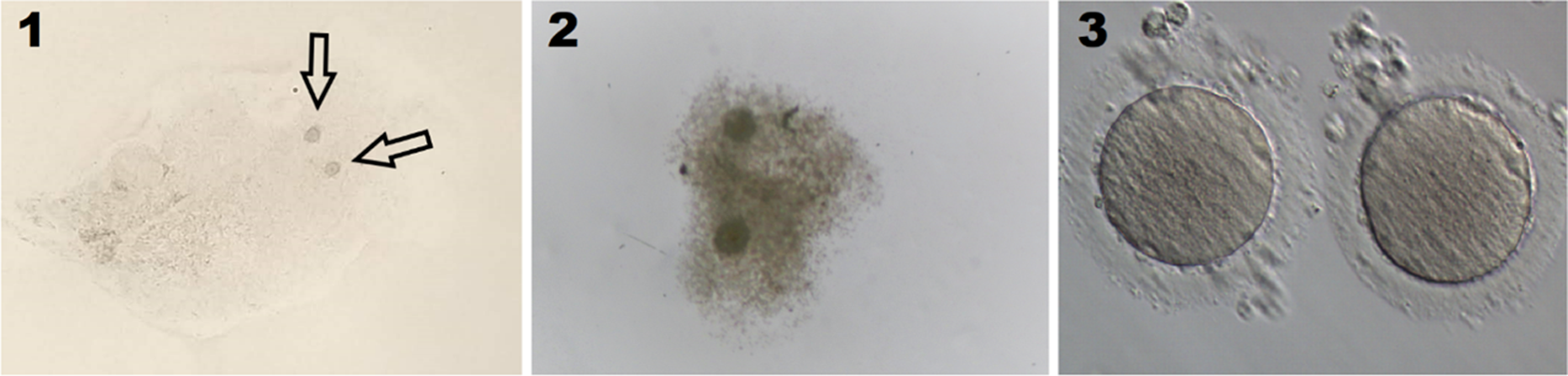
A 23-year-old nulligravid woman attended our IVF programme due to severe male factor. The couples had normal somatic karyotypes. An ICSI procedure was planned to use the preimplantation genetic test for aneuploidy (PGT-A) within trophectoderm biopsy. For ovarian stimulation, the same GnRH antagonist protocol was used as described in Case 1. Seventeen oocytes were retrieved by transvaginal ultrasound-guided needle aspiration after 36 h of hCG administration. In total, 17 oocytes were recovered including two individual oocytes (labelled as twin eggs) in a cumulus cell mass (CCM) (Fig. 2). Before denudation, the CCM with two oocytes was studied by pipetting, to see if the two CCM merged during aspiration. Excess CCM was cut by the tip of the glass pipette for better visualization (Fig. 2–2). The denudation procedure was applied separately for the CCM with the two eggs and others using hyaluronidase (20 IU/ml; Irvine Scientific, USA). Both oocytes in the single CCM were classified as mature (MII) with a slightly fragmented polar body and fragmented perivitelline space (Fig. 2–3). Of the other 15 oocytes, 12 of them were classified as MII stage with similar features as twin eggs and subsequently injected with the partner’s spermatozoa using ICSI. The next day, only one of the twin eggs developed two pronuclei (2PN) (Fig. 3–1). The fertilized twin oocyte then developed to the 4-cell stage on day 2 with unequal cell size, less than 5% central fragmentation (Fig. 3–2). The embryo reached 7-cells on day 3 with unequal cell size and no visible nucleus in some cells (Fig. 3–3). To help herniation, an ∼20 mm hole on the zona pellucida was opened using a laser pulse (OCTAX Navilase) on day 3. On day 5, the embryo was classified as a low-quality hatching blastocyst (Fig. 3–4). Trophectoderm biopsy was performed using the pulling method and 4–6 cells were removed. The biopsied cells were transferred to polymerase chain reaction tubes and kept in a freezer (∼ −20°C) until sent to the genetic laboratory. Due to low quality, this embryo was labelled no. 8 after seven embryos from other oocytes. The couple decided to send the first six good quality embryos for genetic testing at the first round, in which three of them were reported as chromosomally normal but none of them have been transferred to date.
Photographs of two oocytes in a cumulus cell mass at the day of oocyte pick-up. (1) Arrows point to the location of the oocytes. (2) Excess CCM was cut for better visualization. (3) Both oocytes were mature (MII) and inseminated after denudation.

Development of the fertilized oocyte. (1) One of the twin eggs developed two pronuclei. The oocyte reached the 4-cell stage on day 2 (2), 6-cell stage on day 3 (3) and the low-quality hatching blastocyst stage on day 5 (4).

Discussion
In 1926, Hartman was the first to report polyovularity, binovularity and multinuclear follicles based on histological investigation of ovaries, including animals and human (Hartman Reference Hartman1926). Since then, the origin, incidence and importance of polyovularity in reproductive health have been under investigation and attracted great interest.
To date, many possible models explaining the origin of polyovularity have been suggested. Hartman listed three main theories: emergence from polynuclear ova, fusion of adjacent follicles (concrescence theory) and imperfect separation of a portion of Pflieger’s egg tubes (Hartman, Reference Hartman1926). In addition, Collins and Kent (Reference Collins and Kent1964) reported that fluctuation in the FSH and LH ratio may also be related to polyovularity and formation of a polynuclear ova. Similarly, Papadaki (Reference Papadaki1978) suggested that external gonadotrophin use and equal, rather than unequal, division of the primary oocyte may form the secondary oocyte during the first meiotic division. Guillette and Moore (Reference Guillette and Moore2006) discussed the effect of environmental contaminants and gonadotrophin exposure to fertility and linked them to the formation of multi-oocytic follicles. In brief, it is widely accepted that polyovularity is an abnormal follicular development that can easily be affected by gonadotrophin level and environmental contaminants, although there is lack of evidence to show the underlying mechanism (Gougeon, Reference Gougeon1981).
In 1912, Arnold investigated ovaries obtained from 18-year-old women at autopsy and found that 88 larger follicles in ovaries contained more than one oocyte, of which 35 of them contained two oocytes and 53 follicles had more than two oocytes. He quoted Stoeckel (Reference Stoeckel1898) who had previously reported similar findings after evaluating the ovary a 29-year-old woman but that technical conditions at that time should be kept in mind and may have caused bias (Arnold, Reference Arnold1912). It is now well documented that incidences of polyovular and polynuclear follicles are higher in fetuses and young animals, but gradually disappear by undergoing atresia in women up to 40 years of age (Sherrer et al., Reference Sherrer, Gerson and Woodruff1977; Papadaki, Reference Papadaki1978; Gougeon, Reference Gougeon1981). However, there have been reports showing considerable numbers of polyovular follicles in adult ovaries, indicating that some polyovular follicles in younger aged women can even reach to ovulatory phase (Dandekar et al., Reference Dandekar, Martin and Glass1988; Ron-El et al., Reference Ron-El, Nachum, Golan, Herman, Yigal and Caspi1990).
The incidence of polyovularity was evaluated by Gougeon (Reference Gougeon1981) in 36 pairs of ovaries and 81 biopsy samples of adult women (aged between 18–52 years old). It was reported that the relative frequency of polyovular follicles varied between 0.06% and 2.44% and was not age dependent. He also concluded that gonadotrophin hormones, pregnancy or the day of the menstrual cycle did not affect this frequency. In a similar direction, Ron-El et al. (Reference Ron-El, Nachum, Golan, Herman, Yigal and Caspi1990) published the results of 631 oocyte retrievals and reported that 0.3% of oocyte-containing follicles were polyovular. Conversely, Dandekar et al. (Reference Dandekar, Martin and Glass1988) studied the results of 251 laparoscopic egg retrievals following ovarian stimulation during IVF treatment and reported that 8% of aspirated follicles were polyovular, and 24% of laparoscopies contained at least one polyovular follicle. However, the probability for aspiration of more than one follicle by chance was questioned for this study and may explain the higher rates, although the authors claimed that they aspirated each follicle separately. Therefore, the results obtained from histological investigation of ovarian samples and laparoscopic egg retrieval, when considering only the oocytes in an adjacent cumulus–oocyte mass, conjoined oocytes and two oocytes in single common cumulus mass as proof of polyovularity, seem more logical.
In 1983, Zeilmaker et al. (Reference Zeilmaker, Alberda and van Gent1983) suggested that polyovular follicles could cause dizygotic twinning after observation of fertilization and development of two mature oocytes originating from a polyovular follicle. Similarly, as reported in our second case, at least two mature oocytes could be ovulated from a single follicle and possibly lead to a dizygotic twin pregnancy if normally fertilized. It has also been suggested that polyovularity is linked to chimerism (Zeilmaker et al., Reference Zeilmaker, Alberda and van Gent1983), mosaicism, tetraploidy (Hartman, Reference Hartman1926) and teratomas (Sherrer et al., Reference Sherrer, Gerson and Woodruff1977) but the data are limited, and results are inconclusive. At this time, the accepted definition of polyovular follicle is the inclusion of two oocytes within a common zona pellucida or their fusion in the zonal region and the most reliable reason for the formation of binovular follicles is a failure of separation of two individual germ cells in early folliculogenesis (Rosenbusch and Hancke, Reference Rosenbusch and Hancke2012).
Previous cases have revealed asynchronous maturation of conjoined gametes in which the fertilization rate was 61.4% in total (8/13). Unfortunately, none of these fertilized oocytes implanted after transfer. Turkalj et al. (Reference Turkalj, Kotanidis and Nikolettos2013) reported four cases of conjoined oocytes, of which three of them successfully fertilized but embryos arrested at the cleavage stage. The first live birth was reported by Cummins et al. (Reference Cummins, Koch and Kilani2016) after blastocyst transfer of a genetically screened embryo derived from a conjoined oocyte. To our knowledge, a second case resulting in a live birth was reported in 2013 (Yano et al., Reference Yano, Hashida, Kubo, Ohashi, Koizumi, Kageura, Furutani and Yano2017).
The main limitations of the study are the lack of information regarding genetic results of the embryos and pregnancy outcomes. Patients have frozen embryos developed from oocytes with normal morphology as well as oocytes from polyovular follicles. Due to limited data on safety, our first thought was the tendency to use other embryos first. This information is not available at present and may not be in the near future. Moreover, these embryos may not be used at all, depending on the results of previous attempts.
In conclusion, it is clear that more than one oocyte can be obtained from a single follicle and that these are capable of developing into healthy offspring. Although there has been no report showing that both oocytes had successfully fertilized and developed, the possibility of twin pregnancy originated from polyovular follicles cannot be eliminated. It is not known if the oocytes were genetically identical or not. In light of results from published and current cases, we concluded that these oocytes originated from abnormal folliculogenesis and that transfer of these embryos should be avoided unless other embryos were not available and that they must be genetically screened before transfer if possible.
Financial support
This research received no specific grant from any funding agency, commercial or not-for-profit sectors.
Declaration of interest
All authors declare that they have no conflict of interests in the article.
Ethical standards
None