Introduction
The in vitro growth, maturation and fertilization of oocytes from preantral follicles represent a great potential for assisting in the in vitro production of mammalian embryos. The collection of sperm samples, oocytes and embryos is an aspect of great importance in the development of these technologies (García-Álvarez et al., Reference García-Álvarez, Maroto-Morales, Berlinguer, Fernández-Santos, Esteso, Mermillod, Ortiz, Ramon, Pérez-Guzmán, Garde and Soler2011). One of the most important problems is the transport of ovaries to the laboratory from long distances (Evecen et al., Reference Evecen, Cirit, Demir, Ozdas, Tas, Birler and Pabuccuiglu2010). Type of medium used and storage period as well as temperatures used during transportation of the ovaries are among the factors affecting subsequent follicular survival, development and complete maturation.
Techniques for short-term storage of ovaries were developed for several species, such as ovine (Matos et al., Reference Matos, Andrade, Lucci, Báo, Silva, Santos, Ferreira, Costa, Celestino and Figueiredo2004), caprine (Silva et al., Reference Silva, Lucci, Carvalho, Báo, Costa, Santos and Figueiredo2000; Chaves et al., Reference Chaves, Martins, Saraiva, Celestino, Lopes, Correia, Lima Verde, Matos, Báo, Name, Campello and Figueiredo2008), bovine (Celestino et al., Reference Celestino, Santos, Martins, Matos, Figueiredo, Costa, Silva and Rodrigues2007), swine (Wongsrikeao et al., Reference Wongsrikeao, Otoi, Karja, Agung, Nii and Nagai2005) and equine (Love et al., Reference Love, Choi, Love, Varner and Hinrichs2003). Most of these studies evaluated only the morphological features of preantral follicles through histological analysis, after storage of ovarian tissue in different temperatures, periods and media (Matos et al., Reference Matos, Andrade, Lucci, Báo, Silva, Santos, Ferreira, Costa, Celestino and Figueiredo2004; Celestino et al., Reference Celestino, Santos, Martins, Matos, Figueiredo, Costa, Silva and Rodrigues2007). However, additional measurements of follicular viability and developmental competence, such as in vitro preantral follicle culture, were performed only in caprine species (Chaves et al., Reference Chaves, Martins, Saraiva, Celestino, Lopes, Correia, Lima Verde, Matos, Báo, Name, Campello and Figueiredo2008). These authors have shown that goat ovarian tissue transportation in minimal essential medium (MEM) at 4°C for up to 4 h maintained the percentages of morphologically normal follicles similar to those observed in control tissues and also kept follicular viability after in vitro culture.
Although preservation at low temperatures may be recommended for long periods, some authors have used temperatures close to physiological values (30–37°C) to transport ovaries to the laboratory (less than 3 h) for further oocyte harvesting and in vitro maturation (IVM) studies (Shirazi et al., Reference Shirazi, Ostad-Hosseini, Ahmadi, Heidari and Shams-Esfandabadi2009; Wan et al., Reference Wan, Hao, Zhou, Wu, Yang, Cui, Lui and Zeng2009). In swine, oocyte meiotic competence may be maintained when ovaries are stored at 25°C for 2 h and at 35°C for 2 and 4 h (Tellado et al., Reference Tellado, Alvarez, Dalvit and Cetica2014). A recent experiment has demonstrated that the cleavage rate for the oocytes (aspirated from 2 to 6 mm follicles) collected from ovine ovaries stored at 5–8°C for 12 h was significantly lower (23%) in relation to ovaries transported at 30–35°C for 4 h (47%) (García-Álvarez et al., Reference García-Álvarez, Maroto-Morales, Berlinguer, Fernández-Santos, Esteso, Mermillod, Ortiz, Ramon, Pérez-Guzmán, Garde and Soler2011). In other previous studies, some authors have demonstrated that it is possible to maintain about 50–60% of morphologically normal follicles after the transport of ovine ovarian tissue in physiological temperature (39°C) for only 2 or 4 h (Andrade et al., Reference Andrade, Rodrigues, Amorim, Carvalho, Dode and Figueiredo2002; Matos et al, Reference Matos, Andrade, Lucci, Báo, Silva, Santos, Ferreira, Costa, Celestino and Figueiredo2004). However, they did not evaluate whether this storage may affect the apoptosis rate or the ability of ovine preantral follicles to grow in vitro. In addition, MEM, which has been usually used as a basic culture medium for preantral follicles, was not evaluated as a storage medium for ovine ovaries. Moreover, as reported previously, addition of supplements (e.g. ITS, glutamine, hypoxanthine, and BSA) into the medium could maintain ovarian follicle viability (Silva et al., Reference Silva, van den Hurk, Costa, Andrade, Nunes, Ferreira, Lôbo and Figueiredo2004). Therefore, we hypothesized that an enriched medium (MEM with supplements) could help maintain the viability and developmental competence of preantral follicles enclosed in ovaries stored in near-physiological temperatures for longer periods of transportation.
Studies with sheep are important because they are present on all continents and are commercially seen as highly attractive livestock. Moreover, the use of preantral follicles enclosed in ovarian tissue may help to improve our knowledge about the factors that control early folliculogenesis in mammals (Santos et al., Reference Santos, Menezes, Barberino, Macedo, Lins, Gouveia, Barros, Santos, Gonçalves and Matos2014). Thus, the objectives of this study were to evaluate the effect of storing preantral follicles enclosed in ovine ovarian tissue under different media and periods at a temperature close to physiological values, and to test the ability of these follicles to survive and grow in vitro after being stored. Additionally, the effectiveness of the addition of supplements to preservation medium was studied.
Materials and methods
Unless otherwise mentioned, culture media, supplements and chemicals used in the present study were purchased from Sigma Chemical Co. (St. Louis, MO, USA).
Source of ovaries
Ovine ovaries (n = 10) were obtained at a local slaughterhouse from adult mixed-breed sheep (n = 5). Immediately after death, the ovaries were washed once in 70% alcohol (Dinâmica, São Paulo, Brazil) for 10 s and then twice in minimal essential medium (MEM) containing HEPES and antibiotics (100 μg/ml penicillin and 100 μg/ml streptomycin).
Experimental protocol
At the slaughterhouse, the pair of ovaries from each animal was divided into nine slices approximately 3 × 3 mm (1 mm thick). For each animal, one slice of tissue was randomly selected and immediately fixed for histological analysis (fresh control). The remaining eight slices of ovarian cortex were placed individually in cryotubes containing 2 ml of preservation medium (MEM or MEM+) and stored (simulating transport) at 35ºC for 6 or 12 h. The fragments of ovaries were transported to the laboratory using an oocyte transporter12-Compact® (Wtavet, São Paulo, Brazil). After the transportation periods, four fragments were fixed for morphological and TUNEL analysis (non-cultured fragments) and four fragments were cultured in vitro for 5 days. The preservation media were MEM (the same used for washing the ovaries, containing 100 μg/ml penicillin and 100 μg/ml streptomycin) or MEM supplemented with ITS (0.1 mg/ml insulin, 0.055 mg/ml transferrin and 0.5 μg/ml sodium selenite), 2 mM glutamine, 2 mM hypoxanthine, 3 mg/ml BSA, 50 μg/ml ascorbic acid, 50 ng/ml recombinant follicle stimulating hormone (FSH) (Nanocore, São Paulo-Brazil), 100 μg/ml penicillin and 100 μg/ml streptomycin. The latter medium was named MEM+. The pH of the media were monitored before and after the preservation periods (pH meter – PG2000, Gehaka, São Paulo, Brazil).
The fragments were cultured individually in 1 ml of culture medium in 24-well culture dishes for 5 days at 39°C in an atmosphere of 5% CO2 in air. The basic culture medium consisted of α-MEM (pH 7.2–7.4; GIBCO, New York, USA) supplemented with ITS (0.1 mg/ml insulin, 0.055 mg/ml transferrin and 0.5 μg/ml sodium selenite), 0.23 mM pyruvate, 2 mM glutamine, 2 mM hypoxanthine, 1.25 mg/ml BSA, 50 μg/ml ascorbic acid, 50 ng/ml rFSH (Nanocore, São Paulo, Brazil), 100 μg/ml penicillin and 100 μg/ml streptomycin. Each treatment was repeated five times, thus using the ovaries of five different animals. The culture medium was stabilized at 39°C for 2 h, in an atmosphere of 5% CO2 in air, prior to use and was replenished every second day.
Morphological analysis and assessment of in vitro follicular growth
Tissues from all treatments (fresh control, preserved for 6 or 12 h or preserved and then cultured for 5 days) were fixed in 4% buffered formaldehyde (Dinâmica) for 18 h and then dehydrated in increasing concentrations of ethanol (Dinâmica). After paraffin embedding (Dinâmica), the ovine tissue pieces were cut into 5-μm sections, and every section was mounted on glass slides and stained with Periodic Acid Schiff reagent and contrasted with hematoxylin (Dinâmica). Follicle stage and survival were examined by microscopy (Nikon, Tokyo, Japan) at ×400 magnification.
The developmental stages of preantral follicles have been defined previously as follows (Silva et al., Reference Silva, van den Hurk, Costa, Andrade, Nunes, Ferreira, Lôbo and Figueiredo2004): primordial (one layer of flattened granulosa cells around the oocyte) or growing follicles (intermediate: one layer of flattened to cuboidal granulosa cells; primary: one layer of cuboidal granulosa cells, and secondary: two or more layers of cuboidal granulosa cells around the oocyte and no sign of antrum formation). Additionally, these follicles were classified individually as histologically normal when an intact oocyte was present and surrounded by granulosa cells that were well organized in one or more layers and have no pyknotic nuclei. Atretic follicles were defined as those with a retracted oocyte, pyknotic nucleus, and/or disorganized granulosa cells detached from the basement membrane. Overall, 150 follicles were evaluated for each treatment (30 follicles per treatment-replicate × 5 replicates = 150 follicles).
To evaluate follicular activation (transition from primordial to growing follicles, when surrounding flattened granulosa cells become cuboidal and begin to proliferate) and growth, only morphologically normal follicles with a visible oocyte nucleus were recorded, and the proportion of primordial and growing follicles was calculated at day 0 (fresh control), after 6 or 12 h of preservation and after 5 days of culture. In addition, from the basement membrane, major and minor axes of each oocyte and follicle were measured using Image-Pro Plus® software. The average of these two measurements was used to determine the diameters of both the oocyte and the follicle.
Detection of apoptotic cells by TUNEL assay
Terminal deoxynucleotidyl transferase (TdT) mediated dUTP nick-end labeling (TUNEL) assay was used for a more in-depth evaluation of ovine preantral follicle quality before and after preservation and/or culture in vitro. TUNEL was performed using a commercial kit (In Situ Cell Death Detection Kit, Roche Diagnostics Ltd., Indianapolis, USA) following the manufacturer's protocol, with some modifications (Santos et al., Reference Santos, Menezes, Barberino, Macedo, Lins, Gouveia, Barros, Santos, Gonçalves and Matos2014). Briefly, sections (5 μm) mounted on glass slides were deparaffinized and rehydrated through graded alcohols, then rinsed in PBS (pH 7.2). Antigen retrieval by microwave treatment was performed in sodium citrate buffer (pH 6.0; Dinâmica) for 6 min. Endogenous peroxidase activity was blocked by 3% H2O2 (Dinâmica) in methanol (Dinâmica) at room temperature for 10 min. After rinsing in Tris buffer (Dinâmica), the sections were incubated with TUNEL reaction mixture at 37°C for 1 h. Then, the specimens were incubated with Converter-POD in a humidified chamber at 37°C for 30 min. The DNA fragmentation was revealed by incubation of the tissues with diaminobenzidine (DAB; 0.05% DAB in Tris buffer, pH 7.6, 0.03% H2O2) during 1 min. Finally, sections were counterstained with Harry's haematoxylin in a dark chamber at room temperature for 1 min, dehydrated in ethanol, cleared in xylene, and mounted with balsam (Dinâmica). For negative controls, slides were incubated with label solution (without TdT enzyme) instead of TUNEL reaction mixture.
Only follicles that contained an oocyte nucleus were analyzed for apoptotic assay. The number of brown TUNEL positive cells (oocyte and granulosa cells) was counted in 10 randomly fields per treatment using Image-Pro Plus® software. The percentage of TUNEL positive or apoptotic cells was calculated as the number of apoptotic cells out of the total number of cells.
Statistical analysis
Percentages of morphologically normal, primordial and developing follicles were submitted to analysis of variance (ANOVA) test and the Tukey's test was applied for comparison among treatments. Values of pH and apoptotic cells were submitted to chi-squared test and differences were considered to be statistically significant when P < 0.05. The results of follicular survival and growth were expressed as the mean ± standard deviation (SD) and the results of follicular activation were expressed as the mean ± standard error of the mean (SEM).
Results
Effect of preservation conditions on follicular morphology before and after in vitro culture
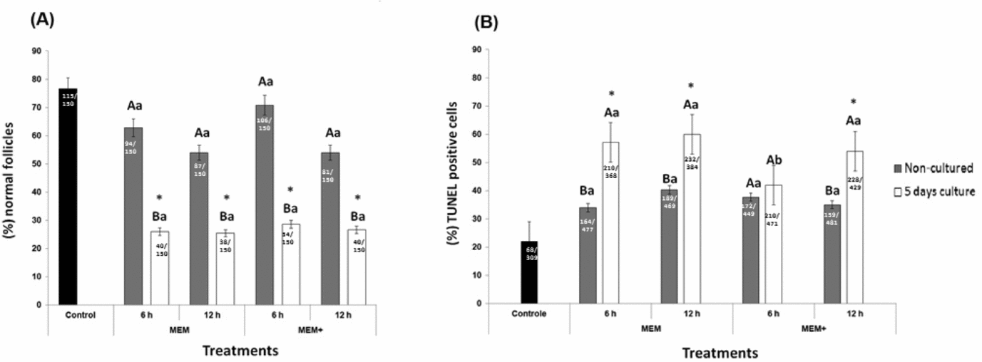
Among the 1350 preantral follicles analyzed after in vitro preservation and culture of ovine ovarian tissue in different media and periods, 572 were primordial, 677 intermediate, 73 primary and 28 secondary follicles. The percentages of morphologically normal preantral follicles in fresh tissues (control) and after preservation in MEM or MEM+ for 6 or 12 h and further culture are shown in Fig. 1 A. Preservation of ovarian slices in MEM or MEM+ (non-cultured), independently of the time period, resulted in similar percentages (P > 0.05) of morphologically normal follicles when compared with the fresh control. Nevertheless, compared with the fresh control tissue, a decrease (P < 0.05) in the percentage of normal follicles was observed in tissues cultured for 5 days after being preserved in both media and for different periods. Additionally, the media did not show differences in follicular survival after 5 days of culture (P > 0.05).
Percentages of (A) morphologically normal ovine preantral follicles, and (B) TUNEL-positive cells in the fresh control, after preservation for 6 or 12 h (non-cultured) and after 5 days of culture. *Differs significantly from fresh control (P < 0.05). A,BDifferent letters denote significant differences between treatments in the same preservation medium and period (non-cultured × cultured) (P < 0.05). a,bDifferent letters denote significant differences between preservation periods in the same media and under the same treatment (P < 0.05).

For pH evaluation, the mean pH values for the MEM or MEM+ after 6 h of preservation were 7.38 ± 0.21 and 7.36 ± 0.32 respectively. The storage of ovarian fragments for 12 h did not result in changes (P > 0.05) of pH in either solution (7.27 ± 0.38 and 7.24 ± 0.31 for MEM and MEM+, respectively).
Apoptotic cell detection
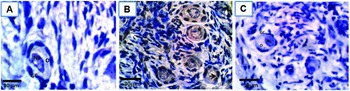
Figure 1 B shows the percentage of TUNEL detection in ovine preantral follicles after preservation and/or culture. TUNEL-positive cells increased (P < 0.05) after 5 days of culture in ovarian tissues preserved in MEM for both periods and in those preserved in supplemented MEM+ for 12 h when compared with fresh control. Only for tissues preserved in supplemented medium (MEM+) for 6 h, was the percentage of TUNEL-positive cells similar (P > 0.05) between non-cultured tissues and tissues cultured for 5 days. In addition, after culture, there was a significant increase in the percentage of apoptotic cells in ovarian tissue preserved for 12 h in MEM+, compared with those preserved for 6 h in this medium. Apoptotic cells were noticed in the oocyte, granulosa, stromal (Fig. 2) and theca cells (data not shown).
Apoptosis detection using TUNEL assay in the different ovine ovarian cell types. Normal primordial follicles in the fresh control (A); apoptotic primordial follicles preserved for 6 h in MEM followed by 5 days of culture (B); negative control (C). In (B), note the apoptotic oocyte (*), and granulosa cell (arrow). GC, granulosa cell; Nu, oocyte nucleus; O = oocyte; ×400 magnification.

Follicular activation and growth during in vitro preservation and culture
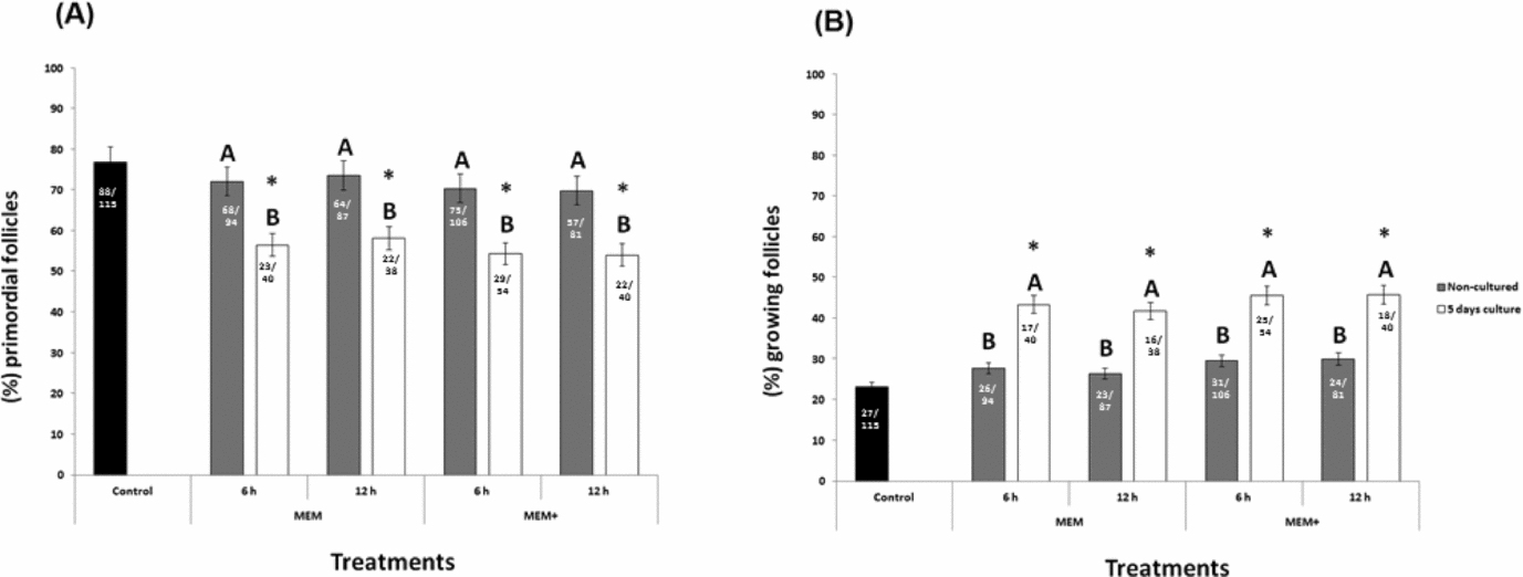
The percentage of primordial and growing follicles (intermediate, primary, and secondary) in fresh tissue or in tissues preserved for 6 or 12 h in MEM or MEM+ (non-cultured), and cultured for 5 days is shown in Fig. 3. Fresh ovarian tissues predominantly contained primordial (76.8%) follicles. No change in the percentage of primordial or growing follicles was observed between fresh and preserved (non-cultured) tissues (P > 0.05). Under all culture conditions, a decrease in the percentage of primordial follicles was observed concomitant with an increase (P < 0.05) in the percentage of developing follicles compared with the fresh control and preserved (non-cultured) tissues.
Percentages (mean ± standard error of the mean (SEM)) of primordial (A) or growing (B) follicles in the fresh control, after preservation for 6 or 12 h (non-cultured) and after 5 days of culture. *Differs significantly from fresh control (P < 0.05). A,BDifferent letters denote significant differences between treatments in the same preservation medium and at the same time period (P < 0.05).

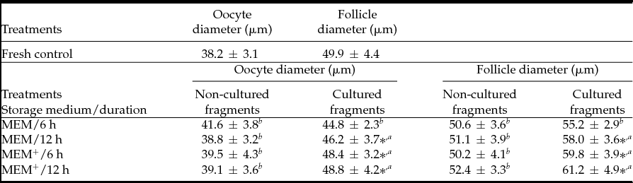
Follicle and oocyte diameters were measured and are shown in Table 1. After 5 days of culture, tissues preserved in MEM for 12 h or in supplemented medium (MEM+) for both preservation periods increased (P <0.05) follicular and oocyte diameters when compared with fresh control and other treatments.
Mean oocyte and follicular diameter (mean ± SD) in the fresh control, after preservation for 6 or 12 h and after 5 days of culture

*Differs significantly from fresh control (P < 0.05).
a,b Different letters denote significant differences within the column (P < 0.05). SD, standard deviation.
Discussion
In the present study, we evaluated for the first time the effect of preserving ovine ovarian cortex in an oocyte transporter, using the temperature of 35°C (close to physiological temperature in the sheep) and in different storage media (with or without supplements) and time periods (6 or 12 h). This study could be very useful in situations where laboratories are distant from the farms or slaughterhouses, allowing the recovery of good quality follicles for cryopreservation and/or in vitro embryo production systems.
In our study, preservation (without culture) of ovarian cortex in MEM or supplemented MEM+ at 35°C for up to 12 h resulted in similar percentages of normal follicles as those found in the fresh control. Conversely, authors have demonstrated previously that short-term storage (4 h) of ovine ovarian tissue in physiological temperatures (39ºC) increases the percentage of atretic preantral follicles (Andrade et al., Reference Andrade, Rodrigues, Amorim, Carvalho, Dode and Figueiredo2002; Matos et al., Reference Matos, Andrade, Lucci, Báo, Silva, Santos, Ferreira, Costa, Celestino and Figueiredo2004). Therefore, the results of our study can be considered better than those reported previously by our team (Andrade et al., Reference Andrade, Rodrigues, Amorim, Carvalho, Dode and Figueiredo2002; Matos et al., Reference Matos, Andrade, Lucci, Báo, Silva, Santos, Ferreira, Costa, Celestino and Figueiredo2004) as about 71% and 54% of the follicles were morphologically normal after storage of ovaries in a supplemented medium at 35°C for up to 6 and 12 h, respectively. It is likely that our preservation medium (MEM) is richer in nutrients, such as amino acids, glucose and vitamins, than those used previously for ovine tissues, which may have improved follicular survival. In addition, the oocyte transporter used for preserving ovarian tissue kept the temperature constant (35ºC) throughout the conservation period, which could help in the maintenance of normal follicle morphology. Furthermore, the satisfactory rates of normal follicles after ovary transport may also be associated with the maintenance of pH values. Andrade et al. (Reference Andrade, Rodrigues, Amorim, Carvalho, Dode and Figueiredo2002) suggested that the resulting drop in pH after storage at 39°C could have caused follicular injuries, with the release of intracellular contents. Our finding is important as some authors have shown that near-physiological temperature is more desirable for the transportation of caprine viable cumulus–oocyte complexes (COCs) and fresh embryos to the laboratory (Shirazi et al., Reference Shirazi, Ostad-Hosseini, Ahmadi, Heidari and Shams-Esfandabadi2009; Quan et al., Reference Quan, Zhiping, Zhixing, Song, Min, Xiao'e and Yong2010). In previous studies, ovaries have also transported been to the laboratory at approximately 30–37°C (Shirazi et al., Reference Shirazi, Ostad-Hosseini, Ahmadi, Heidari and Shams-Esfandabadi2009; Wan et al., Reference Wan, Hao, Zhou, Wu, Yang, Cui, Lui and Zeng2009) and some of the studies used an oocyte transporter for ovarian tissue storage (Byrd et al., Reference Byrd, Flores-Foxworth and Westhusin1997; Love et al., Reference Love, Choi, Love, Varner and Hinrichs2003).
Morphological assessment of follicular integrity has been largely used to evaluate the effectiveness of various treatments (media, temperature and time of preservation) to which ovarian follicles are submitted (Matos et al., Reference Matos, Andrade, Lucci, Báo, Silva, Santos, Ferreira, Costa, Celestino and Figueiredo2004; Celestino et al., Reference Celestino, Santos, Martins, Matos, Figueiredo, Costa, Silva and Rodrigues2007). However, additional measurements such as in vitro follicular development may be used as a reliable evaluation of follicles and/or oocyte viability (Abd-Allah, Reference Abd-Allah2010). In the current study, the percentage of morphologically normal follicles was reduced when ovaries were stored in both media (supplemented or not) and for different preservation periods (6 or 12 h) and then cultured for 5 days. Similar to our results, other authors have shown a decrease in morphological normal follicles after the transport of caprine ovaries at 32°C and then were further cultured for 5 days (Silva et al., Reference Silva, van den Hurk, Costa, Andrade, Nunes, Ferreira, Lôbo and Figueiredo2004; Martins et al., Reference Martins, Van den Hurk, Santos, Silva, Matos, Celestino, Rodrigues, Pessoa, Ferreira and Figueiredo2005). The degeneration rates observed in the present study after 5 days can be attributed partially to the culture conditions. In addition to FSH and ascorbic acid, it is likely that the presence of other components in the culture medium (for example, other hormones and/or growth factors) could help to maintain the viability of the follicles.
Although histological analysis of atresia in follicles remains the most classic and reliable method, the TUNEL assay happens to be a very sensitive method for in situ visualization of apoptosis at the cellular level (Sreejalekshmi et al., Reference Sreejalekshmi, Raghavendra, Subramani, Murthy, Jamuna, Prasad, Ravindra and Selvaraju2011). Our results demonstrated that only ovarian tissues preserved in supplemented medium (MEM+) for 6 h did not increase the percentages of TUNEL-positive cells, which were similar between non-cultured tissues (only preserved) and tissues cultured for 5 days (preserved and cultured). Previous studies have shown that the addition of supplements (Saha et al., Reference Saha, Shimizu, Geshi and Izaike2000; Silva et al., Reference Silva, van den Hurk, Costa, Andrade, Nunes, Ferreira, Lôbo and Figueiredo2004; Peng et al., Reference Peng, Yang, Wang, Tong and Guo2010) into the culture medium could inhibit apoptosis and maintain ovarian follicle viability. Among the supplements tested, selenium improved in vitro development of follicles through enhancement of granulosa cells proliferation, increasing follicular total antioxidant capacity and glutathione peroxidase activity, as well as decreasing reactive oxygen species (Abedelahi et al., Reference Abedelahi, Salehnia, Allameh and Davoodi2010). In addition, glutamine is an effective energy substrate for oocytes (Songsasen et al., Reference Songsasen, Spindler and Wildt2007). Moreover, BSA promoted the maintenance of follicular structure and survival during in vitro culture (Rodrigues et al., Reference Rodrigues, Silva, Faustino, Bruno, Magalhães, Campello and Figueiredo2010). Therefore, the supplementation of the medium may be correlated with the reduction in apoptosis seen in the ovaries preserved in MEM+ for up to 6 h. In addition, it is likely that the increase in the storage period could have caused a depletion in intracellular energy sources, followed by consumption of the nutrients and oxygen available in the preservation medium (Matos et al., Reference Matos, Andrade, Lucci, Báo, Silva, Santos, Ferreira, Costa, Celestino and Figueiredo2004), resulting in the higher apoptotic rates found in tissues preserved for 12 h in supplemented MEM+ followed by 5 days of culture. Preservation of ovarian follicles from apoptotic processes would allow the collection of oocytes with the improved qualities that are necessary for sustaining fertilization and the early phases of embryogenesis.
Follicular activation and growth (follicular and oocyte diameter) were higher in cultured tissues than in fresh control or non-cultured tissues, except those from fragments preserved for 6 h in MEM and then cultured for 5 days, in which no growth was observed. These results demonstrated that storage of ovarian tissue under those conditions did not affect the ability of ovine preantral follicles to grow in vitro. Byrd et al. (Reference Byrd, Flores-Foxworth and Westhusin1997) have transported ovine oocytes at 39°C in a portable incubator, a type of oocyte transporter, and have observed maturation, fertilization, and developmental rates of oocytes similar to those of oocytes matured in a conventional incubator.
In conclusion, ovine ovarian tissue was successfully preserved in supplemented medium (MEM+) at a temperature close to physiological values (35°C) for up to 6 h without affecting the apoptosis rate in the ovarian follicles and their ability to develop in vitro. The lower apoptotic rate in ovarian follicles preserved in supplemented medium is an indicator of good oocyte quality, in terms of a greater capacity to be fertilized and to produce embryos. Therefore, the preservation of ovarian fragments during transportation under these conditions will be useful in the future to optimize the use of oocytes enclosed in preantral follicles for reproductive proposes.
Acknowledgements
This work was supported by Conselho Nacional de Desenvolvimento Científico e Tecnológico (CNPq) and Fundação de Amparo à Ciência e Tecnologia do Estado de Pernambuco (FACEPE). Rodrigo José de Sousa Gonçalves is a recipient of a grant from CAPES (Brazil). We would like to acknowledge the generous donation of rFSH made by Dr. José Ricardo de Figueiredo of the State University of Ceará, Brazil. The authors would also like to especially thank MSc Seldon Almeida de Souza for his assistance in the development of this study.
Declaration of interest
None of the authors has any conflict of interest to declare.