Implications
Mammary stem cells (MaSC) provide for net growth, renewal and turnover of mammary epithelial cells, and are therefore potential targets for strategies to increase production efficiency. Appropriate regulation of MaSC can potentially benefit milk yield, persistency, dry period management and repair of mammary tissue damaged by mastitis. This paper provides an overview of current knowledge of MaSC, addresses the relevance of MaSC biology to the efficiency of milk production in dairy cows and discusses recent attempts to regulate and characterize bovine MaSC.
Introduction
This paper provides an overview of current knowledge of mammary stem cells (MaSC) and addresses the relevance of MaSC biology to the efficiency of milk production in dairy cows. Milk yield is a function of the number and the secretory activity of mammary epithelial cells (MEC). Because proliferation, turnover and regeneration of the mammary epithelium are attributed to the proliferation and differentiation of MaSC and their progenitor progeny, the number and activity of mammary secretory cells are determined by MaSC function (Capuco and Ellis, Reference Capuco and Ellis2005).
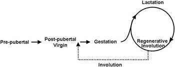
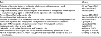
In female mammals, growth and development of the mammary glands occur primarily postnatally, with their function in the mature animal being tightly coupled with reproductive strategy. This dictates cycles of mammary growth, differentiation, lactation and regression (Figure 1). Mice have provided the primary model for study of mammary growth and development for reasons that include: short generation time, low cost and availability of animal models for tissue transplantation and genetic modification (transgenic and gene knockout). Although much can be learned about mammary gland biology from studies with mice, differences between species ultimately necessitate careful evaluation of the applicability of research in one species to other species (Capuco and Akers, Reference Capuco and Akers1999; Capuco et al., Reference Capuco, Ellis, Wood, Akers and Garrett2002a and Reference Capuco, Annen, Fitzgerald, Ellis and Collier2006). Investigations of bovine MaSC biology have been limited, using in vivo and in vitro approaches (Table 1).
Mammary development and lactation cycles. Lactation is linked to reproductive cycles and ceases after weaning or discontinuation of milking. After forced weaning of nonpregnant mice, the murine mammary gland undergoes extensive involution after which it resembles the gland of a virgin mouse (dotted arrow, involution). Milking of dairy cows is typically terminated during the final 2 months of gestation, resulting in extensive mammary cell turnover and minimal regression (termed regenerative involution) in preparation for the onset of the subsequent lactation.

Publications addressing aspects of bovine MaSC or progenitor cells

MaSC = mammary stem cells.
This paper addresses in vivo studies of bovine MaSC and progenitor cells during early postnatal mammary development. Because most of the stem cell and MaSC literature has addressed the biology of murine and human stem cells, research addressing bovine MaSC is discussed within the context of principles reasoned from research in other species.
Stem cells and progenitor cells: types and properties
Stem cells are capable of self-renewal and providing for the generation of multiple cell lineages (stemcells.nih.gov/info/glossary.asp). Potency terms are used to distinguish among stem cells that differ in their capacities to generate cells of multiple lineages. The fertilized egg and the progeny of its first several divisions are said to be totipotent, or omnipotent, in that they are capable of generating all embryonic and extra-embryonic cell types that are necessary to produce a viable organism. Of course, cleavage of these cells (natural or artificial) provides for multiple identical offspring.
The two primary categories of stem cells are embryonic stem cells and adult (somatic) stem cells. Embryonic stem cells have been isolated from the inner cell mass of the blastocyst (Evans and Kaufman, Reference Evans and Kaufman1981; Martin, Reference Martin1981). They are termed pluripotent because they are capable of generating cells of all the three germ layers of the organism: ectoderm, mesoderm and endoderm, and hence all the tissues of the adult organism. Adult stem cells are found in the tissues of an ‘adult’ organism and their regenerative potentials, at least under normal circumstances, are more limited than those of embryonic stem cells. These cells are capable of forming the specialized cell types found within the tissue in which they reside. Consequently, they are typically multipotent (i.e. capable of differentiating into cells of related lineages) and are often named on the basis of the tissue in which they reside (e.g. epithelial, mesenchymal). It is generally accepted that adult stem cells are responsible for growth, differentiation and maintenance of the tissues in which they reside (Jones and Watt, Reference Jones and Watt1993; Cosentino et al., Reference Cosentino, Shaver-Walker and Heddle1996; Fliedner, Reference Fliedner1998). The MaSC are stem cells for the epithelial component of the mammary gland; in addition, there are mesenchymal stem cells that provide for the stromal (fat pad) lineages of this organ.
A third category of artificially produced stem cells, known as induced pluripotent stem cells (iPSC), has received considerable interest since development of procedures for their production from somatic mouse (Takahashi and Yamanaka, Reference Takahashi and Yamanaka2006) and from human fibroblasts (Takahashi et al., Reference Takahashi, Okita, Nakagawa and Yamanaka2007; Yu et al., Reference Yu, Vodyanik, Smuga-Otto, Antosiewicz-Bourget, Frane, Tian, Nie, Jonsdottir, Ruotti, Stewart, Slukvin and Thomson2007). Wilmut et al.(Reference Wilmut, Schnieke, McWhir, Kind and Campbell1997) had previously demonstrated that factors in the oocyte could reprogram the nuclei of differentiated somatic cells to an undifferentiated state. More recent studies inducing pluripotency used retroviral transfections to express key genes that are essential and those that facilitate the production of pluripotent cells. Because iPSC can be derived from adult cells, they provide an alternative to harvesting pluripotent stem cells from embryos. Methods for derivation of the cells without the need for viral transfection, which may cause mutagenesis, are under investigation (Zhou et al., Reference Zhou, Wu, Joo, Zhu, Han, Lin, Trauger, Bien, Yao, Zhu, Siuzdak, Scholer, Duan and Ding2009; Desponts and Ding, Reference Desponts and Ding2010; Jia et al., Reference Jia, Wilson, Sun, Gupta, Huang, Li, Panetta, Chen, Robbins, Kay, Longaker and Wu2010). This research has shed light on critical genes necessary to maintain cells in an undifferentiated state and alterations in the epigenetic landscape associated with various states of cell differentiation.
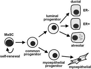
Stem cells undergo two types of mitotic division, symmetric and asymmetric division. When a stem cell undergoes symmetric division, it gives rise to two daughter stem cells and promotes expansion of the stem cell population. When a stem cell undergoes asymmetric division, it gives rise to a stem cell (self-renewal) and a progenitor cell. A progenitor cell has more restricted renewal capacity than a stem cell and has frequently more restricted differentiation potential. Although a somatic stem cell is functionally viable for the life span of the organism, progenitor cells have limited proliferation capacity and eventually undergo senescence. A depiction of a minimalist hypothetical MaSC lineage, based on research in the mouse, is depicted in Figure 2. Salient features include the production of multiple progenitor cells that ultimately differentiate into the various categories of epithelial cells (EC) and myoepithelial cells within the mature gland. Another feature of cell lineage in a variety of tissues is the greater proliferative activity of progenitor cells than the parental stem cell. Frequently, stem cells are relatively inactive and the proliferative activity of the tissue is met by the stem cell activity, leading to an increase in the number of the progenitor cells, which in turn actively proliferate. In the words of one researcher, progenitor cells may ‘work while stem cells sleep’ (Jones and Simons, Reference Jones and Simons2008). Because of the need to expand the MaSC and progenitor cell compartments in proportion to duct growth, MaSC are quite proliferative during periods of rapid duct growth.
Mammary epithelial hierarchy as proposed and adapted from Visvader and Lindeman (Reference Visvader and Lindeman2006). MaSC = mammary stem cells; ER = estrogen receptors.

Enthusiasm for stem cell research frequently relates to the potential use of stem cells in human regenerative medicine (Mimeault et al., Reference Mimeault, Hauke and Batra2007). Proponents envision a time when diseases such as diabetes, Parkinson's, amyotrophic lateral sclerosis and traumatic injuries such as spinal cord damage, are treatable with stem cell-based therapies. This vision is not unreasonable, as bone marrow transplants have been used for many years to regenerate defective hematopoietic tissue.
Regenerative medicine also has a place in animal and veterinary science (Pacini et al., Reference Pacini, Spinabella, Trombi, Fazzi, Galimberti, Dini, Carlucci and Petrini2007; Ribitsch et al., Reference Ribitsch, Burk, Delling, Geissler, Gittel, Julke and Brehm2010). For the dairy sciences, stem cells provide an opportunity for cell-based therapies aimed at altering mammary function, either by repopulating tissue with genetically modified exogenous stem cells or by modifying the function of endogenous MaSC. The latter provides an opportunity for altering management strategies, whereas the former provides a basic research tool for studying mammary function. This review focuses on evaluating the properties of bovine MaSC as a step toward the goal of regulating the function of endogenous MaSC to improve production efficiency.
Evidence for MaSC and progenitor cells
Experimental evidence for the presence of MaSC is tightly coupled with the development of a technique for implantation of tissue fragments or cells into the cleared fad pads (i.e. previously cleared of epithelium) of syngeneic mice (DeOme et al., Reference DeOme, Faulkin, Bern and Blair1959; Faulkin and DeOme, Reference Faulkin and DeOme1960; Daniel et al., Reference Daniel, Young, Medina and DeOme1971). These early studies showed that cells (stem cells) within the mammary epithelium were capable of reconstituting the mammary gland. Serial transplantations showed that normal stem cells senesced and were unable to repopulate the cleared fat pad after 5 to 8 serial transplantations (Daniel et al., Reference Daniel, Aidells, Medina and Faulkin1975). Although it is often stated that adult stem cells are ‘immortal’, it is clear that MaSC have a limited life span, but one that greatly exceeds the animal's life span. MaSC were shown to occur throughout the mammary tree and to be present throughout the mammary life cycle (Smith and Medina, Reference Smith and Medina1988). As reviewed by Smith and Medina (Reference Smith and Medina2008), ‘these early studies suggested the presence of a mammary cell that could repopulate the mammary gland and could undergo a normal and complete morphogenic program (that is, a stem cell). Such cells were spaced throughout the mammary tree, were quiescent and had a finite lifespan.’ Moreover, transplants were effective when taken from donors in various physiological states, including virgin, gestation, involution and lactation (Smith and Medina, Reference Smith and Medina1988). Finally, the capacity of a single genetically marked EC (Kordon and Smith, Reference Kordon and Smith1998), or a cell isolated by multiparameter cell sorting techniques (Shackleton et al., Reference Shackleton, Vaillant, Simpson, Stingl, Smyth, Asselin-Labat, Wu, Lindeman and Visvader2006), to reconstitute the mammary epithelium of a cleared mammary fat pad provided convincing evidence for the presence of MaSC.
Transplantation studies using limiting dilutions of enzymatically dispersed MEC also provided evidence for multipotent cells with lobule-limited or duct-limited regenerating capacity (Smith, Reference Smith1996). These cells were multipotent in that they were capable of generating both EC and myoepithelial cells present in the lobule-limited and duct-limited glands, as well as cells that were positive and cells that were negative for estrogen and progesterone receptors (ER and PR, respectively). Using cells that were genetically marked with lacZ, Smith and colleagues showed that lobule-limited progenitors became prominent during pregnancy (cells that they called parity-identified MEC); however, these progenitors were also present before pregnancy and could be amplified in the mammary gland of virgin mice by administration of nonlactogenic growth factors (Wagner et al., Reference Wagner, Boulanger, Henry, Sgagias, Hennighausen and Smith2002; Booth et al., Reference Booth, Boulanger and Smith2007).
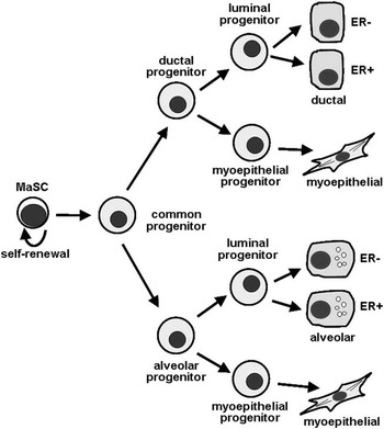
We propose an alternative stem cell/progenitor cell hierarchy (Figure 3) to that commonly described and hypothesized by Visvader and Lindeman (Reference Visvader and Lindeman2006) and Vaillant et al. (Reference Vaillant, Asselin-Labat, Shackleton, Lindeman and Visvader2007; Figure 2). According to both schemes, the MaSC is ER-negative and gives rise to progenitors that are ER-negative and ER-positive. Because both ductal (ductal progenitor) and lobuloalveolar (alveolar progenitor) structures contain myoepithelial cells, we propose that myoepithelial and luminal progenitors are generated downstream from the ductal- and alveolar-limited progenitor cells. This may account for the presence of only complete (containing luminal and myoepithelial cells) ductal or lobuloalveolar structures and the absence of myoepithelial outgrowths in dilution-limiting transplantation studies.
Proposed epithelial lineage in the mouse mammary gland. The mammary stem cells (MaSC) is estrogen receptor (ER)-negative and gives rise to progenitors that are ER− and ER+. Mammary transplantation studies demonstrate that progenitors exist, which are limited to generating either ductal (ductal progenitor) or lobuloalveolar (alveolar progenitor) structures. Each of these structures contain myoepithelial cells. We therefore propose that myoepithelial and luminal progenitor cells are generated downstream from the ductal and alveolar progenitor cells.

Significance of MaSC function to production agriculture
Milk yield is a function of the number and activity of MEC (Capuco et al., Reference Capuco, Wood, Baldwin, McLeod and Paape2001; Boutinaud et al., Reference Boutinaud, Guinard-Flamenta and Jammes2004), and both these parameters are impacted by MaSC function. As described below, various phases of mammary gland physiology involve the accumulation or turnover/regeneration of MEC. We anticipate that the ability to enhance the activity of MaSC or progenitor cells during these phases will have significant impact on the efficiency of milk production.
Overall, the scope of postnatal mammary development in dairy cows is impressive. The mass of mammary parenchyma within each gland (quarter) of the udder is a few hundred milligrams at birth, whereas at the onset of lactation it is 4 to 6 kg for a Holstein cow (Keys et al., Reference Keys, Capuco, Akers and Djiane1989; Capuco et al., Reference Capuco, Akers and Smith1997). This represents >10 000-fold increase in tissue mass.
Prepubertal mammary development is important to the milk-producing ability of an animal, as either excessive energy intake or under-nutrition may impair future milk yield of dairy animals (Sejrsen and Purup, Reference Sejrsen and Purup1997), which may be due to decreased mammary development during this phase of life (Sejrsen et al., Reference Sejrsen, Huber, Tucker and Akers1982). However, increased level of nutrition before weaning may be positively correlated with mammary growth and milk production (Bar-Peled et al., Reference Bar-Peled, Robinzon, Maltz, Tagari, Folman, Bruckental, Voet, Gacitua and Lehrer1997; Brown et al., Reference Brown, Vandehaar, Daniels, Liesman, Chapin, Forrest, Akers, Pearson and Nielsen2005; Meyer et al., Reference Meyer, Capuco, Ross, Lintault and Van Amburgh2006a and Reference Meyer, Capuco, Ross, Lintault and Van Amburgh2006b) or at least without negative impact (Daniels et al., Reference Daniels, Capuco, McGilliard, James and Akers2009). Much of the epithelial growth during this stage involves penetration of the mammary fat pad by the mammary ducts. In the murine mammary gland, structures at the distal extremity of mammary ducts, known as the terminal end buds (TEBs), are responsible for ductal elongation. In ruminants, more arborescent structures known as terminal ductal units (TDU) are responsible for the ductal elongation and branching that occurs prepubertally (Capuco et al., Reference Capuco, Ellis, Wood, Akers and Garrett2002a). Proliferation of EC occurs throughout the TDU, with the greatest rates of growth at the periphery of the TDU and periphery of the gland (Capuco et al., Reference Capuco, Ellis, Wood, Akers and Garrett2002a). Pre- and post-pubertally, there is a progressive increase in the ductal structures upon which alveoli develop during pregnancy (Sinha and Tucker, Reference Sinha and Tucker1969; Sejrsen et al., Reference Sejrsen, Huber, Tucker and Akers1982; Meyer et al., Reference Meyer, Capuco, Ross, Lintault and Van Amburgh2006a). The ability to regulate MaSC, and in turn mammary epithelial growth, may promote increased mammary development during the prepubertal period or may provide a means to reduce or eliminate possible negative impacts of increased nutrition prepubertally. This would enable accelerated heifer growth and a shortening of the time to milk production.
During pregnancy, extensive and exponential mammary growth occurs along with the development of alveoli that emanate from the distal termini of ducts (Akers, Reference Akers2002). The alveoli consist of a single layer of EC overlain and engulfed by a few myoepithelial cells and their processes. During pregnancy, MEC undergo extensive cytological and biochemical differentiation necessary for transition to an organ that is capable of producing copious quantities of milk during lactation. The ability to regulate MaSC and progenitor cells during first gestation may permit cows to enter their first lactation with a greater number of more fully differentiated cells.
It has been established that the decline in milk production during lactation is predominantly because of a steady decline in the number of MEC due to apoptotic cell death (Capuco et al., Reference Capuco, Wood, Baldwin, McLeod and Paape2001; Boutinaud et al., Reference Boutinaud, Guinard-Flamenta and Jammes2004). Slight alterations in the rates of MEC proliferation or apoptosis will have profound effects on the number of MEC during lactation, and hence the persistency of the lactation. Furthermore, the continued replacement of older cells with younger cells can help maintain the secretory activity of the MEC throughout lactation. Thus, the ability to regulate the activity of MaSC and progenitor cells has the potential to increase persistency of lactation.
For dairy cows, a nonlactating period (dry period) between lactations is important for maximizing milk production in the successive lactation. During lactation, the gradual decline in the number of mammary cells is approximately 50% (Capuco et al., Reference Capuco, Wood, Baldwin, McLeod and Paape2001). During the prepartum period (whether lactating or not), the full complement of EC is restored. In addition, during a typical dry period there is extensive turnover of secretory cells, rather than the extensive involution that accompanies forced involution in rodents (Capuco et al., Reference Capuco, Akers and Smith1997). This is likely because cows are in the final months of gestation when milking is terminated, and mitogenic effects of the hormones of pregnancy counterbalance the death-inducing effects of milk stasis (Capuco and Akers, Reference Capuco and Akers1999). This led to the use of the term regenerative involution to distinguish events during the dry period from those occurring during the involution response to forced weaning in nonpregnant mice (Figure 1; Capuco et al., Reference Capuco, Li, Long, Ren, Hruska, Schorr and Furth2002b and Reference Capuco, Ellis, Hale, Long, Erdman, Zhao and Paape2003). We postulated that a dry period promotes replacement of senescent MEC (Capuco et al., Reference Capuco, Akers and Smith1997), with a particular need to replace progenitor cells that are committed to the alveolar lineage (Capuco and Akers, Reference Capuco and Akers1999). As previously described, committed progenitor cells have a limited life span and are subject to senescence. An ability to promote regeneration of these progenitor cells by MaSC should enable a shortening of the dry period or eliminate the need for a dry period (Capuco and Ellis, Reference Capuco and Ellis2005; Capuco et al., Reference Capuco, Annen, Fitzgerald, Ellis and Collier2006; Annen et al., Reference Annen, Fitzgerald, Gentry, McGuire, Capuco, Baumgard and Collier2007). This would reduce nonproductive periods of the mammary cycle, and thus increase the efficiency of milk production.
An ability to regulate the function of MaSC and mammary progenitor cells would increase lactation efficiency. The ability to appropriately regulate MaSC and mammary progenitor cells would provide a means to increase production efficiency by increasing the number of fully functional mammary secretory cells, by replacing senescent or damaged cells and by providing a means to decrease nonproductive periods of the mammary gland cycle without negatively impacting milk yield during lactation periods.
Identification of MaSC
Published in vivo studies of bovine MaSC have used prepubertal heifer calves (Table 1). Reasons for selection of this physiological state are several-fold: (1) The rate of proliferation in the prepubertal mammary gland is impressive. The epithelial 5-bromo-2′-deoxyuridine (BrdU)-labeling index (fraction of EC labeled following a pulse labeling period with BrdU) within the peripheral TDU was found to be approximately 10% (Capuco et al., Reference Capuco, Ellis, Wood, Akers and Garrett2002a), declining as the heifer nears puberty (Meyer et al., Reference Meyer, Capuco, Ross, Lintault and Van Amburgh2006a). (2) It was reasoned that at this early stage of mammary development, MaSC and progenitor cells would be relatively abundant and active (Capuco et al., Reference Capuco, Ellis, Wood, Akers and Garrett2002a) considering the need for both symmetric and asymmetric divisions to provide for duct elongation and lineage differentiation (Ellis and Capuco, Reference Ellis and Capuco2002; Meyer et al., Reference Meyer, Capuco, Ross, Lintault and Van Amburgh2006a). (3) The prepubertal mammary gland is very sensitive to the important mammary mitogen, estrogen (Capuco et al., Reference Capuco, Ellis, Wood, Akers and Garrett2002a; Berry et al., Reference Berry, Jobst, Ellis, Howard, Capuco and Akers2003; Li et al., Reference Li, Meyer, Van Tassell, Sonstegard, Connor, Van Amburgh, Boisclair and Capuco2006; Connor et al., Reference Connor, Meyer, Li, Van Amburgh, Boisclair and Capuco2007), and it is important to understand the relationship between estrogenic stimuli and MaSC/progenitor cell proliferation (Sleeman et al., Reference Sleeman, Kendrick, Robertson, Isacke, Ashworth and Smalley2007). (4) Peripubertal mice and rats have served as an important model for the study of murine stem cells (Chepko and Smith, Reference Chepko and Smith1997; Smith, Reference Smith2005; Vaillant et al., Reference Vaillant, Asselin-Labat, Shackleton, Lindeman and Visvader2007). (5) Prepubertal heifers are significantly smaller than sexually mature heifers and offer advantages of cost and disposal after treatment with BrdU. (6) Duct elongation of the prepubertal mammary gland necessitates the occurrence of both symmetric and asymmetric stem cell divisions. (7) Microanatomy of the prepubertal mammary gland lends itself to the implementation of laser microdissection to isolate and study the characteristics of specific cell types (Choudhary et al., Reference Choudhary, Daniels, Evock-Clover, Garrett and Capuco2010a and Reference Choudhary, Li, Evock-Clover and Capuco2010b). (8) As stated earlier, the prepubertal period is important for ductal growth and for laying the tissue foundation for future milk production. Practices that enhance or diminish prepubertal development have the potential to increase or decrease, respectively, future milk production.
Adult stem cells are characteristically undifferentiated cells within an adult tissue. Histologic analyses have indicated that a pale-staining cell population present during all stages of mammary development and differentiation may serve as MaSC (Chepko and Smith, Reference Chepko and Smith1997). These ‘pale’ or ‘light’ cells have been described in mammary tissue from all species so far examined, including humans (Ferguson, Reference Ferguson1985), mice (Smith and Medina, Reference Smith and Medina1988), rats (Chepko and Smith, Reference Chepko and Smith1997), goats (Li et al., Reference Li, Wilde, Finch, Fernig and Rudland1999), sheep (Ellis et al., Reference Ellis, Edwards and Akers1995) and cattle (Ellis et al., Reference Ellis, Purup, Sejrsen and Akers2000; Ellis and Capuco, Reference Ellis and Capuco2002).
On the basis of ultrastructural analyses, Chepko and Smith (Reference Chepko and Smith1997) hypothesized that small, light-staining cells (SLC) within the mammary parenchyma act as multipotent MaSC. This was based on the mitotic competence, cytologic features, transition species, spatial distribution and frequency of the SLC relative to other MEC types: undifferentiated large light cells (ULLC), differentiated large light cells, LDC and myoepithelial cells. It was suggested that a morphologically indistinguishable subset of SLC act as primary progenitors and give rise to the ULLC. In turn, the ULLC divide multiple times to expand the parenchymal cell population and produce a lineage for more LDC or myoepithelial cells. Within the murine mammary epithelia, a subset of SLC is thought to act as stem cells, whereas ULLC act as transit-amplifying cells, accounting for patches of these cells in rapidly proliferating mammary epithelium (during pregnancy and early lactation). Similar patches were also noted for label-retaining epithelial cells (LREC) and their progeny, which have been referred to as stem cell transitional units (Kenney et al., Reference Kenney, Smith, Lawrence, Barrett and Salomon2001).
We carried out an analysis of MEC proliferation in prepubertal BrdU-injected Holstein heifers to investigate the hypothesis that lightly staining cells served as stem and progenitor cells in the bovine mammary gland. We observed light-, dark- and intermediate-staining cells in histologic sections stained with basic fuchsin and azure II. Light-staining cells comprised 10% of the total parenchymal cell population, but accounted for 50% of the cell proliferation. Intermediate-staining cells comprised 60% of the cell population and 43% of proliferating cells. Dark-staining cells comprised 30% of the parenchymal cell population, but only 7% of proliferating cells. Furthermore, the number of lightly staining cells was directly proportional to the proliferative capacity of the mammary tissue from heifers of different ages. These results strongly supported the concept that lightly staining mammary parenchymal cells are the primary proliferative cell population, and we suggested that the most undifferentiated population of these cells likely contains MaSC or primitive progenitor cells (Ellis and Capuco, Reference Ellis and Capuco2002). However, because this population accounts for approximately 10% of MEC prepubertally, it undoubtedly contains more than MaSC.
In an attempt to more accurately identify putative bovine MaSC, we took advantage of the capacity of stem cells to retain labeled DNA strands for extended periods of time (Capuco, Reference Capuco2007). This retention of label has been attributed to segregation and selective retention of template DNA strands by stem cells undergoing asymmetric division, rather than being due to cell quiescence after incorporation of label (Clarke et al., Reference Clarke, Anderson, Howell and Potten2003; Smith, Reference Smith2005; Booth and Smith, Reference Booth and Smith2006; Capuco, Reference Capuco2007). LREC were present in quantities (∼0.2% of MEC; Capuco, Reference Capuco2007) that are consistent with the prevalence of stem cells in mouse mammary gland (Kordon and Smith, Reference Kordon and Smith1998; Shackleton et al., Reference Shackleton, Vaillant, Simpson, Stingl, Smyth, Asselin-Labat, Wu, Lindeman and Visvader2006; Stingl et al., Reference Stingl, Eirew, Ricketson, Shackleton, Vaillant, Choi, Li and Eaves2006).
Smith demonstrated that LREC constitute a major proliferative component in murine mammary gland, because 80% of the hormone-stimulated LREC (3H-thymidine tagged LREC) incorporated BrdU when administered as two injections 24 h apart in estrogen-treated mice. To evaluate the proliferative capacity of LREC in bovine mammary gland, we examined expression of the Ki67 proliferation antigen and proliferating cell nuclear antigen (PCNA), a cofactor of DNA polymerase delta. Approximately 4% of the LREC were Ki67-positive (Capuco, Reference Capuco2007) and 14% PCNA-positive (Capuco et al., Reference Capuco, Evock-Clover, Minuti and Wood2009), and were clearly proliferative in absence of exogenous estrogen supplementation. These results imply that LREC in the prepubertal mammary gland are proliferative, but not rapidly cycling in the absence of hormonal stimuli.
Immunohistochemical analysis of LREC was used to determine their localization within the mammary epithelial layer and their ER status. The epithelium of the bovine mammary gland is composed of multiple layers of EC. The basal cells that overlie the basement membrane ultimately gives rise to a layer of myoepithelial cells, over which there are often several layers of EC in the TDU. In a bovine mammary gland of a prepubertal heifer, the basal layer does not express significant quantities of α-smooth muscle actin, and therefore may be comprised of myoepithelial progenitors and/or other stem/progenitor cells (Capuco et al., Reference Capuco, Ellis, Wood, Akers and Garrett2002a; Ballagh et al., Reference Ballagh, Korn, Riggs, Pratt, Dessauge, Akers and Ellis2008). LREC were primarily localized in the basal layer or within one or two cells above the basal layer. Consistent with the proposed MaSC niche (Chepko and Smith, Reference Chepko and Smith1997; Chepko and Dickson, Reference Chepko and Dickson2003), LREC were localized near the basement membrane and not in contact with the ductal lumen (Capuco, Reference Capuco2007; Capuco et al., Reference Capuco, Evock-Clover, Minuti and Wood2009). Approximately 75% of the LREC were ER-negative and localized within the basal epithelial layer or in close proximity, whereas ER-positive LREC were not luminal, but were typically more distant from the basement membrane than were the ER-negative LREC (Capuco et al., Reference Capuco, Evock-Clover, Minuti and Wood2009).
The ER status of MaSC has been the subject of considerable investigation in the biomedical field because of the importance of estrogen in mammary growth and because of implications to mammary tumorigenesis and therapy. Although there have been conflicting reports (Clarke et al., Reference Clarke, Anderson, Howell and Potten2003 and Reference Clarke, Spence, Anderson, Howell, Okano and Potten2005; Booth and Smith, Reference Booth and Smith2006), the preponderance of evidence indicate that MaSC are ER-negative. Multiparameter sorting of murine mammary cell suspensions showed that a single cell of the epithelial population expressing high levels of the heat stable antigen (CD24) and β1- or α6-integrin (CD29 or CD49f, respectively) was able to generate a functional mammary gland when implanted into cleared mammary fat pads (Shackleton et al., Reference Shackleton, Vaillant, Simpson, Stingl, Smyth, Asselin-Labat, Wu, Lindeman and Visvader2006; Stingl et al., Reference Stingl, Eirew, Ricketson, Shackleton, Vaillant, Choi, Li and Eaves2006). These sorted stem cells (Asselin-Labat et al., Reference Asselin-Labat, Shackleton, Stingl, Vaillant, Forrest, Eaves, Visvader and Lindeman2006) were ER-negative. Furthermore, the ability to repopulate a cleared mammary fat pad was essentially restricted to a population of cells that were CD24-low expressors, but these too were ER-negative (Sleeman et al., Reference Sleeman, Kendrick, Ashworth, Isacke and Smalley2006 and Reference Sleeman, Kendrick, Robertson, Isacke, Ashworth and Smalley2007). In human breast, Dontu and colleagues reported that increased aldehyde dehydrogenase-1 activity was associated with a population of human primary MEC that exhibited stem cell properties and were ER-negative (Ginestier et al., Reference Ginestier, Hur, Charafe-Jauffret, Monville, Dutcher, Brown, Jacquemier, Viens, Kleer, Liu, Schott, Hayes, Birnbaum, Wicha and Dontu2007; Liu et al., Reference Liu, Ginestier, Charafe-Jauffret, Foco, Kleer, Merajver, Dontu and Wicha2008). These data are consistent with our previous suggestion that the population of ER-negative LREC in bovine mammary gland represent MaSC (Capuco, Reference Capuco2007; Capuco et al., Reference Capuco, Evock-Clover, Minuti and Wood2009).
Proliferating MEC appear to be ER-negative in cow (Capuco et al., Reference Capuco, Ellis, Wood, Akers and Garrett2002a), mouse (Zeps et al., Reference Zeps, Bentel, Papadimitriou, D'Antuono and Dawkins1998) and human (Anderson and Clarke, Reference Anderson and Clarke2004). Moreover, proliferation of MEC is seemingly mediated by a variety of paracrine factors derived from cells of the epithelial or stromal compartments of the mammary gland (Hovey et al., Reference Hovey, McFadden and Akers1999; Akers et al., Reference Akers, McFadden, Purup, Vestergaard, Sejrsen and Capuco2000; Bocchinfuso et al., Reference Bocchinfuso, Lindzey, Hewitt, Clark, Myers, Cooper and Korach2000; Capuco et al., Reference Capuco, Ellis, Wood, Akers and Garrett2002a; Mallepell et al., Reference Mallepell, Krust, Chambon and Brisken2006; Hovey and Aimo, Reference Hovey and Aimo2010), and our microarray studies support the importance of paracrine factors and cell–cell communication in driving prepubertal growth of the bovine mammary epithelium (Li et al., Reference Li, Meyer, Van Tassell, Sonstegard, Connor, Van Amburgh, Boisclair and Capuco2006). Similarly, estrogenic stimulation of MaSC appears to be mediated by paracrine factors emanating from ER-positive MEC, because duct elongation and development fails in ER-null mice (Mallepell et al., Reference Mallepell, Krust, Chambon and Brisken2006) and in mice that are devoid of the estrogen mediator, amphiregulin (Ciarloni et al., Reference Ciarloni, Mallepell and Brisken2007). To fully populate a cleared fat pad, a single ER-negative stem cell presumably must undergo asymmetric division to provide a population of ER-positive progenitors, which in turn regulate proliferation of MaSC (Dontu et al., Reference Dontu, El-Ashry and Wicha2004; Lamarca and Rosen, Reference Lamarca and Rosen2008).
To further our understanding of bovine MaSC and evaluate the efficacy of label retention as a method for identifying these cells, we conducted transcriptome analysis of putative LREC within the prepubertal bovine mammary gland. Molecular profiles of four categories of cells within the bovine mammary epithelium (two subpopulations of putative stem cells and two subpopulations of control cells) were obtained, with the goal of localizing and characterizing MaSC in situ. LREC were identified in mammary cryosections by an immunostaining procedure that retained RNA quality (Choudhary et al., Reference Choudhary, Daniels, Evock-Clover, Garrett and Capuco2010a). Using laser microdissection, LREC from basal (LRECb) and embedded (LRECe) layers of mammary epithelium were isolated along with adjacent control EC. For each heifer, cells (6 to 13 cells) in each category were lysed, cDNA synthesized, amplified and labeled for hybridization (Choudhary et al., Reference Choudhary, Li, Evock-Clover and Capuco2010b) to custom microarrays (Li et al., Reference Li, Meyer, Van Tassell, Sonstegard, Connor, Van Amburgh, Boisclair and Capuco2006). There were 592 genes differentially expressed (P ⩽ 0.05; ⩾ twofold change) between LRECb and basal EC, and 110 genes differentially expressed between LRECe and embedded EC. Of these, 387 genes with enriched expression in LRECb were involved in cell growth and proliferation, cell cycle and post-translational modifications. Low expression of ER-α and high expression of aldehyde dehydrogenase 3B1 in LRECb were consistent with stem cell character. We found high expression of NR5A2, a pluripotency transcription factor (Heng et al., Reference Heng, Feng, Han, Jiang, Kraus, Ng, Orlov, Huss, Yang, Lufkin, Lim and Ng2010), and no expression of XIST, an X-chromosome inactivation factor (Savarese et al., Reference Savarese, Flahndorfer, Jaenisch, Busslinger and Wutz2006; Lagarkova et al., Reference Lagarkova, Shutova, Bogomazova, Vassina, Glazov, Zhang, Rizvanov, Chestkov and Kiselev2010), in LRECb. Comparison between LRECb and LRECe showed downregulation of cell survival and proliferation factors (IGF2, HSPB6, LAMC1), nestin (stem cell marker), epigenetic modifiers (JRID2, METTL33, SMARCC2) and upregulation of apoptotic genes (SFRS5, THAP3) and XIST in LRECe. We conclude that BrdU label retention identifies stem and progenitor cells, wherein MaSC (LRECb) are located in the basal region of the mammary epithelium and progenitor cells (LRECe) are localized in more apical layers. Furthermore, our data provide molecular signatures that may provide markers for MaSC and progenitor cells. Such markers will permit tracking of MaSC function as affected by physiological state or treatment. This represents the first transcriptome characterization of putative MaSC and progenitor cells excised from known locations within the mammary epithelium.
The MaSC niche
The stem cell niche is the microenvironment in which a stem cell resides. Signals from nearby cells and the extracellular stroma appear instrumental in regulating stem cell activity. There is strong evidence that stem cell activity in the mammary gland is largely dictated by the stem cell niche. Research by Gilbert Smith and colleagues demonstrated that the normal lineages of neural stem cells (Booth et al., Reference Booth, Mack, Androutsellis-Theotokis, McKay, Boulanger and Smith2008) and spermatogonia (Boulanger et al., Reference Boulanger, Mack, Booth and Smith2007) are redirected when placed in a mammary environment, permitting them to repopulate a cleared mammary fat pad. This reaffirms the concept of plasticity among somatic stem cells and points to the importance of the stem cell niche. More subtly, transplantation of mammary cells from ERα-null mice into cleared mammary fat pads demonstrated that mammary ductal growth occurred only if ERα is expressed in the MEC (Mallepell et al., Reference Mallepell, Krust, Chambon and Brisken2006). However, the work additionally demonstrated that genetically marked ERα-null MEC could effectively regenerate the mammary tree when initially mixed with wild-type EC. Thus cell–cell interactions permitted the ERα-null stem cells to provide progeny for regenerating the mammary gland.
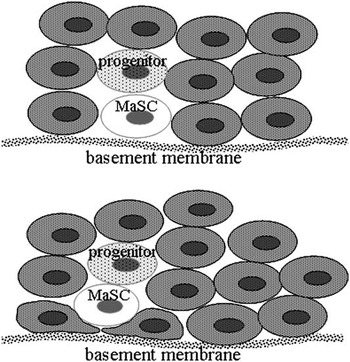
Initial morphological analysis indicated that putative murine MaSC (SLC) were localized in close proximity to the epithelial basement membrane, but separated from the basement membrane by thin cytoplasmic extensions of the luminal and myoepithelial cells (Chepko and Smith, Reference Chepko and Smith1997; Chepko and Dickson, Reference Chepko and Dickson2003). Similarly, in prepubertal bovine mammary gland, our laser microdissection data suggest that the MaSC are localized in the basal region of the mammary epithelium (Choudhary et al., Reference Choudhary, Li, Evock-Clover and Capuco2010b). However, limitations of our method do not permit determination of whether MaSC are in direct contact with the epithelial basement membrane within the mammary glands of prepubertal heifers (Figure 4).
Localization of mammary stem cells (MaSC) within the mammary epithelium of the prepubertal heifer. Data indicate that MaSC are present within the basal layer (upper image) of terminal ductular units (Choudhary et al., Reference Choudhary, Li, Evock-Clover and Capuco2010b). However, the methodology used cannot distinguish between localization in the basal layer and localization above processes of an underlying cell (lower image). The later is the hypothesized locale of MaSC in the murine epithelium during the peripubertal period.

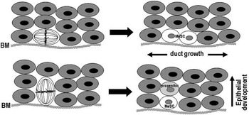
During periods of mammary gland expansion, there is a need for symmetrical division of MaSC to maintain a sufficient number of MaSC within the mammary epithelium and a distribution of MaSC throughout the mammary tree. Analogous to the situation in epidermis (Jones and Simons, Reference Jones and Simons2008; Blanpain and Fuchs, Reference Blanpain and Fuchs2009), one might envision the plane of symmetrical division being parallel to the basement membrane so that the two resulting daughter MaSC remain in the basal region and the division promotes elongation of the mammary duct (Figure 5). Within this same context, asymmetric division of MaSC promotes establishment of multiple epithelial layers and generation of progenitor cells in an apical position, more distal from the basement membrane than are the parental MaSC (Figure 5).
Hypothetical orientation of symmetric and asymmetric divisions of mammary stem cells (MaSC) in mammary epithelium of the prepubertal heifer. Symmetric division is hypothesized to occur within a plane that is parallel to the basement membrane (BM), facilitating ductal growth and expansion of the MaSC population. Asymmetric division is hypothesized to occur in a plane that is perpendicular to the BM, accounting for the generation of progenitor cells in position that is apical to the MaSC. Similar orientations have been described for epithelial stem cells in the skin (Blanpain and Fuchs, Reference Blanpain and Fuchs2009).

At the macroscopic level it is clear that MaSC are present throughout the murine mammary gland, and by extension in the bovine mammary gland. However, at the microscopic level, additional study is necessary to determine the localization of these cells within mammary structures that are evident during various phases of mammary gland development.
In the pubertal mouse, cap cells, which are evident at the distal face of the TEB, appear to be multipotent MaSC because they can provide for myoepithelial and luminal cell lineages (Esmailpour and Huang, Reference Esmailpour and Huang2008). However, because they are no longer evident after the ducts reach the margins of the mammary fat pad, this niche appears to be transient (Woodward et al., Reference Woodward, Chen, Behbod and Rosen2005). In the mammary gland of a mature virgin mouse, the MaSC seem to reside in a basal position, as described earlier (Chepko and Smith, Reference Chepko and Smith1997; Smith and Chepko, Reference Smith and Chepko2001), in a niche underlain by cytoplasmic extensions of adjacent EC or myoepithelial cell(s) and surrounded by EC, some of which are ER-positive (Brisken and Duss, Reference Brisken and Duss2007). This stem cell niche appears to persist throughout maturity in virgin mice and in mice that are treated with ovarian steroids to induce lobuloalveolar growth (Joshi et al., Reference Joshi, Jackson, Beristain, Di Grappa, Mote, Clarke, Stingl, Waterhouse and Khokha2010). Evaluation of the expression of cell lineage markers and in vitro propagation studies in human breast tissue from reduction mammoplasty led to the conclusion that a regional stem cell lineage is present in the human breast, with MaSC present in mammary ducts and lobule progenitor cells prevalent in the terminal ductal lobules (Villadsen et al., Reference Villadsen, Fridriksdottir, Ronnov-Jessen, Gudjonsson, Rank, LaBarge, Bissell and Petersen2007). Whether stem cells are present within the alveoli of lactating mammals remains a pertinent question. To our knowledge, this has yet to be evaluated, most likely because of methodological issues. Recent investigations of MaSC have involved flow cytometric sorting of mammary suspensions based on a number of lineage-dependent markers (Welm et al., Reference Welm, Tepera, Venezia, Graubert, Rosen and Goodell2002; Asselin-Labat et al., Reference Asselin-Labat, Shackleton, Stingl, Vaillant, Forrest, Eaves, Visvader and Lindeman2006; Shackleton et al., Reference Shackleton, Vaillant, Simpson, Stingl, Smyth, Asselin-Labat, Wu, Lindeman and Visvader2006; Stingl et al., Reference Stingl, Eirew, Ricketson, Shackleton, Vaillant, Choi, Li and Eaves2006; Joshi et al., Reference Joshi, Jackson, Beristain, Di Grappa, Mote, Clarke, Stingl, Waterhouse and Khokha2010). Obtaining enzymatically dispersed cell suspensions from lactating tissue is problematic (low yield and disproportionate representation of cell types) and lactating MEC express increased levels of ATP-binding cassette transporters (potential markers or contributors to the side population; Capuco and Ellis, Reference Capuco and Ellis2005; Jonker et al., Reference Jonker, Merino, Musters, van Herwaarden, Bolscher, Wagenaar, Mesman, Dale and Schinkel2005). Label retention studies are feasible in lactating rodents where there is significant mammary proliferation during early lactation (Baldwin and Milligan, Reference Baldwin and Milligan1966; Traurig, Reference Traurig1967; Tucker et al., Reference Tucker, Paape and Sinha1967), but difficult in ruminants where there is insufficient proliferation during lactation (Capuco et al., Reference Capuco, Wood, Baldwin, McLeod and Paape2001) to permit dilution of the BrdU label in non-MaSC. Expression of marker proteins is the most promising, but perhaps a less specific, approach.
Altering MaSC function and kinetics
It is logical to assume that MaSC function can be altered. Changes in progenitor populations within the mammary gland are evident during the course of mammary development and differentiation (Booth et al., Reference Booth, Boulanger and Smith2007; Joshi et al., Reference Joshi, Jackson, Beristain, Di Grappa, Mote, Clarke, Stingl, Waterhouse and Khokha2010) and these changes can be induced by appropriate hormonal treatment (Booth et al., Reference Booth, Boulanger and Smith2007 and Reference Booth, Boulanger, Anderson, Jimenez-Rojo, Brisken and Smith2010; Joshi et al., Reference Joshi, Jackson, Beristain, Di Grappa, Mote, Clarke, Stingl, Waterhouse and Khokha2010). At the extreme end of the spectrum, the epigenetic landscape of somatic cells can be altered to induce pluripotency (Takahashi and Yamanaka, Reference Takahashi and Yamanaka2006). A number of genetic pathways have been implicated as regulators of MaSC proliferation and differentiation. These have included Wnt, NOTCH, Hedgehog, BRCA1, p21, p63, Pten and p53 (Sherley, Reference Sherley2002; Dontu et al., Reference Dontu, Abdallah, Foley, Jackson, Clarke, Kawamura and Wicha2003a; Lindvall et al., Reference Lindvall, Evans, Zylstra, Li, Alexander and Williams2006; Liu et al., Reference Liu, Ginestier, Charafe-Jauffret, Foco, Kleer, Merajver, Dontu and Wicha2008).
In vitro experiments with rat hepatocytes indicated that p53 promotes asymmetric proliferation of somatic stem cells through downregulation of inosine-5′-monophosphate dehydrogenase (Sherley et al., Reference Sherley, Stadler and Johnson1995; Rambhatla et al., Reference Rambhatla, Ram-Mohan, Cheng and Sherley2005), the rate limiting enzyme for guanine nucleotide synthesis. This can be circumvented and symmetric division can be promoted by treatment with xanthosine, a naturally occurring ribonucleoside, causing expansion of the stem cell population (Lee et al., Reference Lee, Crane, Merok, Tunstead, Hatch, Panchalingam, Powers, Griffith and Sherley2003). Analogously, in vivo treatment of bovine MEC by infusion of xanthosine into the mammary gland resulted in an apparent expansion of the MaSC population (Capuco et al., Reference Capuco, Evock-Clover, Minuti and Wood2009). This effect was evidenced by an increase in the number of LREC and an increase in tissue telomerase activity. The method provides a possible means to manage mammary growth, cell turnover and tissue regeneration, thereby improving dairy cow productivity and udder health. Studies are in progress to evaluate the ability of this treatment to influence production parameters and efficiency of production.
In vitro studies
The focus of this paper has been on in vivo studies to characterize bovine MaSC, progenitor cells and their potential interactions. The cleared fat pad model, which has provided a means to assess the regenerative properties of selected cell populations within the murine mammary gland, is not directly applicable to studies of bovine MaSC and progenitor cells. However, altering the murine fat pad in a manner that is analogous to the adaptations made to support transplantation of human mammary cells (Kuperwasser et al., Reference Kuperwasser, Chavarria, Wu, Magrane, Gray, Carey, Richardson and Weinberg2004) may provide a suitable host environment for transplantation of bovine mammary cells.
Fortunately, in vitro approaches provide an important complementary approach to in vivo studies. Of particular importance for studying cell lineage, in vitro studies provide a means to characterize the regenerative properties of selected populations. These approaches, including the use of in vitro approaches to evaluate the differentiation potential of a cell population (Dontu et al., Reference Dontu, Abdallah, Foley, Jackson, Clarke, Kawamura and Wicha2003a and Reference Dontu, Al-Hajj, Abdallah, Clarke and Wicha2003b), have been used for bovine studies (Table 1) and will likely be used more widely in the future. Not only do they provide a means to evaluate the characteristics of putative stem cells and progenitor cells, in vitro approaches also provide a means to evaluate methods for altering the proliferation kinetics and differentiation of MaSC and progenitor cells, and for assessing genomic alterations that regulate these processes. Recent development of a computational learning-based algorithm to predict the immediate lineage outcome of a stem cell division, from in vitro time-lapse digital images, offers the promise of another powerful tool to evaluate stem cell function (Cohen et al., Reference Cohen, Gomes, Roysam and Cayouette2010).
Future
Additional studies are necessary to more fully characterize EC lineage and the regulation of epithelial growth, turnover and differentiation. Development of markers for stem cells and progenitor cells will facilitate evaluation of the potential impacts of traditional management strategies and strategies designed to impact MaSC and progenitor cell function. The challenge is ‘to comprehend the interaction among these components that allows the long-term maintenance of MaSC activity’ (Smith and Medina, Reference Smith and Medina2008) and develop effective strategies to impact this activity.
Acknowledgment
The USDA is an equal opportunity provider and employer. This work was funded by the United States Department of Agriculture, Agricultural Research Service, CRIS 1265-31000-083-00D and National Research Initiative Competitive Grant no. 2008-35206-18825 from the USDA National Institute of Food and Agriculture.