Introduction
Cavernous sinus thrombosis secondary to sphenoid sinusitis is a well-known phenomenon in which delayed diagnosis can have potentially catastrophic outcomes. In the post-antibiotic era, the incidence of cavernous sinus thrombosis has drastically decreased. However, medical providers should remain astute to this complication, because the mortality rate has been reported to be as high as 30 per cent.Reference Eweiss, Mukonoweshuro and Khalil1–Reference Cannon, Antonio, McCloskey, Hines, Tobin and Shetty4 This case report presents a rare complication of contralateral cavernous sinus thrombosis secondary to acute, unilateral sphenoid sinusitis without preceding orbital complications.Reference Eweiss, Mukonoweshuro and Khalil1, Reference Bhatia and Jones2
Case report
An otherwise healthy 16-year-old white male with a history of recurrent sinusitis presented to our medical centre with suspected meningitis. His clinical history revealed ‘helmet-like’ headaches and spiking fevers lasting for 5 days. Cerebral spinal fluid studies revealed an elevated white blood cell count with predominant neutrophils, normal protein and low glucose. Blood tests revealed leukocytosis with a left shift. Initial cultures were negative. A computed tomography (CT) scan revealed mucosal thickening in the posterior ethmoid sinuses and opacification of the right sphenoid sinus (Figure 1). On physical examination, there were no abnormal findings, except for positive Brudziński's and Kernig's signs.
Axial computed tomography scan demonstrating opacification of the right sphenoid sinus (arrow) and mucosal thickening in the posterior ethmoid sinus (arrowhead).

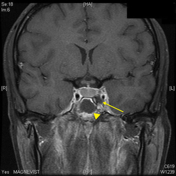
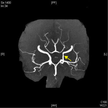
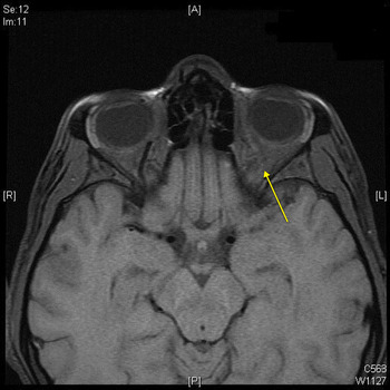
The patient was admitted and treated empirically with vancomycin, ceftriaxone and dexamethasone. On day 2, he developed diplopia with left abducens nerve palsy. Magnetic resonance imaging (MRI) and magnetic resonance angiography/magnetic resonance venography revealed bilateral, posterior, ethmoid mucosal thickening, right-sided sphenoid sinus fluid opacity with basal meningitis, thrombosis of the left cavernous sinus, narrowing of the left internal carotid artery and thrombosis of the left superior ophthalmic vein (Figures 2–4).
Coronal, T1-weighted magnetic resonance image demonstrating thrombosis of the left cavernous sinus (arrow) and associated sphenoid disease (arrowhead).

Magnetic resonance angiogram demonstrating narrowing of the left internal carotid artery (arrow).

Axial, T1-weighted magnetic resonance image demonstrating thrombosis of the left superior ophthalmic vein (arrow).

The otolaryngology department at the Children's Hospital of Michigan (Detroit Medical Center, Michigan, USA) promptly performed bilateral, endoscopic, total ethmoidectomies and bilateral, endoscopic sphenoidotomies that revealed polypoid mucosa within the right anterior and posterior ethmoid sinus and frank pus within the right sphenoid sinus; the left sphenoid sinus was free of disease. All specimens showed no growth, likely a result of antibiotic therapy.
On post-operative day 1, resolution of diplopia, headache, fever and meningeal irritation was noted. The patient improved and by day 8 was discharged on ceftriaxone, vancomycin and oral metronidazole.
Discussion
To our knowledge, this was the first reported case of acute, unilateral sphenoid sinusitis with contralateral cavernous sinus thrombosis in an adolescent. This patient presented with headaches, fever and meningismus but lacked specific sinus complaints. In view of masked symptoms associated with sinusitis, it is imperative to have a suspicion for sinus infections as a cause of meningitis progressing to intracranial complications.
The pathophysiology of this phenomenon cannot be understood without considering anatomy. The cavernous sinuses are formed by folds of dura along the superolateral aspect of the sphenoid sinus, extending from the superior orbital fissure to the petrous apex. Both cavernous sinuses are connected via anterior and posterior intercavernous veins and have an intimate relationship with the sella turcica. Blood flows in anterograde fashion into the cavernous sinus by way of the ophthalmic veins and the valveless superficial middle cerebral, inferior cerebral and emissary veins. Drainage from the cavernous sinus is through the superior and inferior petrosal sinuses into the superior sagittal sinus and internal jugular vein. Along the wall of the cavernous sinus rest the oculomotor, trochlear, ophthalmic (V1 branch of the trigeminal nerve) and maxillary nerves. Travelling medially within the cavernous sinus, are the abducens nerve and the internal carotid artery. This anatomical significance is illustrated by the early involvement of the abducens nerve, resulting in lateral gaze palsy.Reference Bhatia and Jones2
Isolated, acute sphenoid sinusitis is an uncommon finding characterised largely by significant retro-orbital, frontal or temporal headaches.Reference Cannon, Antonio, McCloskey, Hines, Tobin and Shetty4 This vague presentation may delay diagnosis, thereby leading to complications.Reference Cannon, Antonio, McCloskey, Hines, Tobin and Shetty4 The mechanism for the intracranial advancement of sphenoid sinusitis is either via direct lateral extension through the thin diploic bone and nasal mucosa, or by thrombophlebitis of the emissary veins draining into the cavernous sinus.Reference Bhatia and Jones2
Traditionally, the most common cause of cavernous sinus thrombosis has been staphylococcal skin infection of the midface.Reference Cannon, Antonio, McCloskey, Hines, Tobin and Shetty4, Reference DiNubile5 However, an increase in sinusitis as a prominent cause of cavernous sinus thrombosis has also been reported.Reference Bhatia and Jones2, Reference Lew, Southwick, Montgomery, Weber and Baker6 Studies reviewing the bacteriology of acute sphenoiditis have shown streptococcus as the most common organism associated with sphenoidal disease.Reference DiNubile5, Reference Southwick, Richardson and Swartz7 When analysing paranasal, sinus-associated cavernous sinus thrombosis, a shift toward streptococcal organisms as a common aetiology should be considered.
There are many signs and symptoms associated with cavernous sinus thrombosis. Preseptal oedema, chemosis and proptosis are early signs of venous congestion and have been frequently observed.Reference Bhatia and Jones2, Reference Cannon, Antonio, McCloskey, Hines, Tobin and Shetty4, Reference DiNubile5, Reference Southwick, Richardson and Swartz7 Subsequently, patients may develop diplopia from limitations of extraocular muscle movement. Meningismus is common and has been documented in 40 per cent of patients.Reference DiNubile5 After 48 hours, bilateral ocular findings become evident as the communicating intercavernous veins allow the propagation of thrombophlebitis.Reference Cannon, Antonio, McCloskey, Hines, Tobin and Shetty4, Reference DiNubile5 Late complications include pituitary insufficiency, cortical venous thrombosis and epidural, subdural or parenchymal abscesses.Reference Younis and Lazar8 Carotid artery involvement can lead to a carotid-cavernous fistula or carotid aneurysm.Reference Younis and Lazar8 Lemierre's syndrome as a result of septic emboli has also been described.Reference Cannon, Antonio, McCloskey, Hines, Tobin and Shetty4
Currently, CT and MRI are used to confirm the suspected clinical diagnosis of cavernous sinus thrombosis. Magnetic resonance imaging using flow parameters and magnetic resonance venography are more sensitive than CT for the evaluation of cavernous sinus thrombosis. Magnetic resonance imaging modalities will generally confirm cavernous sinus thrombosis with the following radiographic findings: deformity of the cavernous portion of the carotid artery; heterogeneous signal from the affected cavernous sinus; and hyperintense signal in all thrombosed vascular sinuses.Reference Cannon, Antonio, McCloskey, Hines, Tobin and Shetty4
The primary treatment for cavernous sinus thrombosis is empirical, intravenous, antibiotic therapy until culture-directed therapy is available.Reference Price, Hameroff and Richardson9 Appropriate initial choices may include nafcillin sodium, ceftriaxone and metronidazole.Reference Eweiss, Mukonoweshuro and Khalil1–Reference Cannon, Antonio, McCloskey, Hines, Tobin and Shetty4 If methicillin-resistant staphylococcus is suspected, vancomycin should be substituted for nafcillin sodium.Reference Eweiss, Mukonoweshuro and Khalil1–Reference Cannon, Antonio, McCloskey, Hines, Tobin and Shetty4
• Cavernous sinus thrombosis secondary to sphenoid sinusitis is a phenomenon in which delayed diagnosis can have catastrophic outcomes
• We describe a case of acute, unilateral sphenoid sinusitis with contralateral cavernous sinus thrombosis in an adolescent
• This case had no preceding orbital complications
Most surgical therapies are directed at eradicating the inciting infection. When dealing with sinusitis-associated cavernous sinus thrombosis, functional endoscopic sinus surgery directed at the affected sinus has been shown to improve outcomes when performed early, as shown in this case report.Reference Younis and Lazar3, Reference Younis and Lazar8
The use of corticosteroid treatment for cavernous sinus thrombosis is controversial and should only be considered with adjunct antibiotics. Corticosteroids may blunt the immune response and potentiate sepsis, so a risk–benefit analysis must be used. Steroid therapy is indicated in cases where hypopituitarism develops due to associated pituitary gland ischaemia.Reference Younis and Lazar8
Conclusion
Although cavernous sinus thrombosis is a rare clinical entity in the antibiotic era, acute and persistent sphenoid sinusitis may progress to cavernous sinus thrombosis despite maximal medical treatment. The clinician must maintain a differential diagnosis of sinusitis when evaluating patients who present with nondescript neurological or ocular findings with cavernous sinus thrombosis, even in the absence of changes due to orbital cellulitis. The mainstay of treatment includes early diagnosis, aggressive intravenous antibiotics and appropriate surgical management.
Acknowledgements
Walter Belenky, an astute clinician, contributing author, teacher and friend.