Introduction
Cystic echinococcosis (CE) is considered as one of the most severe helminthic diseases in humans, caused by the metacestode stage of the species complex Echinococcus granulosus sensu lato (s.l.). The adult form of these cestodes is found in the intestines of the Canidae family. A number of mammals, including humans, can become infected by ingesting eggs excreted in the faeces of canids, which grow into hydatid cysts, the larval stage of the parasite (Thompson, Reference Thompson2017). Based on the molecular studies, the E. granulosus s.l. complex consists of a collection of ten different genotypes of the parasite, of which some have been considered as separate species, such as the E. granulosus sensu stricto (s.s.) complex (G1-G3 genotypes), E. canadensis complex (G6-G8 and G10 genotypes), E. ortllepi (G5 genotype) and E. equinus (G4 genotype) (Nakao et al., Reference Nakao, Lavikainen, Yanagida and Ito2013); however, the status of G2 as a valid genotype (Kinkar et al., Reference Kinkar, Laurimäe and Sharbatkhori2017) and the G6-G10 genotypes as a unique species is a matter of controversy (Addy et al., Reference Addy, Wassermann and Kagendo2017; Laurimäe et al., Reference Laurimäe, Kinkar and Moks2018).
CE usually involves the liver (68.8–80%) and lung (10–22.4%) of humans (Salamone et al., Reference Salamone, Licari and Randisi2016), although hydatid cysts can develop in almost all organs of the body. The spleen, kidney, peritoneum, brain, cardiac and skeletal muscles are unusual cyst locations reported across different geographical areas of the world (Geramizadeh, Reference Geramizadeh2013; Salamone et al., Reference Salamone, Licari and Randisi2016). Most of these unusually located hydatid cysts are reported as secondary cysts, while extrahepatic and extrapulmonary localization of primary hydatid disease is very rare (Hamamci et al., Reference Hamamci, Besim and Korkmaz2004; Aksakal et al., Reference Aksakal, Kement, Okkabaz, Altuntaş and Öncel2016; Geramizadeh, Reference Geramizadeh2017).
The diagnosis and treatment of hydatidosis in humans can be affected by the location of hydatid cysts. Cerebral cysts cannot be diagnosed using serological techniques with specific immunodominant antigens such as antigen B and antigen 5 (Shafiei et al., Reference Shafiei, Teshnizi, Kalantar, Gholami, Mirzaee and Mirzaee2016), hence the necessity of magnetic resonance imaging (MRI) for preoperative diagnosis (Abbassioun & Amirjamshidi, Reference Abbassioun and Amirjamshidi2001). The outcome of the surgical treatment of extrahepatic hydatid disease varies according to the location of the cyst (Tsaroucha et al., Reference Tsaroucha, Polychronidis, Lyrantzopoulos, Pitiakoudis, Karayiannakis, Manolas and Simopoulos2005). For example, Ciurea et al. (Reference Ciurea, Fountas, Coman, Machinis, Kapsalaki, Fezoulidis and Robinson2006) reported the successful removal rate of intact brain hydatid cysts to be approximately 60–70%.
Over the recent years, several studies have been conducted to investigate the geographical distribution of genotypes of E. granulosus causing CE in human and animal hosts in different parts of the world (Cao et al., Reference Cao, Chen, Li, Ma, Xiao, Wang and Gao2018; Laatamna et al., Reference Laatamna, Ebi, Brahimi, Bediaf, Wassermann, Souttou and Romig2019; Moudgil et al., Reference Moudgil, Moudgil, Asrani and Agnihotri2019); however, there is limited information regarding the relationship between parasite genotypes and their phenotypic characteristics, such as pathogenesis or the localization of their larva in unusual organs of the hosts.
In the present study, we compared the genotypes and sequence types (STs) of extra-hepatopulmonary CE isolates in north-east Iran; furthermore, reliable haplotypes were retrieved from GenBank with common hydatid cysts of the liver to investigate the genetic relationship between hydatid cyst strains and cyst location in host organs.
Materials and methods
Study area and samples
North Khorasan Province (28,434 km2) is located in north-east Iran, and Bojnord city is the capital of the province, with a steppe climate. This province, with a large number of livestock (sheep, cattle, goats and camels), is one of the main centres of animal husbandry in Iran, where many cases of human hydatidosis have been reported (Salehi et al., Reference Salehi, Adinezade, Khodajo, Saberi and Yousefi2013).
A total of 12 human CE cysts were included in this study, collected from patients who underwent surgery in the hospitals of North Khorasan Province between 2003 and 2016. Among these cysts, ten pathologically confirmed, paraffin-embedded CE samples were from unusually infected organs: brain (n = 4), kidney (n = 1), spleen (n = 1), peritoneum (n = 1), pelvic region (n = 1), armpit (n = 1) and left leg (n = 1). The two remaining cysts were alcohol-preserved cysts recently isolated from the liver of the patients. Demographic and diagnostic data were obtained from patients' hospital records.
DNA extraction and polymerase chain reaction (PCR) amplifications
The paraffin-embedded (FFPE) blocks were cut into sections with a thickness of 5–10 µm. These sections were deparaffinized using xylene, and rehydrated using 100%, 90%, 80% and 70% ethanol (Schneider et al., Reference Schneider, Gollackner, Edel, Schmid, Wrba, Tucek, Walochnik and Auer2008). The rehydrated samples were kept at an ambient temperature for 2–3 h; DNA was then extracted via the FFPE Tissue DNA Extraction Micro Kit (Favorgen, Taiwan).
For the alcohol-preserved samples, the germinal layers were subjected to DNA extraction using the Genomic DNA Extraction Tissue DNA Mini Kit (Favorgen, Taiwan), according to the manufacturer's instructions. PCR amplification of the mitochondrial cytochrome c oxidase I (cox1) gene (446 base pairs (bp)-long fragment) was performed through primers JB3 (5′-TTTTTTGGGCATCCTGAGGTTTAT-3′) and JB4.5 (5′-TAAAGAAAGAACATAATGAAAATG-3′) (Bowles et al., Reference Bowles, Blair and McManus1992).
The primer pairs CO1F6 (5′-TTTGTYTCTTTRGATCATAAGCG-3′) and Cox1_schist_3′ (5′-TAATGCATM GGAAAA AAACA-3′) were further employed for the amplification of a 1180-bp-long fragment of the mitochondrial cox1 gene in Echinococcus spp. (Lockyer et al., Reference Lockyer, Olson and Østergaard2003; Morgan et al., Reference Morgan, DeJong, Kazibwe, Mkoji and Loker2003). Amplification was carried out in a final volume of 50 µl, containing 250 µM of each dNTP, 30 pmol of each primer, 2.5 units of Hi-Fid DNA polymerase (Kawsar Biotech Company, Iran; this enzyme also has the proofreading 3′–>5′ exonuclease activity), 5 µl of PCR buffer containing MgCl2 and 7 µl of DNA. The reaction was cycled using a step-down program, according to Karamian et al. (Reference Karamian, Haghighi, Hemmati, Taylor, Salehabadi and Ghatee2017). PCRs were performed in a MasterCycler gradient thermal cycler (Eppendorf, Germany). A 100 bp ladder (Jena Bioscience, Germany) was used as the DNA size standard. The PCR products were separated on ethidium-bromide-stained agar gel (1.7%) and visualized by a transilluminator.
DNA sequencing
The obtained PCR amplicons were directly purified from the reaction mixtures using the Favorgen GEL/PCR Purification Kit (Favorgen, Taiwan), and were then sent to Bioneer Company (South Korea) and sequenced using the Applied Biosystems automated DNA sequencer (3730 XL). Sequencing was performed in both directions using the same PCR primers by the BigDye Terminator v3.1 cycle sequencing kit. The representative sequences for cox1 gene were deposited in the GenBank database under accession numbers MH010304–MH010313, MH025946 and MH025947.
Phylogenetic analysis
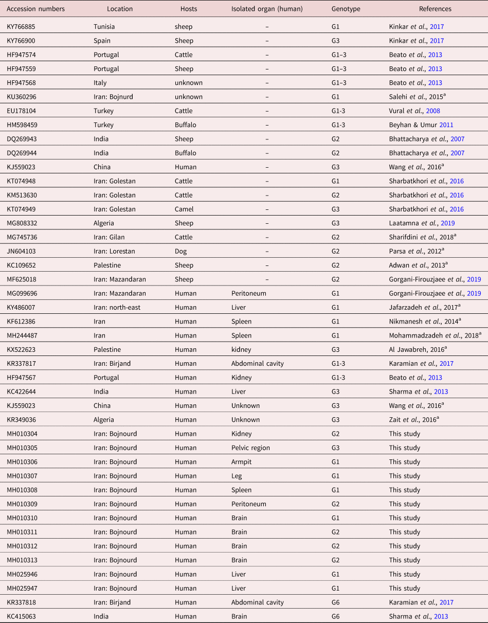
The obtained cox1 sequences (table 1) were edited using Chromas Lite 2.01 (http://technelysium.com.au/wp/). BLAST software (http://www.ncbi.nlm.nih.gov/blast/) was used to compare the obtained cox1 sequences with sequences available at GenBank to primarily identify the genotypes of the isolated hydatid cysts. A phylogenetic tree was generated using E. granulosus s.s. (G1-3) STs in this study and those from GenBank (table 1). Two G6 STs were used as out-group. Using ClustalW, the alignment process was performed, and the aligned sequences were then manually refined in BioEdit software version 7.2.5 (http://www.mbio.ncsu.edu/bioedit/bioedit.html) (Hall et al., Reference Hall, Biosciences and Carlsbad2011). The maximum likelihood (ML) tree was inferred by MEGA 6 software (Tamura et al., Reference Tamura, Stecher, Peterson, Filipski and Kumar2013). Nodal support was assessed by bootstrapping with 1000 replicates.
GenBank accession numbers of cox1 of Echinococcus genotypes used in the phylogenetic analysis in the present study.

a Unpublished sequences in GenBank.
Results
The genotypes of the samples
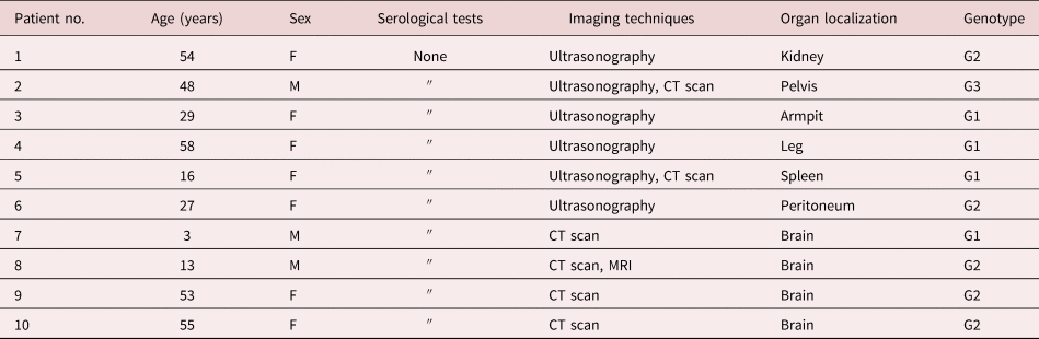
The mean age of patients with unusually located cysts was 35.6. Two of the four patients with brain hydatid cysts were a child and a teenager, and 70% of unusually located cysts belonged to females. The ultrasonography and computed tomography (CT) scan imaging methods were the main para-clinically diagnostic techniques, while no serological tests were performed in the diagnosis and treatment of the patients (table 2).
Diagnostic methods, organ localization and identified genotypes of extra-hepatopulmonary hydatid cysts.

Sequence analysis of PCR products (446 bp size) using JB3 and JB4.5 primers, in five of ten paraffin-embedded CE samples showed parts of the phosphatidylglycerophosphatase A (pgpA) and aldo/keto reductase genes of Escherichia coli instead of the Echinococcus cox1 gene. To solve this problem, all samples were primarily amplified with CO1F6 and Cox1_schist primers, and the amplified band of 1180 bp was extracted from the agarose gel. In the next step, PCR was performed on this fragment using JB3 and JB4.5 primers, and sequencing in both directions was done using the same primers. Two alcoholic liver cysts were also amplified and sequenced using the same primers. Results showed that all the ten paraffin-embedded tissue blocks and two alcohol-preserved liver cysts belonged to the Echinococcus granulosus s.s. (G1-G3) complex.
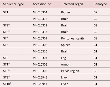
A total of ten different cox1 STs were identified among all 12 samples. Eight STs were among ten FFPE hydatid cyst blocks collected from unusually infected organs (table 3).
The cox1 STs and genotypes, infected organ and GenBank accession numbers of hydatid cyst isolates from North Khorasan Province included in the study.

aNew sequence types.
A brain CE sample (MH010312) and the renal cyst (MH010304) had a very similar sequence (ST1). These sequences were quite similar to the hydatid cysts isolated from Buffalo in India (DG269944; Bhattacharya et al., Reference Bhattacharya, Bera, Bera, Maity and Das2007), which exhibited maximum similarities with the G2 genotype of E. granulosus s.s.
Furthermore, two other brain cysts plus a cyst isolated from the peritoneum belonged to genotype G2. These isolates showed three new STs (ST2–ST4). One of these brain cysts (MH010311, ST2) and the cyst isolated from the peritoneum (MH010309, ST4) of a patient differed in one nucleotide from an Indian isolate (DG269944). Moreover, the third cerebral cyst (MH010313, ST3) and an Indian isolate (DG269944) had one nucleotide difference. The sequences of the fourth brain cyst (MH010310), in addition to the cyst isolated from a patient's spleen (MH010308), were completely identical (ST5) with the sequences of hydatid cysts isolated from cattle (EU178104; Vural et al., Reference Vural, Baca, Gauci, Bagci, Gicik and Lightowlers2008), buffalo from Turkey (HM598459; Beyhan & Umur, Reference Beyhan and Umur2011) and sheep from Portugal (HF947559; Beato et al., Reference Beato, Parreira, Roque, Gonçalves, Silva, Maurelli, Cringoli and Grácio2013). These isolates were identified as genotype G1. The sequence of a cyst isolated from a patient's leg (MH010307, ST6) was identical to a cyst isolated from sheep in Portugal (HF947555) from genotype G1. Although the genotype of the armpit cyst (MH010306, ST7) was also G1, it differed in one nucleotide from a number of isolated hydatid cysts from sheep in Algeria (Laatamna et al., Reference Laatamna, Ebi, Brahimi, Bediaf, Wassermann, Souttou and Romig2019), as well as one Tunisian isolate (KY766885) from the research conducted by Kinkar et al. (Reference Kinkar, Laurimäe and Sharbatkhori2017). The cyst isolated from the pelvis in this study (MH010305, ST8) was most similar to the haplotype EgAlg51 (belonging to the genotype G3 of the E. granulosus s.s.) in the research conducted by Laatamna et al. (Reference Laatamna, Ebi, Brahimi, Bediaf, Wassermann, Souttou and Romig2019).
Two alcoholic cysts of the liver belonged to G1 genotype of E. granulosus and had two new distinct STs.
Phylogenetic analysis
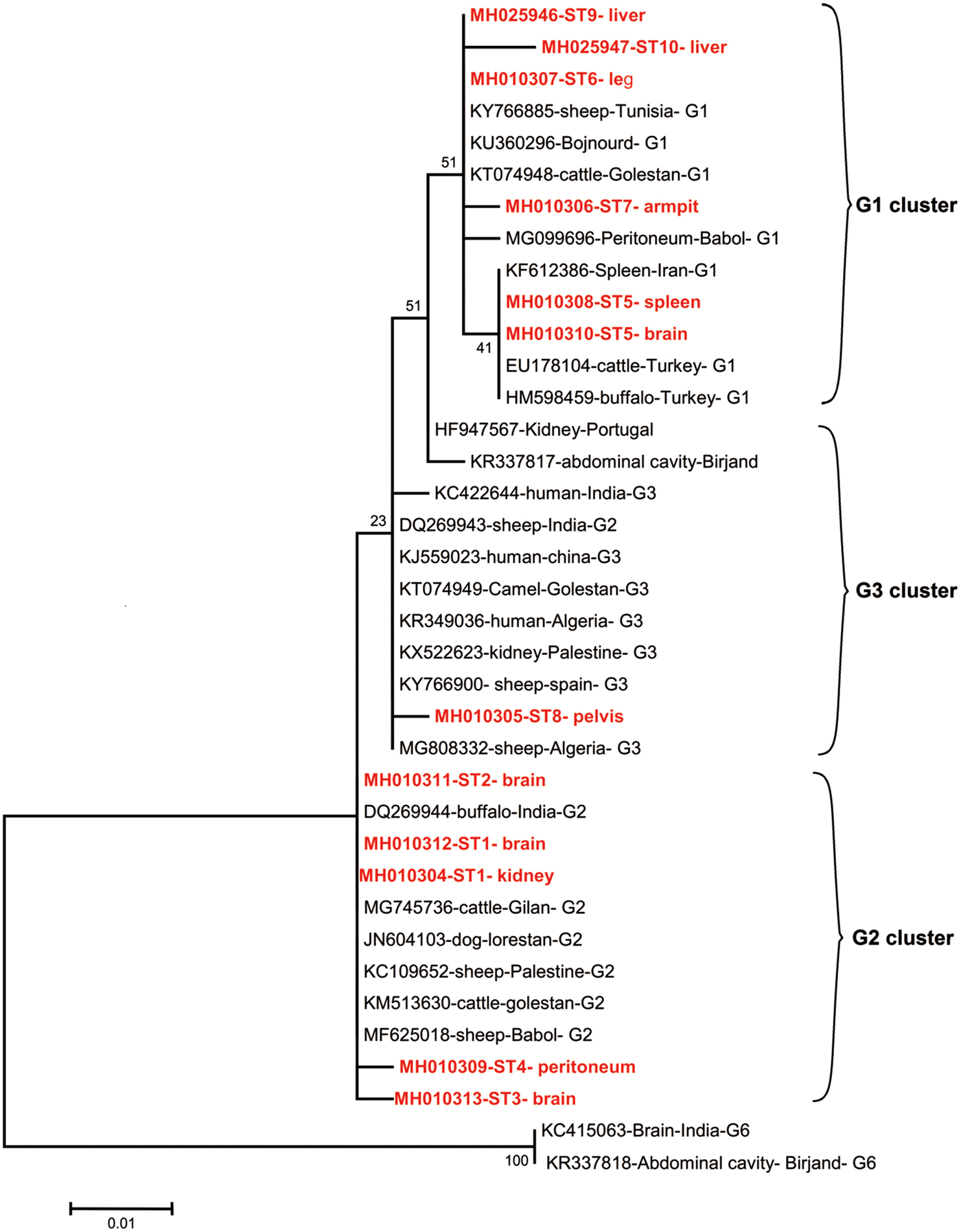
The phylogenetic association between the STs from usually and unusually located hydatid cysts of the current and other studies was investigated by generating an ML tree (fig. 1). The E. granulosus s.s. isolates consisted of one clade, where all G1 genotype STs were grouped in a main cluster in which we observed STs from armpit (ST3), leg (ST4), brain and spleen (ST5) and both liver (ST9–10) samples in our study. The brain and spleen hydatid samples from the current study and a spleen sample from another province of Iran and two samples from Turkey consisted of a subcluster in the G1 genotype main cluster. A peritoneum hydatid cyst isolated from another northern province in Iran (Mazandaran), and the armpit and liver hydatid isolates from the current study, showed different phylogenetic positions compared with other sequences in the main G1 genotype cluster. All defined G3 genotype sequences from GenBank, including a pelvic hydatid isolate of the current study (ST8) and a kidney isolate (Palestine), were grouped. The pelvic isolate showed a different position from most isolates in the G3 genotype group. One G2 genotype isolate was among the G3 genotype group, and two undefined-genotype isolates, namely an abdominal cavity (East Iran) and a kidney cyst, were positioned between the G1 and G3 clusters. Most G2 isolates showed similar positions in the tree, and five isolates of our study, comprising three hydatid cysts of the brain (STs 1-3) and the cysts isolated from the kidney (ST1) and the peritoneum (ST4), were in the genotypes G2 cluster. Two G6 genotype STs were used as out-group, forming a separate clade from all E. granulosus s.s. STs.
Maximum likelihood tree model generated using partial cytochrome c oxidase subunit I mitochondrial nucleotide sequences of Echinococcus granulosus sensu stricto and E. canadensis G6 genotype from FFPE blocks of extra-hepatopulmonary hydatid cysts and two liver cysts from north-east Iran, together with GenBank-retrieved haplotypes.

Discussion
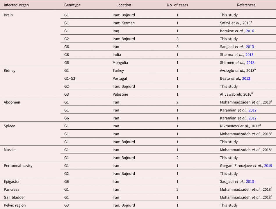
In this study, all unusually located human hydatid cysts in north-east Iran belonged to E. granulosus s.s. The kidney and peritoneum cysts and most of the brain cysts (three of four) were identified as the G2 genotype. The G1 genotype was found within the muscles of the limbs, spleen and one brain case, while G3 was only reported in a cyst belonging to the pelvic region. Based on the sequence data from our study and those retrieved from GenBank, all spleen and limb isolates were G1 genotypes (table 4), while the cysts from the peritoneum belonged to G1 and G2 genotypes. The kidney cysts' STs were found in G2 and also in the area between the G2 and G3 genotypes (probably G1/G3 genotype) based on the tree topology.
The genotypes of extra-hepatopulmonary hydatid cysts isolated from different organs of humans, reported from different parts of the world.

Data obtained by searching the scientific databases as well as genes registered in NCBI GenBank.
a Unpublished sequences in GenBank.
Until now, the G6 genotype of E. canadensis has been reported as the main cause of cerebral CE (Sadjjadi et al., Reference Sadjjadi, Mikaeili, Karamian, Maraghi, Sadjjadi, Shariat-Torbaghan and Kia2013; Sharma et al., Reference Sharma, Sehgal, Fomda, Malhotra and Malla2013; Shirmen et al., Reference Shirmen, Batchuluun and Lkhamjav2018). As far as the authors of this research are concerned, the present study reported the first case of the E. granulosus s.s. G2 genotype in the brain of humans. Furthermore, G2 was the dominant genotype of brain cysts, while G1 genotype was reported from one remaining cyst in the current study. The only case of cerebral hydatid cysts due to the G1 genotype was previously reported by Karakoc et al. (Reference Karakoc, Kasimcan, Pipia, Tore, Alberti, Varcasia, Sav and Oruçkaptan2016). The sequence of one of the hydatid cysts in the brain (MH010312), and the isolate from kidneys (MH010304) of our patients, were completely identical to a micro-variant of the G2 genotype (DQ269944) from India (Bhattacharya et al., Reference Bhattacharya, Bera, Bera, Maity and Das2007).
Because of the rare occurrence of unusually located hydatid cysts, FFPE blocks of pathobiological laboratory banks are usually the only available material for molecular investigations. The problems associated with DNA extraction from these blocks (Schneider et al., Reference Schneider, Gollackner, Edel, Schmid, Wrba, Tucek, Walochnik and Auer2008; Rostami et al., Reference Rostami, Torbaghan, Dabiri, Babaei, Mohammadi, Sharbatkhori and Harandi2015) are assumed as another cause of the limited data on the unusually located cyst genotypes. In the present study, in the first stage of cox1 fragments sequencing with JB3 and JB4.5 primers, parts of the phosphatidyl glycerol phosphatase A (pgpA) and aldo/keto reductase genes of E. coli were sequenced in half of the FFPE blocks instead of the Echinococcus cox1 gene. The problem was solved using the method described in the Results, which seems to be useful in similar circumstances.
So far, several studies have been conducted in Iran to determine the genotypes of parasites causing hydatidosis in different final and intermediate hosts. As a result of these investigations, in most cases, G1 and G6 genotypes were observed in different hosts, while G3 and G2 genotypes showed the lowest percentages, respectively (Khademvatan et al., Reference Khademvatan, Majidiani, Foroutan, Tappeh, Aryamand and Khalkhali2019). In the case of human hydatid cysts, most patients were infected with the G1 and, subsequently, G6 genotype (Rostami et al., Reference Rostami, Torbaghan, Dabiri, Babaei, Mohammadi, Sharbatkhori and Harandi2015; Karamian et al., Reference Karamian, Haghighi, Hemmati, Taylor, Salehabadi and Ghatee2017); only one case of human hydatid cysts with the G2 genotype was reported prior to the present study (Rostami et al., Reference Rostami, Torbaghan, Dabiri, Babaei, Mohammadi, Sharbatkhori and Harandi2015). The reported cases of human hydatid cysts with the G3 genotype were limited to, at most, six cases (Pezeshki et al., Reference Pezeshki, Akhlaghi, Sharbatkhori, Razmjou, Oormazdi, Mohebali and Meamar2013; Rostami et al., Reference Rostami, Torbaghan, Dabiri, Babaei, Mohammadi, Sharbatkhori and Harandi2015). On the other hand, regarding the studies carried out in North Khorasan and its adjacent provinces, Golestan and Khorasan Razavi, most hydatid cysts from different hosts belonged to the G1 genotype (Fadakar et al., Reference Fadakar, Tabatabaei, Borji and Naghibi2015; Moghaddas et al., Reference Moghaddas, Borji, Naghibi, Shayan and Razmi2015; Gholami et al., Reference Gholami, Jahandar, Abastabar, Pagheh, Mobedi and Sharbatkhori2016; Kamelli et al., Reference Kamelli, Borji and Naghibi2016; Sharbatkhori et al., Reference Sharbatkhori, Tanzifi, Rostami, Rostami and Harandi2016). One explanation for the notably higher frequency of G2 (41.6%) and G3 (8.3%) in unusually located hydatid isolates in the current study, in comparison to the prevalence of the mentioned genotypes in Iran, is the high tendency of these genotypes to extra-hepatopulmonary tissues.
Oudni-M'rad et al. (Reference Oudni-M'Rad, Cabaret, M'rad, Chaâbane-Banaoues, Mekki, Zmantar, Nouri, Mezhoud and Babba2016) observed a genetic differentiation between the isolates of E. granulosus s.s. cysts by comparing the genotypes of hepatic and pulmonary hydatid isolates. Based on the current results, there may be such differences between the genotypes of common liver and lung cysts and the unusually located hydatid cysts in humans.
As shown in table 4, it is to be noted that concerning the reports on the genotyping of Echinococcus from different parts of the world, G6, G3 and G2 genotypes have a higher tendency towards non-hepatopulmonary organs, whereas only 30.7% of hydatid cysts of brain, kidneys and abdominal and peritoneal cavities have been identified as G1 genotypes of E. granulosus (Beato et al., Reference Beato, Parreira, Roque, Gonçalves, Silva, Maurelli, Cringoli and Grácio2013; Sadjjadi et al., Reference Sadjjadi, Mikaeili, Karamian, Maraghi, Sadjjadi, Shariat-Torbaghan and Kia2013; Sharma et al., Reference Sharma, Sehgal, Fomda, Malhotra and Malla2013; Karamian et al., Reference Karamian, Haghighi, Hemmati, Taylor, Salehabadi and Ghatee2017; Shirmen et al., Reference Shirmen, Batchuluun and Lkhamjav2018). Brain and kidneys were the organs with the highest variety of parasite genotypes. More investigations are required to clarify the reasons for the higher tendency of certain genotypes to these organs. On the other hand, a high genetic heterogeneity was seen among G1 genotype STs from unusually located hydatid isolates belonging to haplotypes with lower prevalence in the current study area and its adjacent areas.
It should also be mentioned that half of the extra-hepatopulmonary hydatid cysts in this study (ST1, ST5, ST6) and the hydatid cysts isolated from India, Turkey and Portugal (Bhattacharya et al., Reference Bhattacharya, Bera, Bera, Maity and Das2007; Vural et al., Reference Vural, Baca, Gauci, Bagci, Gicik and Lightowlers2008; Beyhan & Umur, Reference Beyhan and Umur2011) shared identical cox1 haplotypes. In contrast, there were no identical STs found in genotyped hydatid cysts in Iran. The rest of the STs (ST2–4 and 7–8) were reported for the first time in the world, and were more similar to haplotypes outside Iran. Meanwhile, all these identical haplotypes from other countries were collected from non-human intermediate hosts of E. granulosus. Indeed, all of the STs of extra-hepatopulmonary hydatid cysts in the present study were reported in humans for the first time.
Accordingly, the replacement of Echinococcus larvae in organs other than the liver and lung can be related to the genotype of the parasite. Genotypes G2 and G3 are more commonly found in unusual organs.
We hypothesized that less frequent genotypes in each geographic area are less likely to be consistent with placement in the host's usual organs (liver and lung), while they tend to create unusual infections that may be more severe for the host. As shown by Sadjjadi et al. (Reference Sadjjadi, Mikaeili, Karamian, Maraghi, Sadjjadi, Shariat-Torbaghan and Kia2013), the G6 genotype of Echinococcus, with a lower prevalence comparison with the G1 genotype in Iran, tends to infect the human central nervous system.
Financial support
This work was supported by the Vice-Chancellors of the Bojnourd University of Medical Sciences (project number 915-P-95) and the Birjand University of Medical Sciences (project number 4269).
Conflicts of interest
None.
Ethical standards
The current study was approved by the Ethical Committee of the Faculty of Medicine, Birjand University of Medical Sciences, Birjand, Iran (IR.BUMS.REC.1397.053). All participants gave their written informed consents for participating in this study, and subjects remained nameless, with no individual names revealed.