Implications
Ovum pick-up (OPU) is the only technique which enables studies on cattle in countries like India, where cattle slaughter is banned. This work reflects the importance and feasibility of using OPU to retrieve oocytes from cattle and producing embryos in vitro. This study confirms that the developmental competence of oocytes retrieved through OPU bears relation to their morphological appearance and the observed oocyte morphologies were comparable to slaughterhouse-derived cumulus–oocyte complexes (COCs), indicating that the quality of oocytes is not affected by procedure or instruments used in OPU which is confirmed by evaluating developmental potential of different grade COCs and supplemented by studying relative gene expression of the associated genes.
Introduction
Ovum pick-up (OPU) is increasingly used in cattle both as an alternative to superovulation as well as an opportunity to increase the maternal contribution to genetic improvement. The technique of OPU-IVF assumes additional importance in Indian subcontinent due to prevailing ban on cow slaughter. It, thus, is an important assisted reproductive technique for production of large number of offspring from cattle of high genetic merit, besides facilitating an increase in selection intensity and shortening of generation interval by the use of young heifers. Though the linking of this oocyte retrieval approach with in vitro embryo production proved to be a valuable new development in cattle breeding and embryo transfer industry (Duszewska and Reklewski, Reference Duszewska and Reklewski2000), the variation in the quality of recovered oocytes and their developmental potential remains a major concern. This variation in oocyte quality might be attributed to OPU devices such as needle (Bols et al., Reference Bols, Van Soom, Ysebaert, Vandenheede and de Kruif1996 and Reference Bols, Ysebaert, Van Soom and de Kruif1997) and negative aspiration pressure (Bols et al., Reference Bols, Van Soom, Ysebaert, Vandenheede and de Kruif1996 and Reference Bols, Ysebaert, Van Soom and de Kruif1997) that are used for oocyte retrieval, in addition to major animal effect (if any) such as age which has been shown to bear a considerable effect on both cleavage and blastocyst formation rates (Su et al., Reference Su, Yang, He, Li, Ma, Wang, Presicce and Ji2012). Since, morphological grading system, based on cumulus–oocyte complexes (COCs) aspirated from slaughter house obtained ovaries, seems ambiguous in OPU retrieved oocytes, owing to their harsher retrieval as compared with the follicular aspiration (Bols et al., Reference Bols, Jorssen, Goovaerts, Langbeen and JLMR2012; Merton et al., Reference Merton, Knijn, Flapper, Dotinga, Roelen, Vos and Mullaart2013), the relative expression of various genes associated with cumulus–oocyte interaction, fertilization, genome reprogramming and development would serve as an important criterion to do the same irrespective of, or in addition to, the morphological appearance of the COCs. In order to justify or negate this notion, the present study was carried out to examine the developmental competence of OPU-derived COCs by studying the expression profile of a panel of genes, related to oocyte proper, cumulus–oocyte interaction, fertilization, embryonic development and apoptosis, both at germinal vesicle (GV) stage as well as after in vitro maturation (IVM). The panel of genes selected for expression analysis included genes associated with: follicular development and ovulation like, CONNEXIN37, CONNEXIN43, BMP15, GDF9, bFGF (Gospodarowicz et al., Reference Gospodarowicz, Cheng, Lui and Bohlen1985; Goodenough et al., Reference Goodenough, Goliger and Paul1996; Fair et al., Reference Fair, Hulshof, Hyttel, Greve and Boland1997; Simon et al., Reference Simon, Goodenough, Li and Paul1997; Elvin et al., Reference Elvin, Clark, Wang, Wolfman and Matzuk1999; Otsuka et al., Reference Otsuka, Yao, Lee, Yamamoto, Erickson and Shimasaki2000; Ackert et al., Reference Ackert, Gittens, O’Brien, Eppig and Kidder2001; Nilsson et al., Reference Nilsson, Parrott and Skinner2001; Granot and Dekel, Reference Granot and Dekel2002; Gittens et al., Reference Gittens, Mhawi, Lidington, Ouellette and Kidder2003; Moore and Shimasaki, Reference Moore and Shimasaki2005); Fertilization like, ZP2 and ZP3 (Chen et al., Reference Chen, Costa and Beja-Pereira2011; Shah et al., Reference Shah, Saini, Ashraf, Zandi, Manik, Suresh, Palta and Chauhan2015); Embryonic-genome activation like, ZAR1, HSF1 (Christians et al., Reference Christians, Davis, Thomas and Benjamin2000; Wu et al., Reference Wu, Viveiros, Eppig, Bai, Fitzpatrick and Matzuk2003); and Embryonic survival like, BAX, BID, BCL-XL, MCL1 (Chang and Yang, Reference Chang and Yang2000; Jin et al., Reference Jin, Li, Choi, Choi, Kim, Cui and Kim2007; Goovartaes et al., Reference Goovaerts, Leroya, Rizos, Bermejo-Alvarez, Gutierrez-Adan, Jorssen and Bols2011). The developmental competence was further elucidated by comparing embryonic developmental potential of Group 1 and Group 2 oocytes, following in vitro fertilization (IVF) and culture of the presumed zygotes.
Material and methods
Reagents and media
All chemicals were procured from Sigma Chemical Co., St. Louis, MO, USA, unless otherwise indicated. Fetal bovine serum (FBS) was from Hyclone (Logan, UT, USA). For total RNA extraction, RNAqueous-Micro Kit (Ambion, Austin, TX, USA) was used, while Superscript III first strand cDNA synthesis kit (Invitrogen, Waltham, Massachusetts, USA) was used for cDNA preparation. SYBR green was purchased from Bio-Rad, Hercules, California, USA, and the primers were purchased from Integrated DNA Technologies (IDT, Skokie, Illinois, USA). All the disposable plastic ware was from Nunc, Rosklide, Denmark and Thermo Fisher Scientific, MA, USA, whereas the 0.22 µm filters were from Millipore Corporation, Bedford, MA, USA. The ultrasound scanner (SSD-500) was from Aloka, Mitaka, Tokyo, Japan, transvaginal transducer from Corometrics Medical Systems, Inc., Wallingford, Connecticut, USA, vacuum pump from Karl Storz Endoskpe, West Germany, and needles (single lumen, 18-G, 55 cm long and with an echogenic tip) used for collection of oocytes were from ALPS Surgico, Ambala, India.
Donor cows and scheme of OPU
Healthy, cross-bred Karan Fries cows (n=6) aged between 13 and 15 years, which were otherwise put to auction, were used for OPU. They were evaluated by transrectal palpation and ultrasonography before each procedure. No exogenous hormones were given to stimulate folliculogenesis or synchronize follicular development. OPU from all the animals was performed on twice-weekly basis based on the assumption that COCs in this schedule will be from growing healthy follicles (3 to 8 mm in diameter). A total of 25 sessions were conducted on the six cows, over a period of 3 months.
In vitro production of embryos
Collection of oocytes by ovum pick-up
Follicular aspiration was performed using an ultrasound machine (Aloka SSD-500, Japan) with a 5 MHz transvaginal convex transducer, equipped with a needle guide, single lumen 18 G 55-cm long sterile needle with an ultrasound echo tip and a vacuum pressure of 90 mm Hg. The oocyte retrieval was performed under general (Rompun, 0.8 ml/animal) and epidural (2% Xylocaine, 3 to 5 ml/animal) anaesthesia, administered 15 min before the start. The contents (oocytes, follicular fluid, granulosa cells and tissue debris) retrieved were allowed to settle down for 10 to 15 min in 50 ml falcon tube, the sediment was collected and searched for oocytes under a zoom stereomicroscope at 20× magnification (Manik et al., Reference Manik, Chauhan, Singla and Palta2002).
Grouping of the retrieved oocytes
The collected oocytes were classified into four categories based on surrounding cumulus cell layers and homogeneity of ooplasm, as per the criterion established by Chauhan et al. (Reference Chauhan, Singla, Palta, Manik and Madan1998) for slaughterhouse-derived COCs: grade A (COCs, with an unexpanded cumulus mass having five or more layers of cumulus cells surrounding the zona pellucida and with homogenous cytoplasm); grade B (COCs with an unexpanded cumulus mass having two to four layers of cumulus cells and with homogenous cytoplasm); grade C (oocytes partially denuded of cumulus cells and with regular cytoplasm) and grade D (oocytes completely denuded of cumulus cells and with regular or irregular shrunken cytoplasm). The graded oocytes were further categorized into two groups: Group 1 – Good quality which included A and B grade COCs, and Group 2 – Poor quality which included C and D grade COCs. A total of 285 COCs were graded as of good quality while the rest (334) as of poor quality. Since, the number of COCs retrieved per session was not fairly good, so the graded oocytes from each session were pooled together and either exclusively used for: (i) total RNA extraction at GV stage (IMM A+B/C+D); (ii) total RNA extraction after IVM (MAT A+B/C+D); and (iii) IVF for embryo production.
The COCs retrieved from seven sessions were pooled into their respective groups (60 to 70 in each group), based on their morphology and used for total RNA extraction at GV-stage for qPCR gene expression analysis.
IVM of oocytes
The COCs retrieved from another 11 sessions (12 to 20 COCs per session) were subjected to IVM. Group 1 and Group 2 COCs, from each session, were washed separately, four to six times in washing medium (TCM-199+10% FBS+0.81 mM sodium pyruvate+50 µg/ml gentamicin sulfate), followed by two washes in maturation medium (TCM-199+10% FBS+5 µg/ml pFSH+10 µg/ml LH+1 µg/ml estradiol-17β+50 µM cysteamine+0.81 mM sodium pyruvate+10% buFF+50 µg/ml gentamicin sulfate) and placed in 100 µl IVM droplets, in a 35 mm Petri dish and overlaid with sterile mineral oil, followed by incubation at 38.5°C in a humidified CO2 incubator (5% CO2 in air, ⩾95% humidity). After 22 to 24 h of incubation, the matured oocytes were collected separately and washed thrice with DPBS to remove the media components. The respective Group 1 and Group 2 matured COCs were stored in RNA later at 4°C. A total of 60 to 70 COCs, pooled from 11 separate IVM experiments for each group, were employed for total RNA extraction for qPCR gene analysis.
IVF and culture of embryos
The respective grade oocytes retrieved from the rest of the sessions (seven sessions), were employed separately for IVF and subsequent culture for in vitro embryo production. The semen used for IVF throughout the study was from the same proven cattle bulls, obtained from Animal Breeding Research Center of the institute. The processing of spermatozoa for IVF was carried out as per the protocol described by Chauhan et al. (Reference Chauhan, Singla, Palta, Manik and Madan1998). Briefly, the frozen straws were thawed at 37°C and centrifuged twice at 1000×g for 8 min in BO (Brackett and Oliphant’s) medium; 50 µl of the capacitated spermatozoa, at a concentration of 1 to 2×10⁶ motile spermatozoa per ml, were added to 1% fatty acid free bovine serum albumin (BSA) supplemented BO droplets (50 µl) containing the COCs, on a sterile 35 mm Petri dish. The contents of the droplet were mixed by gentle shaking and overlaid with sterile embryo non-toxic mineral oil. After 18 to 20 h incubation, the cumulus cells were washed off the presumptive zygotes by gentle pipetting, followed by three washes in RVCL® (Cook, Queensland, Australia) medium supplemented with 1% fatty acid free BSA. The presumed zygotes were cultured in this medium for 8 days post insemination on original beds of granulosa cells with 50% of the media being replaced every 48 h. The developmental potential of the presumed zygotes obtained from Group 1 and Group 2 COCs was evaluated from their respective cleavage rates and progression through 2-, 4-, 8- to 16-cell, morulae and up to blastocyst stages. A total of eight such IVF and IVC experiments were performed to investigate embryonic developmental potential of the COCs from each Group.
Real time quantitative PCR (qPCR)
PCR primers were designed based on published nucleotide sequences when and where available or were based on sequence homology analysis (using ClustalW) between murine, human, sheep, goat and bovine sequences obtained from NCBI (www.ncbi.nlm.nih.gov) and ENSEMBL genome browser, for the respective genes. Primers were designed and verified using the web-based software PRIMER-3/Beacon designer. Total RNA was extracted separately from the respective Group 1 and Group 2 pools, both at GV stage and after IVM using RNAqueous Micro Kit (Ambion, Foster City, California, USA), as per the manufacturer’s instructions. The extracted RNA was quantified in a Nanodrop and used for cDNA preparation when 260/280 absorbance ratio was in the range of 1.9 to 2.0. A 20 ng of the total RNA (extracted from 15 to 20 COCs) was used for cDNA preparation with Superscript III first strand cDNA synthesis kit (Invitrogen), using oligodT primers following manufacturer’s instructions. cDNA formation was confirmed by PCR reactions set up in a final volume of 25 μl having 10× PCR buffer, 10 mM dNTPs, 1.0 U of DreamTaq DNA polymerase and 10 pmol/μl each of forward and reverse primers for Glyceraldehyde-3-phosphate dehydrogenase (GAPDH), at 56°C annealing temperature. The PCR reactions for other gene specific primers (Supplementary Table S1) were standardized by employing gradient PCR to find the most suitable annealing temperature for the corresponding primers. Real-time PCR, after optimization of the reactions, was performed in CFX96 system (Bio-Rad) using SYBR green master mix (Bio-Rad). The reactions were performed in triplicates in a final volume of 10 μl using specific primers for each gene (Supplementary Table S1) under the following amplification conditions: initial denaturation at 95°C for 3 min; followed by 40 PCR cycles (denaturation: 95°C for 15 s; annealing temperature: respective for each gene given in Table 1)) for 15 s; extension: 72°C for 15 s, melting cycle starting from 65°C up to 95°C with a 0.5°C/s transition rate. GAPDH and β-ACTIN were taken as the endogenous control gene and the assay was repeated four times. The real time PCR specificity was further confirmed by the melt curve analysis. The results are the average
 $${\rm 2}^{{{\minus}{\rm ddC}_{{\rm t}} }} $$
values±s.e.m. of four such qPCR assays (n=4).
Number of oocytes of different grades of crossbred cattle (Karan fries) obtained through ovum pick-up

Table represents the data of 25 sessions, carried on six animals.
1 Data is presented as mean ±s.e. of mean.
Statistical design
Statistical analysis was performed using SPSS version 17.0 (SPSS Inc., Armonk, New York, USA). The relative gene expression was compared by
 $${\rm 2}^{{{\minus}{\rm ddC}_{{\rm t}} }} $$
method (Schmittgen and Livak, Reference Schmittgen and Livak2008), taking the geometric mean of GAPDH and β-ACTIN as the calibrator. The gene expression data was analyzed between status (immature v. mature), grade (Group 1 v. Group 2) and interactions by using two-way ANOVA. P-values were corrected according to Bonferroni and the difference was considered statistically significant at P<0.05. The embryonic developmental rates were analyzed for significance using student’s t-test.
Results
Embryonic development of the retrieved oocytes
A cumulative total of 619 COCs were collected (130 A-grade, 155 B-grade, 117 C-grade and 217 D-grade COCs) which amounts to 24.76±2.4 COCs per session (Table 1). A total of 94 Group 1 and 105 Group 2 COCs retrieved from seven separate sessions were subjected separately to IVM, fertilization and in vitro culture. Embryonic development in Group 2 oocytes was significantly lower as compared with Group 1 oocytes (Table 2). The cleavage rate in Group 2 was found to be significantly lower than that of Group 1 COCs, as observed 48 h post-insemination (Table 2). The development of the presumed zygotes from Group 2 oocytes got arrested at 8- to 16- cell stage, (Figure 1) while Group 1 oocytes developed up to morula and blastocyst stages, 8 days after IVF (Figure1; Panel 1 and 2), indicating the better developmental competence of the latter. The results were found to be significantly different at all stages. This reflects on the initial quality of oocytes used in the culture, as oocyte quality was the only major variable involved.
In vitro embryo production of ovum pick-up (OPU)-derived cumulus–oocyte complexes (COCs) Panel 1; (a) A/B grade (Group 1) COCs, (b) In vitro matured COCs, (c) 2-cell stage, (d) 4-cell stage, (e) 8- to 16-cell stage, (f) Morulae, (g) Blastocyst. Panel 2: Corresponding stages of C/D grade (Group 2) COCs (a’ to e’).

Embryonic developmental competence of crossbred Karan Fries cattle oocytes obtained through ovum pick-up

Data from seven sessions.
Given values are percentage (mean±s.e.m.) as well as absolute values (n) from total number of respective oocytes taken (n).
a,bValues within a column having different superscripts differ significantly (P<0.05).
1 2-cell%: number of embryos cleaved/number of embryos cultured.
2 4-cell%, 8 to 16 cell%, morula%, blastocyst%: number of embryos of respective stage/number of embryos cleaved.
Expression of genes related to COCs
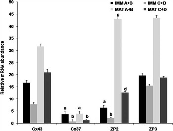
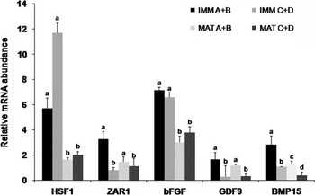
Among the genes that mediate interaction between cumulus cells and oocyte proper, expression analysis of CX43 and CX37 was examined. Group 2 oocytes showed a non-significant and significant decrease in expression of CX43 and CX37, respectively, both before (Figure 2) and after IVM (Figure 2), when compared with Group 1 COCs. Group 2 immature as well as in vitro matured COCs showed significantly lesser expression of GDF9 than corresponding Group 1 COCs (Figure 3). This reflects that COC quality affects the GDF9 expression and not the maturation stage (GV stage or after IVM). BMP15 showed significantly increased expression in immature as well as mature Group 1 COCs. The expression in both Group 1 and Group 2 COCs, however, decreased significantly following IVM, indicating the effect of maturation on BMP15 expression (Figure 3). However, no statistically different interaction was found between oocyte quality and state of maturation on the expression of these genes.
qPCR expression analysis of genes associated with cumulus–oocyte interaction and fertilization (CX43, CX37, ZP2 and ZP3) in immature and in vitro matured A/B (Group 1) and C/D grade (Group 2) COCs. The values represent the mean±s.e.m. of 4 (n=4) separate qPCR experiments. Bars with different superscripts differ significantly (P<0.05).

qPCR expression analysis of genes associated with cumulus–oocyte interaction and embryonic development (HSF1, ZAR1, bFGF, GDF9 and BMP15) in immature and in vitro matured A/B (Group 1) and C/D grade (Group 2) COCs. The values represent the mean±s.e.m. of 4 (n=4) separate qPCR experiments. Bars with different superscripts differ significantly (P<0.05).

Expression of genes related to fertilization
Among the genes that play a significant role in fertilization, the expression of ZP2 and ZP3 was analyzed. It was found that ZP2 exhibited a significantly greater expression in Group 1 oocytes as compared to Group 2 oocytes at GV stage (Figure 2), indicating the better sperm binding potential of the former. However, a significant (P<0.05) increase in expression of ZP2 in both Groups of COCs was observed following maturation, indicating the effect of both COC quality as well as maturation state on ZP2 expression. The same pattern, though statistically non-different was observed for ZP3 expression (Figure 2). No statistically different interaction was found between oocyte quality and state of maturation for the expression of these genes.
Expression of genes related to developmental competence
The expression of HSF1 was not significantly different between Groups 1 and 2 COCs both before and after IVM. Its expression, however, decreased within each group following IVM, indicating probably, a response to some sort of developmental stress (Figure 3). ZAR1, a maternal-effect gene, related to oocyte development beyond cleavage, was expressed in significantly lesser amounts in Group 2 COCs as compared to that of Group 1, both before and after maturation (Figure 3). The expression within each Group was not significantly affected by IVM, suggesting its dependence on COC quality. A pattern somewhat similar to HSF1 expression was observed in case of bFGF expression (Figure 3). From two-way ANOVA analysis, no statistically different interaction was found between oocyte quality and state of maturation for the expression of abovementioned genes.
Genes related to apoptosis and survival
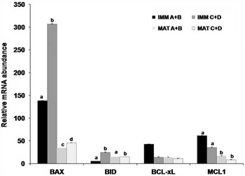
The pro-apoptotic genes, BAX and BID, showed a significantly greater expression in Group 2 oocytes than their counterparts, both at GV-stage as well as after IVM (Figure 4). The expression of BAX, however, showed a significant decrease after IVM (Figure 4) within each group, suggestive of an interplay of both oocyte quality as well as maturation state on its expression. The expression of BID did not alter significantly after IVM. Group 1 COCs showed greater expression of anti-apoptotic gene namely, MCL1, than Group 2 COCS, both before and after maturation (Figure 4). However, MCL1 expression showed a significant decrease within each group following IVM of the COCs. bFGF, in addition to its role in developmental competence, is anti-apoptotic in granulosa cells, showed expression pattern similar to MCL1 Tilly et al., 1992. However, no significant interaction was found between oocyte quality and oocyte state of maturation on BAX and MCL1 gene expression, unlike that of BID which showed a statistically significant interaction.
qPCR expression analysis of pro- and anti-apoptotic genes (BAX, BID, BCL-XL, MCL1) in immature and in vitro matured A/B (Group 1) and C/D grade (Group 2) cumulus–oocyte complexes. The values represent the mean±s.e.m. of 4 (n=4) separate qPCR experiments. Bars with different superscripts differ significantly (P<0.05).

Discussion
The lesser developmental competence and blastocyst formation rate, even of Group 1 COCs in our study, as compared with previous reports (Pontes et al., Reference Pontes, Melo Sterza, Basso, Ferreira, Sanches, Rubin and Seneda2011; Goovaerts et al., Reference Goovaerts, Leroya, Rizos, Bermejo-Alvarez, Gutierrez-Adan, Jorssen and Bols2011) could be due to the advanced age of the donor animals used in our study. A decrease in cleavage rate as well as blastocyst formation rate, following IVF, was observed in COCs retrieved by OPU from old cows (>15 years age) in comparison to middle-aged and young cows in a recent study conducted by Su et al. (Reference Su, Yang, He, Li, Ma, Wang, Presicce and Ji2012). The present study revealed that expression of various genes involved in developmental competence of oocytes is directly related to their quality, vis-a-vis morphological appearance of the retrieved COCs. For example, decreased expression of CX37, as observed in Group 2 COCs, leads to decreased interaction between granulosa cells and oocyte proper, thereby affecting the meiotic competence, as evidenced by the lesser cleavage rate in Group 2 COCs. Our results are in accordance with Fair et al. (Reference Fair, Hulshof, Hyttel, Greve and Boland1997) for their studies on bovine in vitro oocyte maturation. The decreased expression of BMP15 in Group 2 COCs explains for their decreased cleavage rate, as decreased BMP15 expression causes a corresponding decrease in FSH receptor on granulosa cells which in turn leads to inefficient maturation and subsequent embryonic development (Simon et al., Reference Simon, Goodenough, Li and Paul1997). GDF9 has been demonstrated to play an essential role in both early and late folliculogenesis as well as in cumulus expansion (Kovanci et al., Reference Kovanci, Rohozinski, Simpson, Michael, Colin, Bishop, Sandra and Carson2006). This might be the reason for its increased expression in Group 1 COCs which showed better embryonic development, as observed in our study. Furthermore, the decreased cleavage rate after fertilization and presumptive zygote formation, as observed in Group 2 COCs, might be due to decreased expression of ZP2 glycoprotein complex which is involved in spermatozoa–oocyte binding and fertilization. The increased expression of ZP2 gene after IVM might be due to increased activity of Fig-alpha (Factor in the germline-alpha), a helix-loop-helix transcription factor, that regulates expression of ZP1, ZP2 and ZP3 glycoproteins (Soyal et al., Reference Soyal, Amlech and Dean2000). HSF1 and ZAR1 are maternal effect genes that mediate oocyte progression through meiotic metaphase I (Leader et al., Reference Leader, Lim, Carabatsos, Harrington, Ecsedy, Pellman, Mass and Leder2002). The decreased expression of ZAR1 in Group 2 COCs might be the reason for their arrest at the zygotic stage and lack of syngamy between the maternal and paternal pronuclei (Wu et al., Reference Wu, Viveiros, Eppig, Bai, Fitzpatrick and Matzuk2003), hence poorer cleavage rate and subsequent arrest at 8- to 16-cell stage, as observed in our study. The decrease in expression of HSF1 and ZAR1 after IVM, as observed in our study, is in agreement with previous studies conducted in bovine species (Adona et al., Reference Adona, de Bem, Mesquita, Rochetti and Leal2010; Romar et al., Reference Romar, Santis, Papillier, Perreau, Thelie, Dell Aquila, Mermillod and Dalbies-Tran2011). The increased expression of pro-apoptotic genes (BAX and BID) in Group 2 COCs functions as the indicator of their lower survival and developmental ability. The increased expression of the anti-apoptotic genes (bFGF and MCL1), in Group 1 COCs manifests their greater ability to thrive and develop into embryos. bFGF is required for optimal promotion of the primordial to primary oocyte transition (Nilsson et al., Reference Nilsson, Parrott and Skinner2001) and is anti-apoptotic in granulosa cells (Tilly et al., Reference Tilly, Billig, Kowalski and Hsueh1992). The cumulative effect of the increased expression of all these oocyte-specific as well as maternal-effect genes, under study, might have led to progression and development of Group 1 COCs up to morula and blastocyst stage as compared to Group 2 COCs, where development progressed up to 8- to 16- cell stage only, despite of the identical culture conditions. Taking earlier studies into consideration (Liang et al., Reference Liang, Lu, Chen, Zhang, Lu, Zhang, Pang, Huang and Lu2008), it could be concluded that a significant difference does exist in oocyte maturation, fertilizability and developmental competence between different quality oocytes (as classified based on morphological appearance) retrieved through ovum pick-up. Hence, morphological appearance of the COCs could serve as a preliminary and reliable quality indicator for assessing the developmental competence. Despite of assessing the oocyte quality at immature stage (morphological criterion) as well as at matured stage (after GVBD) and over time in culture to blastocyst stage, an urgent need is felt for additional non-invasive quality assessment procedures in which oocyte survives the procedure to re-enter into in vitro production system to develop into the blastocyst and eventually confirm the reliability of the initial quality assessment criterion.
Acknowledgments
Authors acknowledge Department of Biotechnology, Govt. of India, New Delhi for providing financial assistance to carry out the work.
Supplementary material
To view supplementary material for this article, please visit http://dx.doi.org/10.1017/S1751731115001226