Introduction
Species of Echinoparyphium Dietz 1909 infect the gastrointestinal tracts of aquatic birds and mammals worldwide (McCarthy, Reference McCarthy1990; Kostadinova & Gibson, Reference Kostadinova, Gibson, Fried and Graczyk2000). Echinoparyphium is distinct from other genera of Echinostomatidae Looss, 1899 because the species possess a well-developed head collar with 29–45 spines in a double row, with small spines covering the tegument and extending to the testicular level ventrally and to the anterior margin of the ventral sucker dorsally. The large testes are tandem and located in the centre of the hind body. They are elongated–oval, smooth or irregular, contiguous and post-equatorial. The ovaries are small, spherical, and median or submedian pre-equatorial. Based on these morphological features, the genus currently contains approximately 151 species (Kanev, Reference Kanev1990; Huffman & Fried, Reference Huffman and Fried2012). A total of seven species have been described from the Americas as follows: E. aconiatum Dietz 1909, E. flexum Linton 1892, E. rubrum Cort 1914, E. speotyto Buscher 1978, E. contiguum Barker & Bastron 1915 and E. recurvatum von Linstow 1873 from North America, and E. megacirrus Semenas, Brugni & Ostrowski De Nuñez 1999, from South America. The systematic position of Echinoparyphium within Echinorhynchidae has been examined and revised using morphological and molecular data from a limited number of species. In these early phylogenies, Echinoparyphium represents a monophyletic group and is sister to Echinostoma Rudolphi 1809 (Kostadinova et al., Reference Kostadinova, Herniou, Barret and Litlewood2003) or Hypoderaeum Dietz 1909 (Detwiler et al., Reference Detwiler, Bos and Minchella2010). Detwiler et al. (Reference Detwiler, Bos and Minchella2010) were the first to use sequences from multiple genes to delineate species in the genera Echinoparyphium, Echinostoma and Hypoderaeum from Echinostomatidae. From these results, they found a high level of intraspecific variation within Echinoparyphium and recognized a complex of three cryptic species in E. recurvatum.
The adult of E. recurvatum was described from the intestine of the scaup duck (Aythya marilla) in Germany and this echinostome species has also been reported from a range of waterfowl species and occasionally mammals in North America, Europe and Asia. The first and second intermediate hosts include the freshwater snails Lymnaea peregra, L. ovata and Planorbis planorbis from Europe; Radix auricularia coreana from Korea; and Physa gyrina, P. heterostropha, P. parkeri, Physa sp. and Stangicola emarginata from the United States (Sohn, Reference Sohn1998; Kanev et al., Reference Kanev, Fried and Radev2008).
During a malacological survey, snails identified as Physella cubensis in central Mexico were found to be infected with Echinoparyphium cercariae and metacercariae. The aim of the current study was to describe the life stages of an Echinoparyphium species obtained experimentally from snails and chicks. To confirm the identity of the species, sequences from cercariae, metacercariae and adults representing nuclear ribosomal DNA from the internal transcribed spacers (ITSs) and 5.8S gene were analysed. Samples were obtained from both experimental and natural infections.
Materials and methods
Sampling procedures
Snails identified as P. cubensis were collected, using a nylon net, from Patzcuaro Lake, Michoacan (19°41′00″N, 101°41′00″W) in Central Mexico. A total of 180 snails were collected and transported to the laboratory, where they were rinsed with tap water. They were then placed into individual vials with 10 ml of water each. Snails were left overnight with exposure to light. Live emerging cercariae were examined and then placed in other vials that contained uninfected snails (P. cubensis), with the aim of determining the second intermediate host and to obtain encysted metacercariae. To obtain adult echinostomes, 19 newborn chicks (Gallus gallus domesticus) were infected orally with 50 metacercariae each. Eggs from adult echinostomes were obtained and incubated for 13 days at room temperature in tap water (containing antimycotics) to observe eggs and miracidia. Chicks were necropsied 19 days post-infection. Additionally, adults identified as Echinoparyphium recurvatum were collected from naturally infected ducks (Anas clypeata) from Patzcuaro Lake, Michoacan state in central Mexico. Definitive hosts were captured using a shotgun under permission of FAUT 0202 issued by the Secretaría de Medio Ambiente y Recursos Naturales (SEMARNAT) of Mexico to M.G.V. Ducks and chicks were dissected, and their viscera were placed in Petri dishes containing a 0.75% saline solution and examined using a dissecting microscope. Digeneans were placed in 0.75% saline solution and fixed with a hot 4% formalin solution for morphological studies. Other specimens were preserved in 100% ethanol for DNA analyses.
Morphological and molecular analyses
Unflattened specimens preserved in hot 4% formalin solution were stained with Mayer's paracarmine, dehydrated in a graded ethanol series, cleared with methyl salicylate, and mounted on microscope slides in Canada balsam. Drawings were made with the aid of a drawing tube. Measurements are given in millimetres (mm) with ranges followed by means in parentheses. Voucher specimens were deposited in the Colección Nacional de Helmintos (CNHE), Instituto de Biología, Universidad Nacional Autónoma de México, Mexico City. The species identification was conducted following the key of Yamaguti (Reference Yamaguti1975).
Samples of cercariae, metacercariae and adults were placed individually in tubes and digested overnight at 56°C in a solution containing 10 mm Tris–HCl (pH 7.6), 20 mm NaCl, 100 mm disodium EDTA (pH 8.0), 1% sarkosyl and 0.1 mg/ml proteinase K. Following digestion, DNA was extracted from the supernatant using the DNAzol reagent (Molecular Research Center, Cincinnati, Ohio, USA) according to the manufacturer's instructions. The genes ITS1, 5.8S and ITS2 (internal transcribed spacer of the ribosomal RNA repeat, ~1200 bp) were amplified from nuclear ribosomal DNA using the polymerase chain reaction (PCR). ITS was amplified using the forward primer BD1 5′-GTCGTAACAAGGTTTCCGTA-3′ and the reverse primer BD2 5′-ATCTAGACCGGACTAGGCTGTG-3′ (Luton et al., Reference Luton, Walker and Blair1992). PCR reactions (25 μl) consisted of 10 μm of each primer, 2.5 μl of 10 × buffer, 2 mm MgCl2 and 1 U of Taq DNA polymerase (Platinum Taq, Invitrogen Corporation, São Paulo, Brazil). PCR cycling conditions included denaturation at 94°C for 5 min; followed by 35 cycles of 94°C for 1 min, annealing at 50°C for 1 min and extension at 72°C for 1 min; followed by a post-amplification incubation at 72°C for 10 min.
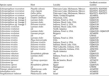
Sequencing reactions were performed using ABI Big Dye (Applied Biosystems, Boston, Massachusetts, USA) terminator sequencing chemistry, and reaction products were separated and detected using an ABI 3730 capillary DNA sequencer. Contigs were assembled and base-calling differences resolved using Codoncode Aligner version 3.5.4 (Codoncode Corporation, Dedham, Massachusetts, USA). Sequences were deposited in GenBank (table 1). Internal transcribed spacer sequences obtained in the current research were aligned with the following species downloaded from GenBank: E. recurvatum, Echinoparyphium sp., Echinostoma revolutum Froelich 1802; Echinostoma trivolvis Cort 1914, Echinostoma robustum Yamaguti 1935, Echinostoma paraensei Lie & Basch 1967, Echinostoma sp., Echinostoma caproni Richard 1964. Petasiger phalacrocoracis Yamaguti 1939, Isthmiophora melis Schrank 1788 and Paryphostomum radiatum Dujardin 1845 were also downloaded from GenBank and used as outgroups (table 1). All these sequences were aligned using the software Clustal W (Thompson et al., Reference Thompson, Higgins and Gibson1994) and adjusted manually with the MacClade program (Maddison & Maddison, 2011).
The occurrence of echinostome species from intermediate and definitive hosts worldwide, including GenBank accession numbers; nd, not known.

Maximum likelihood (ML) and Bayesian inference analyses (BI) were performed for the data set. The ML tree was inferred using RAxML 7.0.4. (Stamatakis, Reference Stamatakis2006). Modeltest program version 3.0 (Posada & Crandall Reference Posada and Crandall1988) was used for inferring the best model of nucleotide substitution for the data set. Tree searches were performed using 1000 (ML) random taxon addition heuristic searches. Clade support was assessed by bootstrap resampling with 10,000 replicates. Bayesian analyses were performed with MRBAYES version 3.1.2 (Huelsenbeck & Ronquist, Reference Huelsenbeck and Ronquist2001). Settings were two simultaneous runs of the Markov chain (MCMC) for 10 million generations, sampling every 200 generations, a heating parameter value of 0.2 and a ‘burn-in’ of 10%. Trees were drawn using FigTree version 1.3.1 (Rambaut & Drummond, Reference Rambaut and Drummond2007). The genetic divergence among taxa was estimated using uncorrected ‘p’ distances with the program MEGA v. 5 (Tamura et al., Reference Tamura, Peterson, Peterson, Stecher, Nei and Kumar2011).
Results
Morphology of larval and adult E. recurvatum
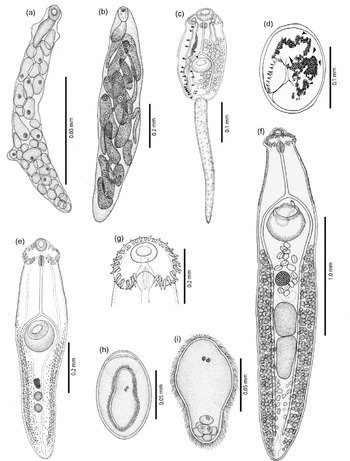
Intramolluscan stages were observed alive from infected snails. Measurements below are in millimetres. Active mother rediae have an elongate pink body 0.866–2.05 (1.27) long by 0.11–0.32 (0.20) wide, pharynx 0.112–0.199 (0.149) long by 0.090–0.165 (0.132) wide, followed by a well-developed caecal sac extending along the anterior third of the body (fig. 1a). Daughter rediae (fig. 1b) have a pink fusiform body containing cercariae. Ten daughter rediae measured 0.958–1.5 long (1.2) by 0.215–0.279 wide (0.25), pharynx 0.059–0.107 (0.081) by 0.057–0.104 (0.078) wide. Rediae have a sac-like caecum.
Morphology of Echinoparyphium recurvatum showing (a) mother rediae; (b) daughter rediae; (c) cercariae; (d) encysted metacercariae; (e) unencysted metacercariae; (f) entire adult worm; (g) anterior end with collar and spines; (h) egg and (i) miracidium.

Each cercaria (fig. 1c) possesses a spindle-shaped body, ovoid to triangular, tapering to the anterior part, 0.23–0.308 (0.272) long by 0.103–0.138 (0.121) wide. Tail long and thin 0.296–0.387 (0.328) long by 0.036–0.046 (0.040) wide. The length of each cercaria is 0.554–0.695 (0.601). The anterior body is armed with a head collar of 45 spines. The main body is packed with cystogenous glands at bladder level, running parallel to the excretory ducts. Oral sucker 0.043–0.052 (0.047) length by 0.035–0.052 (0.040) width, prepharynx 0.021–0.034 (0.026) by 0.011–0.026 (0.016). Intestinal caecae bifurcate anteriorly at the ventral sucker level. The ventral sucker is located post-equatorially 0.043–0.052 (0.047) length by 0.035–0.052 (0.040) width. Excretory system with 23 pairs of flame cells at the anterior level 5 (2[1+2+2]), at ventral sucker level 10 (2[2+1+3+2+2]) and at the rear 8 (2[3+3+2]).
Metacercariae spherical with a thin wall 0.114–0.156 (0.135) (fig. 1d). Ten excysted metacercariae were measured. Total length 0.783–0.844 (0.822) by 0.165–0.193 (0.178) wide. Head collar armed with 45 spines. Oral sucker subterminal 0.047–0.055 (0.050) length by 0.045–0.052 (0.047) width, pharynx 0.038–0.044 (0.040) by 0.019–0.025 (0.023), ventral sucker located equatorially 0.106–0.129 (0.12). Undeveloped reproductive system at the posterior end with two oval testes and one ovary (fig. 1e)
A total of 175 adult worms identified as E. recurvatum were collected from the intestine of 19 chicks (fig. 1f). Adult worms with spindle-shaped elongate body 2.75–3.22 (2.94) long by 0.40–0.55 (0.46) wide. Head collar with 45 spines in two alternating rows including 5 angular spines, 0.049–0.066 (0.057) long by 0.009–0.012 (0.010) wide, the remaining 35 spines 0.045–0.062 (0.054) by 0.006–0.012 (0.009) (fig. 1g). Oral sucker subterminal and spherical 0.078–0.120 (0.096) long by 0.093–0.110 (0.10) wide. Ventral sucker pre-equatorial and spherical 0.26–0.38 (0.32) by 0.24–0.32 (0.29); short prepharynx and pharynx 0.08–0.10 (0.09) long by 0.05–0.06 (0.055); caecae extend nearly to the posterior end. Testes are tandem and ovoid, located in the posterior third of the body. Anterior testis 0.20–0.36 (0.30) long by 0.09–0.22 (0.15) wide, smaller than posterior testis 0.28–0.45 (0.37) by 0.11–0.18 (0.14). Cirrus sac 0.19–0.26 (0.23) length by 0.09–0.15 (0.15) width located at the first part of the body containing the seminal vesicle. Genital pore located at ventral sucker level. Ovary spherical 0.11–0.14 (0.12) by 0.10–0.13 (0.12). Seminal receptacle ovoid and located between testis and ovary. Vitelline glands extend from the middle of the body (pre-ovaric area) until the end of the body. Eggs within uterus are unembryonated 0.070–0.096 (0.085) by 0.040–0.056 (0.047) (fig. 1h).
The miracidium (fig. 1i) was completely formed 13 days after egg deposition, and was very active, rolling on its axis. The body was elongated ovoid 0.095 by 0.035, and completely covered in cilia. Each miracidium had two anterior eye-spots, and a group of fat vesicles at the posterior end.
Molecular characterization
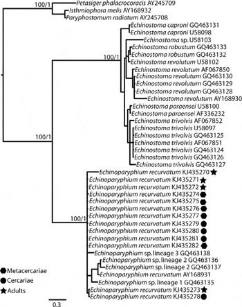
Sequences from ITS1, 5.8S and ITS2 were obtained from two experimental adults and two adults from the natural definitive host (A. clypeata). Additionally, sequences were obtained from four metacercariae and five cercariae isolated from nine intermediate hosts that were identified as E. recurvatum. These sequences were aligned with a data set that contained 27 echinostomatids obtained from GenBank (table 1). Maximum likelihood (ML) analysis yielded a single tree that was identical in topology to the Bayesian inference (BI) consensus tree (fig. 2). The ML and Bayesian consensus trees showed that all the new sequences from E. recurvatum (four adults, four metacercariae and five cercariae) formed a clade with strong bootstrap support and Bayesian posterior probability (100/1.0). This clade also included samples diagnosed morphologically as Echinoparyphium sp. from the USA and the UK. The genetic divergence among the 13 samples of E. recurvatum ranged from 0 to 0.6%.
Maximum likelihood (ML) and Bayesian consensus tree inferred from ITS data set. The topology of the ML and Bayesian consensus trees were identical; numbers near internal nodes show ML bootstrap clade frequencies (given first) followed by Bayesian posterior probabilities. The ML tree has a –ln likelihood of 3302.0363.

Discussion
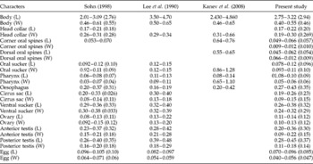
In the current study, the life-cycle stages of E. recurvatum under experimental conditions were characterized. This included non-adult stages from the snail P. cubensis, which serves as the first and second intermediate host. Adult worms were obtained experimentally from chicks and naturally from the shoveller duck (A. clypeta) in central Mexico. Morphologically, the adults obtained both experimentally and naturally had head collars armed with 45 spines in two alternating rows. End-group spines were larger than other head-collar spines. Other features of adults, including suckers, prepharynx, oesophagus and caecae, testes, cirrus sac, ovary and vitelline glands were identical to those described previously for E. recurvatum by other authors (Lee et al., Reference Lee, Sohn and Chai1990; Sohn, Reference Sohn1998; Kanev et al., Reference Kanev, Fried and Radev2008) (table 2).
Comparative morphometrics (in mm) of adult worms of Echinoparyphium recurvatum obtained in the present study with those of Sohn (Reference Sohn1998), Lee et al. (Reference Lee, Sohn and Chai1990) and Kanev et al. (Reference Kanev, Fried and Radev2008); length (L), width (W) and mean values given in parentheses after ranges.

To confirm the conspecificity among cercariae, metacercariae and adults obtained experimentally and naturally, sequences of the ITS1, 5.8S and ITS2 were obtained and compared to published sequences. The genetic divergence estimated among sequences obtained from four adults, five metacercariae and four cercariae from nine snails ranged from 0 to 0.6%. The level of genetic divergence among samples of E. recurvatum is similar to that found within other echinostomid species. For example, the genetic divergence of ITS among E. trivolvis is 0–0.8% and for E. revolutum, 0–1.8%. Within E. robustum, E. paraensei and E. caproni the ITS divergence is 0–0.1%. Maximum likelihood and Bayesian trees showed that the 13 isolates of E. recurvatum formed a monophyletic group with 100% ML bootstrap support and a Bayesian posterior probability of 1.0 (fig. 2). In another comparative study, Saijuntha et al. (Reference Saijuntha, Tantrawatpan, Sithithaworn, Andrews and Teney2011) analysed the first internal transcribed spacer (ITS1) of E. recurvatum from isolates collected in Thailand and the UK, and three isolates of Echinoparyphium sp. from the United States. In their phylogenetic analysis, E. recurvatum was paraphyletic, suggesting that this species could be a complex of species. Saijuntha et al. (Reference Saijuntha, Tantrawatpan, Sithithaworn, Andrews and Teney2011) suggested that the taxonomic status of E. recurvatum requires revision based on more comprehensive analysis of morphological and molecular data. The ML and Bayesian analyses inferred from additional ribosomal DNA data (both internal transcribed spacers plus 5.8S rDNA) recovered the three isolates of Echinoparyphium sp. from the United States plus E. recurvatum from the UK in a single clade with all other E. recurvatum, including the 13 isolates obtained experimentally and naturally from central Mexico. The marked low level of genetic divergence found among all isolates in addition to their monophyly in ML and Bayesian trees, concomitant with the morphological data of adults, metacercariae and cercariae, clearly demonstrates that all these specimens belong to the same evolutionary lineage. Within this clade, there were two subclades, one including individuals of lineages 1 and 2 as classified Detwiler et al. (Reference Detwiler, Bos and Minchella2010) plus E. recurvatum AY168931; the second subclade included two E. recurvatum specimens from this study. Neither of these subclades was supported as assessed by bootstrap resampling or Bayesian posterior probability.
In this study we report for the first time the freshwater snail P. cubensis as the first and second intermediate host of E. recurvatum, expanding its distribution range in the Americas. The adult of E. recurvatum has been recorded in two species of dabbling ducks in Mexico (see Farias & Canaris, 1986; Pérez Ponce de León et al., Reference Pérez Ponce de León, García-Prieto and Mendoza-Garfías2007). In the current study, adults of E. recurvatum were found in the intestines of the shoveller duck (A. clypeata), with metacercariae and cercariae found in the snail P. cubensis. These results indicate that the life cycle is completed in wetlands of central Mexico.
Financial support
This research was supported by the Programa de Apoyo a Proyectos de Investigación e Inovación Tecnológica (PAPIIT No. IN207213) and by Consejo Nacional de Ciencia y Tecnología (CONACyT No. 179048).
Conflict of interest
None.
Ethical standards
The authors assert that all procedures contributing to this work comply with the ethical standards of the national guides on the care and use of laboratory animals (Secretaría de Medio Ambiente y Recursos Naturales) and have been approved by the Instituto de Biología, UNAM.