Introduction
Significant progress has been made during the past three decades on enhancing juvenile body weights in broiler chickens. Along with faster growth (2.2-2.4 kg at 40d), feed efficiency (1.6-1.7) and the proportion of breast meat have been improved in commercial broilers. However, the faster growth has a negative effect on the immune system; thereby increasing the incidence of early chick mortality, probably due to reduced resistance to infectious diseases (Yunis et al., Reference YUNIS, BEN-DAVID, HELLER and CAHANER2000; Juul-Madsen et al., Reference JUUL-MADSEN, SU and SORENSEN2004). The immune system provides the body with the ability to mount a defence against foreign organisms and antigenic substances. Many reports have suggested that chicks in the first week of hatch lack adequate immune responses and remain highly susceptible to infections (Mast and Goddeeris, Reference MAST and GODDEERIS1999; Juul-Madsen et al., Reference JUUL-MADSEN, SU and SORENSEN2004). This has been attributed to the inadequacy of the immune systems development and its functional inability to effectively respond to the diversity of antigens required for protecting the chick during the early part of life (Panda et al., Reference PANDA, RAJU, RAMA RAO, SHYAM SUNDER and REDDY2010). Development of systemic immune competence in the chickens occurs during the post hatch period (Erf et al., Reference ERF, BOTTJE and BESRI1998; Mast and Goddeeris, Reference MAST and GODDEERIS1999).
With the continued increase in the economic importance of broiler chickens, a better understanding of the development and functioning of the avian immune system in these birds is essential. The avian immune system begins in the embryo and continues for several weeks after hatch. Several factors influence the early development of immune system in newly hatched chick, and one of the important factors is initial feed intake (Dibner et al., Reference DIBNER, KNIGHT, KITCHEL, ATWELL, DOWNS and IVEY1998). The transition from use of yolk sac derived nutrients to those present in exogenous feeds provides the correct platform for rapid functional and structural development of the gut (Geyra et al., Reference GEYRA, UNI and SKLAN2001). Feed provides nutrient substrates, which are essential for the growth and development of both primary and secondary lymphoid organs (Dibner et al., Reference DIBNER, KNIGHT, KITCHEL, ATWELL, DOWNS and IVEY1998). The immune system of the hatchling, particularly the mucosal immune system, also requires oral feed intake for rapid development (Bar Shira et al., Reference BAR SHIRA, SKLAN and FRIEDMAN2005). However, in commercial poultry operations, poultry hatch is delayed by over 2-days and chicks are only moved from the hatchery after the majority of chicks have emerged from their shells. Following the release, other hatchery practices including sexing, vaccination and packaging are carried out before they are boxed for transportation. Thus, in practice, chickens from a hatchery may often spend considerable time (48-72 h) without any access to feed or water. It has been reported that early feed deprivation not only affects bird growth (Noy and Sklan, Reference NOY and SKLAN1999; Bhanja et al., Reference BHANJA, ANJALI DEVI, PANDA and SHYAM SUNDER2009; Panda et al., Reference PANDA, RAJU, RAMA RAO and SHYAM SUNDER2009) but also immune system development (Juul-Madsen et al., Reference JUUL-MADSEN, SU and SORENSEN2004; Panda et al., Reference PANDA, RAJU, RAMA RAO, SHYAM SUNDER and REDDY2010). Early dietary restriction was reported to produce metabolic changes leading to impaired systemic immune competence (Nathan et al., Reference NATHAN, HELLER and PEREK1977; Hulsew et al., Reference HULSEW, VAN ACKER, VON MEYENFELDT and SOETER1999) possibly due to depleted lymphoid organ growth (Dibner et al., Reference DIBNER, KNIGHT, KITCHEL, ATWELL, DOWNS and IVEY1998; Panda et al., Reference PANDA, RAJU, RAMA RAO, SHYAM SUNDER and REDDY2010). It has been reported that the primary lymphoid organ, the thymus, is very sensitive to the duration of feed deprivation, and causes a rapid decline in its cellularity and weight gain (Klasing, Reference KLASING2007). Feed deprivation beyond 48 hours in chicks impaired the activity of gut associated lymphoid tissue in the hind gut and delayed the development of lymphocyte populations in the bursa during the first two weeks of life (Bar Shira et al., Reference BAR SHIRA, SKLAN and FRIEDMAN2005; Panda et al., Reference PANDA, RAMA RAO, RAJU, REDDY and SHYAM SUNDER2008). Thus, the time from hatching to the onset of receiving food is the critical period for their development and subsequent performance of the hatchlings. Although the effect of early post hatch nutrition has been widely studied and reviewed, most of the information published remains confined to broiler performance in terms of body weight gain, feed conversion, GI tract development and meat yield, and not on the immune system or immune competence on long-term scale. The present review aims specifically on the development and functioning of immune system and the role of early post hatch nutrition on the maturation of the immune system in broiler chickens.
Avian immune system
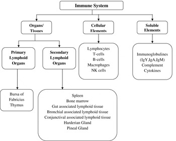
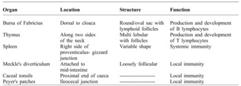
Among the avian species, the immune system of the chicken has been the most extensively studied. Because of its widespread function, the immune system is a complex system, both structurally (Figure 1) and functionally (Table 1). The immune system is widely dispersed throughout the body and it is composed of organs, cellular elements, and soluble (humoral) elements. Like in mammals, immunity in birds is developed through the lymphoid system. The organs of the immune system can be classified into primary (central) and secondary lymphoid organs (Figure 1).
Avian immune system.

The bursa of Fabricius and thymus are the primary lymphoid organs in which the lymphocyte precursors develop into immune competent native lymphocytes. Secondary lymphoid tissues are the spleen, bone marrow, harderian gland, pineal gland and organised lymphoid tissues associated with mucosal surfaces (MALT), including bronchial-associated lymphoid tissues (BALT), gut-associated lymphoid tissues (GALT), conjunctival associated lymphoid tissues (CALT) and other less-prominently organised clusters of lymphoid cells in various organs. These lymphoid tissues are located at strategic sites where foreign antigens entering the body from either the skin or a mucosal surface can be trapped and concentrated. Lymphocytes, antigen transporting and presenting cells and other regulatory cells are located in these anatomically defined tissues and are thought to be organised into structures that optimise cellular interactions to support the efficient removal of unwanted pathogens (McCorkle and Glick, Reference MCCORKLE and GLICK1980; Klasing, Reference KLASING2007).
Avian immune system, its related organs and their structure and functions.

Source: Panda and Reddy (Reference PANDA and REDDY2007)
Chickens basically have two types of defence systems to counter infectious agents; innate (non-specific) immunity and adaptive immunity (specific immunity). Non-specific immunity comprises of physical and chemical barriers (to prevent invasion) and cellular components (macrophages, neutrophils, natural killer cells, etc.). Heterophils are the avian equivalent to mammalian neutrophils. Specific immunity is made up of two parts; humoral immunity (carried out by antibodies) and cell-mediated immunity (carried out by T cell activity). The cellular components of specific immunity are lymphocytes. Chicken lymphocytes consist of B cells, which develop in the bursa of Fabricius and, the T cells in the thymus. Both, the B and T cells express antigen receptors on their surface. It has been reported that each B cell and T cell has a homogenous set of antigen receptors specific for a given antigen (Dibner et al., Reference DIBNER, KNIGHT, KITCHEL, ATWELL, DOWNS and IVEY1998).
Ontogeny of the avian immune system
An understanding of immune system development in neonate broiler chicks and its capacity to respond effectively to the diversity of antigens during the early part of their life is a priority. The development of the immune system in the embryonic and neonatal stages is a dynamic process involving the migration of lymphocyte stem cells to primary immune glands, the rapid and extensive proliferation of lymphocytes in the absence of any antigen within the bursa and thymus, the concurrent differentiation of bursal and thymic lymphocytes, and the eventual seeding of differentiated lymphocytes to the peripheral lymphoid organs such as the spleen and caecal tonsils (Bucy et al., Reference BUCY, CHEN, CIHAK, LOSCH and COOPER1988). The immune system in poultry is partially developed at hatch. The primary immune organs such as the bursa and thymus are present and are populated by lymphoid tissue. However, the cell mediated immune system has a fairly high degree of development during embryogenesis as compared to the humoral immune system (Perey and Bienenstock, Reference PEREY and BIENENSTOCK1973; McCorkle et al., Reference MCCORKLE, LUGINBUHL, SIMMONS, MORGAN and THAXTON1983).
The stem cell migration to chick thymus occurs in three waves during embryogenesis at 6.5, 12 and 18 days of incubation, and these cells pass through thymus to populate peripheral tissues (Bucy et al., Reference BUCY, CHEN, CIHAK, LOSCH and COOPER1988). The thymocytes are CD3+ (avian homologue) and develop to CD4 or CD8 antigens during embryogenesis (Lillehoj and Trout, Reference LILLEHOJ and TROUT1996). The seeding of bursa by lymphocytes occurs between 7.5 and 14 days of embryogenesis (Peault et al., Reference PEAULT, DIETERLEN-LIEVRE and LE DOURARIN1987). At this stage, these cells are committed B cells but are capable of expressing IgM at hatch only (Leslie and Martin, Reference LESLIE and MARTIN1973). Dibner et al. (Reference DIBNER, KNIGHT, KITCHEL, ATWELL, DOWNS and IVEY1998) reported that the dominant immunoglobulin in the bursa of the hatchling is IgM. The IgM-bearing lymphocytes are the precursor cells for the expression of IgG or IgA. The hatchlings do not have the capacity to produce IgG at this stage, and are entirely dependent upon maternal antibodies for humoral immune protection (Martin and Leslie, Reference MARTIN and LESLIE1973; Shawky et al., Reference SHAWKY, SAIF and SWAYNE1993).
The secondary immune organs such as the spleen, caecal tonsils and Meckel's diverticulum are incomplete at hatch (Schat and Myers, Reference SCHAT and MYERS1991). IgM-secreting B-cells are found in the spleen at three days of age whereas IgG-secreting B-cells are not detected until six days of age (Lawrence et al., Reference LAWRENCE, ARNAUD-BATTANDIER, GRAYSON, KOSKI, DOOLEY, MUCHMORE and BLAESE1981). Although B cells are detected in caecal tonsils at hatch, they only express IgM. There are T cells in the lamina propria and epithelium of the gut and other secondary immune organs, but these do not develop cytotoxic ability until some period after hatch. The ability to mount a secondary response, as indicated by the presence of germinal centres or circulating IgG and IgA, begins to appear between the first and fourth week post hatch in broiler chicks (Leslie, Reference LESLIE1975). Bar Shira et al. (Reference BAR SHIRA, SKLAN and FRIEDMAN2003) reported that gut associated lymphoid tissue contained functionally immature T and B lymphocytes at hatch and their functional maturation occurred during the first two weeks of life.
Residual yolk-sac and immunity in neonates
Towards the end of the incubation, residual yolk is internalised into the abdominal cavity and is the only nutrient source to the newly hatched chick until exogenous feeds are supplied (Romanoff, Reference ROMANOFF1960). At hatch, the yolk constitutes 20% of the chick's weight and provides immediate post hatch energy and protein for growth (Sklan and Noy, Reference SKLAN and NOY2000). Although the protein fraction is partly from albumen, a large content of the egg protein in hatchlings is made up of maternal antibodies (Dibner et al., Reference DIBNER, KNIGHT, KITCHEL, ATWELL, DOWNS and IVEY1998). In general, the maternal antibodies are not digested during the incubation process, leaving those immunoglobulins intact and fully functional at the time of hatch, indicating that the protein component of the yolk sac is used for passive immunity of the chicks. Thus, the chick should utilise the residual yolk sac faster to provide early immunity to the newly hatched chick. It is reported that residual yolk is used up more quickly by chicks that have early access to feed compared to those that are fasted for 48 hours (Noy et al., 1996, Bhanja et al., Reference BHANJA, ANJALI DEVI, PANDA and SHYAM SUNDER2009). The reason being that anti-peristaltic movement of yolk from yolk-stalk to the duodenum is stimulated by the presence of feed in the gut (Noy and Sklan, Reference NOY and SKLAN1999; Panda et al., Reference PANDA, SHYAM SUNDER, RAMA RAO and RAJU2006). However, hatchery operations in commercial poultry practices delay the access of feed and water and under practical conditions newly hatched chicks generally receive their first food and water 48-72 hours after hatching. This delay in access to nutrients may limit the utilisation of the yolk sac and result in poor immunity in the newly hatched chick (Bhanja, Reference BHANJA2008).
Furthermore, despite the residual yolk being sufficient to keep the birds alive for the first three to four days, it does not support the expression of the bird's genetic potential for body weight gain, development of the gastro-intestinal tract or immune system (Dibner et al., Reference DIBNER, KNIGHT, KITCHEL, ATWELL, DOWNS and IVEY1998). The same researchers studied the effect of early feeding on lymphocyte proliferation in bursa compared to fasting for 48 hours. It was observed that early feeding led to extensive proliferation of lymphocytes compared to fasted chicks, and that the nutrients of residual yolk may not be a substitute for oral intake in maintaining lymphocyte proliferation, as seen in chicks receiving oral nutrition. This could be attributed to the priority of use of the nutrients by the gut (Ricklefs, Reference RICKLEFS1987) and may be related to the presence of nutrient substrates and antigen exposure (Rao et al., Reference RAO, MCDUFFIE and GLICK1978). Consequently, prolonged feed restriction not only reduced yolk sac utilisation but also affected the development of the immune system.
Early nutrition and the development of immune organs and function
Early feeding has a significant effect on bursa and spleen development of chicks. Dibner et al. (Reference DIBNER, KNIGHT, KITCHEL, ATWELL, DOWNS and IVEY1998) reported significantly higher bursa weight on the 3rd day of broiler chicks fed immediately after hatch compared to those fasted for 48 hours, and the effect remained up to 21days of age. In addition, the lymphocyte proliferation in bursa was faster in the immediately fed chicks. In another study, Juul-Madsen et al. (Reference JUUL-MADSEN, SU and SORENSEN2004) observed that feed deprivation for 24 hours was acceptable for normal immunological response, but further delay in feeding up to 48 hours was unfavourable for immune response and growth in broilers, suggesting that newly hatched broilers can cope with a short duration of starvation. However, the results of Bhanja et al. (Reference BHANJA, ANJALI DEVI, PANDA and SHYAM SUNDER2009) and Panda et al. (Reference PANDA, RAJU, RAMA RAO, SHYAM SUNDER and REDDY2010) indicated that even the shorter duration of 24 hours feed deprivation post hatch depressed development of bursa and spleen, while feed deprivation up to 48 hours post hatch affected broiler growth. Later studies inferred that the immune system was more sensitive and susceptible to nutrient deprivation than growth and development. In a recent study, Rammouz et al. (Reference RAMMOUZ, SAID, ABBOUD, YAMMINE and JAMMAL.2011) reported that even 12-hour feed deprivation post hatch was harmful for the development of the immune system in poultry. Therefore the conflicting information reported by various researchers on immune system development due to delayed feed access requires further investigation, and the exact duration of post hatch deprivation that would not adversely affect the immune system maturation and its functioning needs to be determined.
Approximately 70% of the body's immune system is found in the digestive tract of chickens. One of the key stimulators of intestinal development in the chick is physical exposure to feed, while feed deprivation delays the onset of gut development (Geyra et al., Reference GEYRA, UNI and SKLAN2001; Panda et al., Reference PANDA, RAMA RAO, RAJU, REDDY and SHYAM SUNDER2008). Further, intestinal development occurs in continuation with that of gut associated lymphoid tissue (GALT), which is a component of the mucosal immune system (Thompson et al., Reference THOMPSON, MAYRHOFER and CUMMINS1996; Bar Shira et al., Reference BAR SHIRA, SKLAN and FRIEDMAN2003). GALT is made up of several types of lymphoid tissue, the caecal tonsils, Peyer's patches, Meckel's diverticulum and the bursa of Fabricius that produce and store immune cells to attack and defend against pathogens. GALT in chickens is responsible for the initiation of immune responses against bacterial, viral and parasitic challenges. The development of GALT in the hindgut of broiler chicks was delayed due to early feed deprivation, which persisted for two weeks of life (Bar Shira et al., Reference BAR SHIRA, SKLAN and FRIEDMAN2005). Delay in the onset of feeding results in delay of the maturation and expression of the functional activity of T lymphocytes in the hindgut and also delays lymphocyte proliferation in the bursa of fabricius. It should be emphasised here that the initial first week of life is very critical for the chick's survival and subsequent performance, as delayed access to feed takes nearly two weeks to achieve normalcy. Therefore, chicks with no access to feed immediately after hatch may be more vulnerable to environmental pathogens than those fed immediately after hatch. This vulnerability needs attention particularly in situations where chicks are in transit from hatchery to farms for extended periods.
The mechanism involving the effect of feed deprivation on the recession of lymphoid organ weights is not known, but one possible explanation for the negative effects on weight could be due to the rise in glucocorticoids associated with fasting (Dibner et al., Reference DIBNER, KNIGHT, KITCHEL, ATWELL, DOWNS and IVEY1998). Gulcocorticoids have been reported to hamper the involution of primary lymphoid organs, even in young birds (Nir et al., Reference NIR, NISTAN, DUNNINGTON and SIEGEL1996). Bigot et al. (Reference BIGOT, TESSERAUD, TAOUIS and PICARD2001) reported that fasting induced stress leading to the secretion of corticosterone hormones which inhibited the proliferation of lymphocytes and affected the immune system of birds. Further, he suggested that chicks fasted for more than 24 hours post hatch; degraded the immunoglobulins present in the residual yolk sac to produce proteins for their survival. The development of secondary immune organs, e.g. spleen, depends on the primary immune organs, e.g. bursa and thymus (Glick, Reference GLICK1997). If the development of the bursa is affected, spleen growth will be reduced. Another important factor on the development of immune organs appears to be due to antigen exposure (Rao et al., Reference RAO, MCDUFFIE and GLICK1978). Feeding immediately after hatch provides an early antigen stimulus, facilitating rapid differentiation of the immune organs (Ekino et al., Reference EKINO, MATSUNO, HARADA, FUJI, NAWA and KOTANI1979; Bar Shira et al., Reference BAR SHIRA, SKLAN and FRIEDMAN2005). Additionally feed provides nutrient substrates that are essential for the growth and development of secondary lymphoid organs (spleen) as they are not matured at hatch (Dibner et al., Reference DIBNER, KNIGHT, KITCHEL, ATWELL, DOWNS and IVEY1998).
The newly hatched chick can only express IgM, but not IgG and IgA. Though, IgG was detected in bursa of newly hatched chicks, this represented the maternal IgG deposited by the residual yolk sac (passive immunity) (Bhanja, Reference BHANJA2008). Thus, the humoral immune system of hatchlings consists of IgM and maternal IgG only. IgA is a part of the mucosal immune system and is the last of the major isotypes to appear. Thus, the appearance and levels of biliary IgA is an indicator of humoral immune system maturation and functioning, as the hatchlings on their own have no ability to synthesize IgA. It was reported that early feeding resulted in more rapid appearance of biliary IgA and higher levels of Igs than those fasted for 48 hours (Dibner et al., Reference DIBNER, KNIGHT, KITCHEL, ATWELL, DOWNS and IVEY1998). Germinal centres are the local concentrations of lymphocytes in which T cells, B cells and antigens presenting cells form an organised structure associated with the development of immune memory to antigen, as required for a vaccine response. Thus, early appearance of germinal centres is an indication of immune maturation (Schat and Myers, Reference SCHAT and MYERS1991). Dibner et al. (Reference DIBNER, KNIGHT, KITCHEL, ATWELL, DOWNS and IVEY1998) observed that early feeding resulted in significantly higher numbers of germinal centres in the caecal tonsils of neonates compared to those fasted for 48 hours and the carryover effect was observed up to 21 days of age. Juul-Madsen et al. (Reference JUUL-MADSEN, SU and SORENSEN2004) reported that prolonged feed restriction up to 48 hours was harmful for the development of the immune system. These researchers observed lower proportions of CD4 and CD8 cells on the seventh day post hatch in chicks deprived of feed for 48 hours compared to those with access to feed immediately after hatch. Bar Shira et al. (Reference BAR SHIRA, SKLAN and FRIEDMAN2005) also found impaired development of the functionality of GALT in the hind gut due to early feed deprivation in hatchlings. They reported that the colonisation of the hind gut (caecum and colon) by B and T lymphocytes was delayed and expression of IL-2 m RNA (indicates functional maturation of T lymphocytes) in hindgut T lymphocytes was delayed relatively to that expressed by T lymphocytes in the early fed chicks. It has been observed that thymus is very sensitive to the duration of feed deprivation, energy or amino acid deficiencies, which cause a rapid decline in cellularity and weight (Klasing, Reference KLASING2007). Longer duration of nutrient deprivation may result in fewer CD4+ T cells lowering IgG production and fewer CD8+ cells results in diminished delayed-type of hypersensitivity (Glick et al., Reference GLICK, DAY and THOMPSON1981).
Early post hatch nutrition and immune response
Nutrients are the major decisive factors that determine the expression of genetic potential of birds in terms of growth and immunity (Katanbaf et al., Reference KATANBAF, DUNNINGTON and SIEGEL1988; Klasing and Barnes, Reference KLASING and BARNES1988). Nutrients influence the magnitude of antibody responses and nature of responses (Latshaw, Reference LATSHAW1991). Early feed restriction in poultry has been reported to influence cellular and humoral response to sheep red blood cells (Klasing, Reference KLASING1988). Bhanja (Reference BHANJA2008) reported that feed deprivation beyond 24 hours has a negative effect on both cellular and humoral immune response of broiler chickens. In another study, Kadam et al. (Reference KADAM, NIKAM, PATODKAR, MUGLIKAR, LONKAR, YADAV, MAINI, RAVIKANTH and MESHAM2009) showed that delay in post hatch feeding by 48 hours resulted in poor haemagglutination titre (HI) (a measure of humoral immune response) to Newcastle disease (ND) vaccine at 14th and 21st day of age. A study was conducted on the effect of early post hatch feed privation on humoral immune response, as measured by antibody titre to sheep red blood cell (SRBC) inoculation and ND vaccine during the third week of age in broilers (Panda et al., Reference PANDA, RAJU, RAMA RAO, SHYAM SUNDER and REDDY2010). Antibody titre (both SRBC and ND) was significantly higher in the chicks with access to feed immediately or after 12 h post hatch, compared to those deprived of feed for either 24 or 48 hours. Similarly, the lymphocyte proliferation ratio (LPR) was decreased significantly in the birds deprived of feed for 24 and 48 hours compared to those with access to feed immediately or placement within 12 hours period.
Generally, antibody responses are generated in two sites in the gut, namely the cloacal bursa and the hindgut. Bar Shira et al. (Reference BAR SHIRA, SKLAN and FRIEDMAN2005) reported impaired antibody response (both systemically and locally in the gut) to a rectally administered antigen in the hatchlings that were deprived of feed for 72 hours after hatching. The decrease in antibody production by intestinal cells was not only an indication of impaired local response, but demonstrated decreased migration of plasma cells generated elsewhere (Von Andrian and Mempel, Reference VON ANDRIAN and MEMPEL2003). Nnadi et al. (Reference NNADI, EZE and EZEMA2010) reported that early feeding significantly improved the antibody titre (HI titre) to ND vaccine (at 21 and 27 days of age) compared to cockerel chicks fasted for 72 hours. Similarly, Rammouz et al. (Reference RAMMOUZ, SAID, ABBOUD, YAMMINE and JAMMAL.2011) observed significantly lower antibody response against ND and Infectious Bronchitis virus in the chicks that received feed early, compared to 12 hour delays, at 11 days of age. This higher immunity in the immediately fed chicks could be due to early availability of immunoglobulin due to faster utilisation of yolk sac and early developmental and functional maturation of the gut immune system (Nathan et al., Reference NATHAN, HELLER and PEREK1977; Dibner et al., Reference DIBNER, KNIGHT, KITCHEL, ATWELL, DOWNS and IVEY1998; Panda et al., Reference PANDA, RAMA RAO, RAJU, REDDY and SHYAM SUNDER2008).
Mechanisms of immune development
Three mechanisms have been proposed by Dibner et al. (Reference DIBNER, KNIGHT, KITCHEL, ATWELL, DOWNS and IVEY1998) for the effect of oral nutrition on the immune system of hatchlings. Firstly, early nutrition provides substrates that may otherwise be limited. Secondly, feeding may influence endogenous levels of hormones or other immunomodulators and thirdly, the presence of antigens (feed) in the gastrointestinal tract is necessary to trigger full differentiation of the primary immune cells, particularly the B lymphocytes. The complete differentiation of B lymphocytes is essential for the eventual development of secondary immune structures such as germinal centres or caecal tonsils, along with the associated ability to respond to a vaccine with the development of immune memory. Another proposed mechanism of action is that early nutrition results in faster development of mucosal immune system (hind gut GALT) in chickens (Bar Shira et al., Reference BAR SHIRA, SKLAN and FRIEDMAN2005). Gut development occurs in concert with the development of GALT, and early feeding results in rapid functional and structural development of the gut along with GALT (Thompson et al., Reference THOMPSON, MAYRHOFER and CUMMINS1996; Geyra et al., Reference GEYRA, UNI and SKLAN2001).
Challenge models
Early feeding is reported to improve bird performance in disease challenged conditions (Dibner et al., Reference DIBNER, KNIGHT, KITCHEL, ATWELL, DOWNS and IVEY1998). Using commercial coccidiosis vaccines (challenged orally on the 14 day) to evaluate disease resistance, the above researchers reported poor weight gain and FCR at 18 days of age in the broiler chicks deprived of feed for initial 24 hours of hatch compared to those fed immediately after hatch. The above findings suggest that if chicks are given the optimum nutrient formulation immediately after hatch, they respond to the physiological and environmental challenges in a better way. A similar observation in turkey poults exposed to a challenge model for poultry enteritis and mortality syndrome was observed (Edens et al., Reference EDENS, DOEDER, CAIN and GIESEN1998).
Conclusions
The development of the avian immune system, though initiated very early during embryogenesis, is incomplete at hatch. The residual yolk-sac does not provide the required level of nutrients to fully support immune system maturation during the early hatchling period. Early post hatch feed deprivation not only affected the growth of immune organs like the bursa and spleen, but also reduced lymphocyte proliferation, thereby restricting the function of the immune system. The mucosal immune system in particular is more affected due to early feed deprivation, and needed almost a two week period for the chick to recover. This vulnerability should be considered seriously in situations where hatchlings are in transit for extended periods during transportation from hatchery to farm. Narrowing down the holding period in hatchlings could alleviate most of the problems associated with the early post hatch nutrient deprivation and allows the bird to perform optimally.