The mitral-aortic intervalvular fibrosa makes a part of the cardiac central skeleton, representing the so-called “left trigon”, where the insertion of the left coronary aortic cusp and the left subaortic curtain fuses into the anterior mitral leaflet.Reference Valero, Garcia, Pinto and Délcan 1 This is a very dense fibrous area apparently intact and resistant to dissection, but that might be disrupted by infection, surgery, and trauma, leading to the formation of a pseudoaneurysm of the mitral-aortic intervalvular fibrosa.Reference Espinosa-Caliani, Montijano, Melero and Montiel 2 – Reference Bolcal, Baysan, Doganci, Uzun, Demirkilic and Tatar 5 Rarely, no aetiology can be found and a congenital origin has been claimed. This is, however, a very rare anomaly, seldom described in children. In this paper, we describe the cases of four children referred for treatment to a paediatric cardiology centre and review the published literature.
Case 1
A 12-year-old girl, from Cape Verde, with a history of Staphylococcus aureus endocarditis of the mitral valve since the age of 6 years, was referred for evaluation and treatment. Clinically, she had mild symptoms of heart failure (Class II New York Heart Association) controlled with anti-congestive medication. On physical examination, she showed a holosystolic murmur, best heard at the apex.
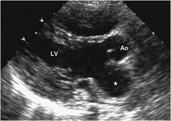
Transthoracic echocardiography identified a pseudoaneurysm of the mitral-aortic intervalvular fibrosa, the diagnosis being confirmed by trans-oesophageal echocardiography as a sac formation at the mitral-aortic transition (Fig 1) showing a distinct dynamic nature, expanding in systole and collapsing in diastole with a diameter of 39 by 29 ml. The pseudoaneurysm protruded into the left atrium and showed a large opening allowing free systolic inflow and diastolic outflow into and off the left ventricle, next to the anterior hinge point of the mitral valve. There was also mild mitral regurgitation.
Case 1: pseudoaneurysm of the mitro-aortic intervalvular fibrosa in the transthoracic echocardiogram long-axis view. Ao = aortic valve; LV = left ventricle, *pseudoaneurysm.

She underwent cardiac surgery on circulatory bypass and cardioplegic arrest. The diagnosis was confirmed and closure of the defect opening was performed, through an oblique aortotomy, using a heterologous pericardial patch and interrupted “U-shaped” nylon sutures.
The post-operative period was uneventful. The initially large thrombosed pseudoaneurysm progressively involuted to a small hyper-echogenic unremarkable image attached to the atrial aspect of the mitral valve, along the 1st year of follow-up. Currently, the patient is asymptomatic with trivial mitral and aortic regurgitation, under medication only with aspirin 100 ml daily and is back in Cape Verde.
Case 2
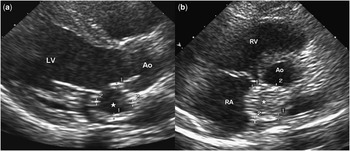
A 12-year-old boy was referred to our outpatient clinic for evaluation of atypical chest pain. There was no history of chest trauma, cardiac surgery, or endocarditis; he only had a history of asthma and frequent respiratory infections. Physical examination was unremarkable. Transthoracic echocardiogram and trans-oesophageal echocardiogram showed a pseudoaneurysm of the mitral-aortic intervalvular fibrosa measuring 23 by 13 ml with characteristic systolic and diastolic flow without any other structural or functional anomaly.
He underwent surgical closure of the pseudoaneurysm opening with a patch of heterologous pericardium through an oblique aortotomy (Fig 2). There were no post-operative complications.
Case 2: ( a ) Pre-operative image of the pseudoaneurysm in the transthoracic echocardiogram long-axis view. ( b ) Transthoracic echocardiogram short-axis view in the post-operative period, showing the thrombosed pseudoaneurysm. Ao = aorta, LV = left ventricle, RV = right ventricle, *pseudoaneurysm.

After 30 months of follow-up, the patient remains asymptomatic, and on echocardiography there is no residual defect. He is actually under no medication.
Case 3
A 9-year-old boy with rheumatic heart disease and signs and symptoms of congestive heart failure (Class III New York Heart Association) was evacuated from Africa for medical care. In the past, he had undergone two cardiac surgeries to correct rheumatic mitral lesions: extensive mitral commissurotomy and implantation of a Carpentier-Edwards® ring at the age of 5 years, and mitral valve replacement by a mechanical valve, 1 year later, owing to failure of repair. The patient returned to his home country in satisfactory condition, but most likely suffered more relapses of rheumatic activity and was finally readmitted to our centre with signs of congestive heart failure.
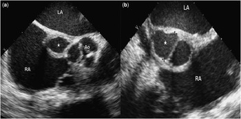
The transthoracic echocardiogram and trans-oesophageal echocardiogram showed a pseudoaneurysm of the mitral-aortic intervalvular fibrosa measuring 18 by 32 ml (Fig 3), major aortic regurgitation, and a well-functioning prosthetic mitral valve.
Case 3: ( a ) Trans-oesophageal echocardiogram image of the mitro-aortic pseudoaneurysm; ( b ) Trans-oesophageal echocardiogram measure of the pseudoaneurysm. LA = left atrium, RA = right atrium, Ao = aortic valve, *pseudoaneurysm.

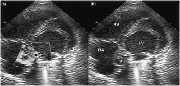
Reoperation (third) consisted of aortic valve replacement by a mechanical prosthesis and closure of the opening of the pseudoaneurysm with direct suture. He was discharged with small persistent residual flow at the pseudoaneurysm (Fig 4), which persists after 18 months of follow-up. He remains asymptomatic, under warfarin and on anti-congestive therapy.
Case 3: ( a and b ) Trans-oesophageal echocardiogram, short-axis view: pseudoaneurysm with ( a ) residual flow inside and without colour Doppler flow ( b ). RV = right ventricle, LV = left ventricle, RA = right atrium, *pseudoaneurysm.

Case 4
The fourth patient was a 13-year-old girl who had undergone three previous cardiac surgeries: the first for ventricular septal defect and the second for aortic regurgitation, a Ross procedure was performed, owing to early endocarditis. The third surgery, at the age of 13 years, was then complicated by Staphylococcus epidermidis endocarditis of the neoaortic valve, leading to a pseudoaneurysm of the mitral-aortic intervalvular fibrosa during active infection, despite well-directed antibiotic coverage. Emergency surgery was performed because of rapid expansion and enlargement of the pseudoaneurysm. Closure of its opening with a patch of homograft was performed; after a protracted post-operative period, the patient was eventually discharged home. Unfortunately, she was readmitted 2 months later for congestive heart failure and recurrence of the pseudoaneurysm of the mitral-aortic intervalvular fibrosa with gigantic dimensions communicating with the posterior wall of the aortic valve measuring 65 by 30 ml, confirmed by magnetic resonance, and a large ventricular septal defect resulting from disruption of the surgical patch. The patient was re-operated, but died during surgery because of uncontrolled rupture of the pseudoaneurysm.
Discussion
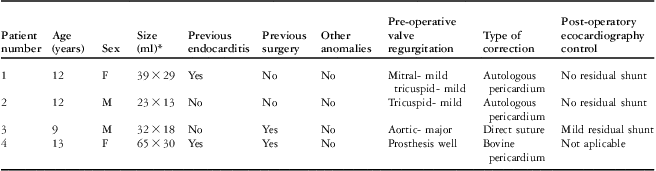
The mitral-aortic intervalvular fibrosa is a thin and apparently strong fibrous tissue that separates the posterior portion of the aortic root from the insertion of the anterior mitral leaflet.Reference Valero, Garcia, Pinto and Délcan 1 First described by catheterisation in 1996,Reference Waldhausen, Petry and Kurlander 6 the pseudoaneurysm of the mitral-aortic intervalvular fibrosa remains a rare pathological finding, whose recognition recently increased with the widespread use of trans-oesophageal echocardiogram and better image quality of modern transthoracic equipment. To the best of our knowledge, this is the largest single-centre series of pseudoaneurysms of the mitral-aortic intervalvular fibrosa in children ever reported (Table 1).
Patient information summary.

F = female; M = male
*Size was measured by echocardiography
True aneurysm and pseudoaneurysms have different pathologies in that the former has a large opening and its walls are made of both fibrous tissue and myocardium – complete structured wall – whereas the later has a small opening, that tends to enlarge, and walls composed of fibrous tissue without any myocardium, and hence more prone to rupture and especially uncertain to predict.Reference Bansal, Moloney, Marsa and Jacobson 7
Pseudoaneurysms of the mitral-aortic intervalvular fibrosa are, mostly an acquired form of disease, secondary to aortic or mitral valve endocarditis, cardiac surgery, or blunt trauma.Reference Espinosa-Caliani, Montijano, Melero and Montiel 2 – Reference Tak 4 , Reference Bolcal, Baysan, Doganci, Uzun, Demirkilic and Tatar 5 , Reference Yokoyama, Tamaki, Kato, Yokote and Mutsuga 8 Rare “congenital” cases have been described. In our series, three of the four patients had previous causative events, endocarditis or valvular surgery, but one patient had no previous disease or aetiological episode in his clinical history.
The mitral-aortic intervalvular fibrosa is a structure deprived of blood supply, offering very little resistance to infection, which can then facilitate the seeding of infection, namely, by aggressive bacteria and formation of abscesses.Reference Bolcal, Baysan, Doganci, Uzun, Demirkilic and Tatar 5 , Reference Tsai, Fu, Lin, Lin and Jan 9 – Reference Kassim, Lowery and Nasur 11 In addition, its thin structure renders it susceptible to progressive enlargement.
The diagnosis of this entity can be made by several imaging modalities such as transthoracic and trans-oesophageal echocardiogram, computerised tomography scan, magnetic resonance, and angiography. However, trans-oesophageal echocardiogram remains the gold standard for diagnosis, as it allows sufficient detailed anatomic and functional assessment of the pseudoaneurysm and related cardiac structures.Reference Kassim, Lowery and Nasur 11 , Reference Serkan 12 In our series, the diagnosis was achieved in all patients by transthoracic echocardiogram, but trans-oesophageal echocardiogram was also performed pre-operatively to better characterise the structures, visualise abscesses or vegetations, and plan surgery. Parasternal long- and short-axis views are the most helpful views, showing a blood filled sac behind the aortic root and post-superior to the anterior mitral leaflet, with a specific dynamic behaviour, expanding in systole with inflow shunt and collapsing in diastole, but without identifiable flow shunt to the left ventricular chamber.Reference Bansal, Moloney, Marsa and Jacobson 7 , Reference Ghersin, Litmanovich, Agmon and Milo 13 This pulsatile expanding–collapsing pattern is indeed characteristic of this pathology. The differential diagnosis should be made to aortic ring abscess, aortic-left ventricular tunnel, dissecting aortic aneurysm, and aneurysms of the sinus of Valsalva.Reference Bansal, Moloney, Marsa and Jacobson 7
All of our patients had non-specific symptoms at the time of diagnosis; nevertheless, pseudoaneurysms of the mitral-aortic intervalvular fibrosa are usually asymptomatic in the absence of complications.Reference Sudhakar, Sewani, Agrawal and Uretsky 14 , Reference Xie, Li and Cheng 15 It is, occasionally, seen in autopsy specimens or as an incidental finding during echocardiography in patients with no risk factors for mitral-aortic intervalvular fibrosa pseudoaneurysm formation, which are then considered as being congenital. Rarely, it can present with a devastating clinical picture, with rupture into neighbouring cardiac structures,Reference Valero, Garcia, Pinto and Délcan 1 , Reference Agirbasli and Fadel 16 coronary compression by the pseudoaneurysm itself during systole causing angina and angina-like symptoms,Reference Parashara, Jacobs and Kotler 10 , Reference Almeida, Pinho and Torres 17 , Reference Zoffoli and Gherli 18 embolisation of thrombotic material from the pseudoaneurysm causing stroke,Reference Koch, Kapoor and Spencer 19 and leading to death.
Our series illustrate well this wide range of clinical presentations, from case 2 – in whom we admit a “congenital” cause – to case 4 – who died because of rupture during surgery.
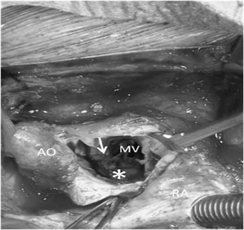
Surgical treatment should be kept simple, with closure of the communication with a patch stopping the flow inside the pseudoaneurysm, ensuring a gradual regression of the pseudoaneurysm and good long-term outcome (Fig 5). Our single case of residual flow occurred after closure of the opening with direct suture, and this technique should be discouraged. A percutaneous approach has also been successfully attempted and might be considered in very particular cases.Reference Valero, Garcia, Pinto and Délcan 1 Rarely, cases may complicate and have a fatal outcome, especially in the presence of active endocarditis,Reference Van Hare, Ben-Shachar, Liebman, Boxerbaum and Riemenschneider 20 , 21 such as was the case for patient 4. Considering the fragile nature of the mitral-aortic intervalvular fibrosa with its potential for severe complications, and a truly unpredictable course, along with improved diagnostic tools and good, low-risk, surgical results, the authors propose that elective surgery should be offered to all children with this diagnosis. Early recognition and treatment of the mitral-aortic intervalvular fibrosa pseudoaneurysm is imperative in order to prevent a potentially fatal outcome.Reference Valero, Garcia, Pinto and Délcan 1 , Reference Yamamoto, Tedoriya, Masuda, Asai, Sano and Kawada 3 , Reference Kassim, Lowery and Nasur 11
Intra-operative view of the surgical technique. Ao = aorta, RA = right atrium, MV = mitral valve, *pseudoaneurysm with patch, arrow, aortic root.

Our work addresses a very rare case of paediatric heart disease that should be increasingly recognised as the non-invasive imaging modalities improve in accuracy.
In our series, the two most frequent and important precipitating factors – endocarditis and valvular surgery – were clearly involved in three patients. The case of the second patient, the only patient without a clearly identifiable aetiological cause – should not be considered “congenital”; in our opinion, because the pathophysiological mechanism of the pseudoaneurysm requires a disruption of the mitral-aortic fibrosa to occur and, on the other hand, for these cases to truly have a congenital aetiology, this anomaly should be disclosed more often in children, without any clear aetiological factor, and this is not the case reported in the literature. Further, it is possible that chest blunt injury or any other previous infection could exist unnoticed in children. In the same patient, significant respiratory symptoms and frequent infections could have an important aetiological role in the so-called “congenital” case. Furthermore, we found no differences in pathophysiology from that reported in the adult population, which makes the probability of a true congenital disease less likely.
Our work also highlights the importance of timely diagnosis and the central role of transthoracic and trans-oesophageal echocardiogram by providing clear and characteristic images and function in children. These methods carry further importance as pseudoaneurysm symptoms are asymptomatic or produce non-specific symptoms. Probably, among children, transthoracic echocardiography provides enough information for surgery planning.
We stress that current surgical treatment and the good predictable results obtained support the indication for early elective repair of the pseudoaneurysm of the left “trigon” with the use of a pericardial patch instead of a direct closure of the defect.