Introduction
This report concerns a familial ossicular anomaly, only a few cases of which have been reported worldwide, diagnosed by clinical examination, family history, audiometrical and radiological evaluation. The findings in three patients – a female proband, her daughter and her mother – were essentially identical, and consisted of a history of hearing loss since birth, no history of ear infections, normal tympanic membranes, marked conductive deafness in both ears and similar findings on temporal bone computed tomography (CT).
Case report
A 32-year-old Korean woman was admitted to the otorhinolaryngology department of the Chuncheon Sacred Heart Hospital, South Korea, with non-progressive hearing disturbance in both ears. She had no history of head injury, ear infection or other otological symptoms.
On examination, the patient had no obvious signs of any problem. Her auricles and external auditory canals were considered to be within normal limits. The tympanic membranes were intact.
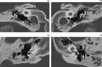
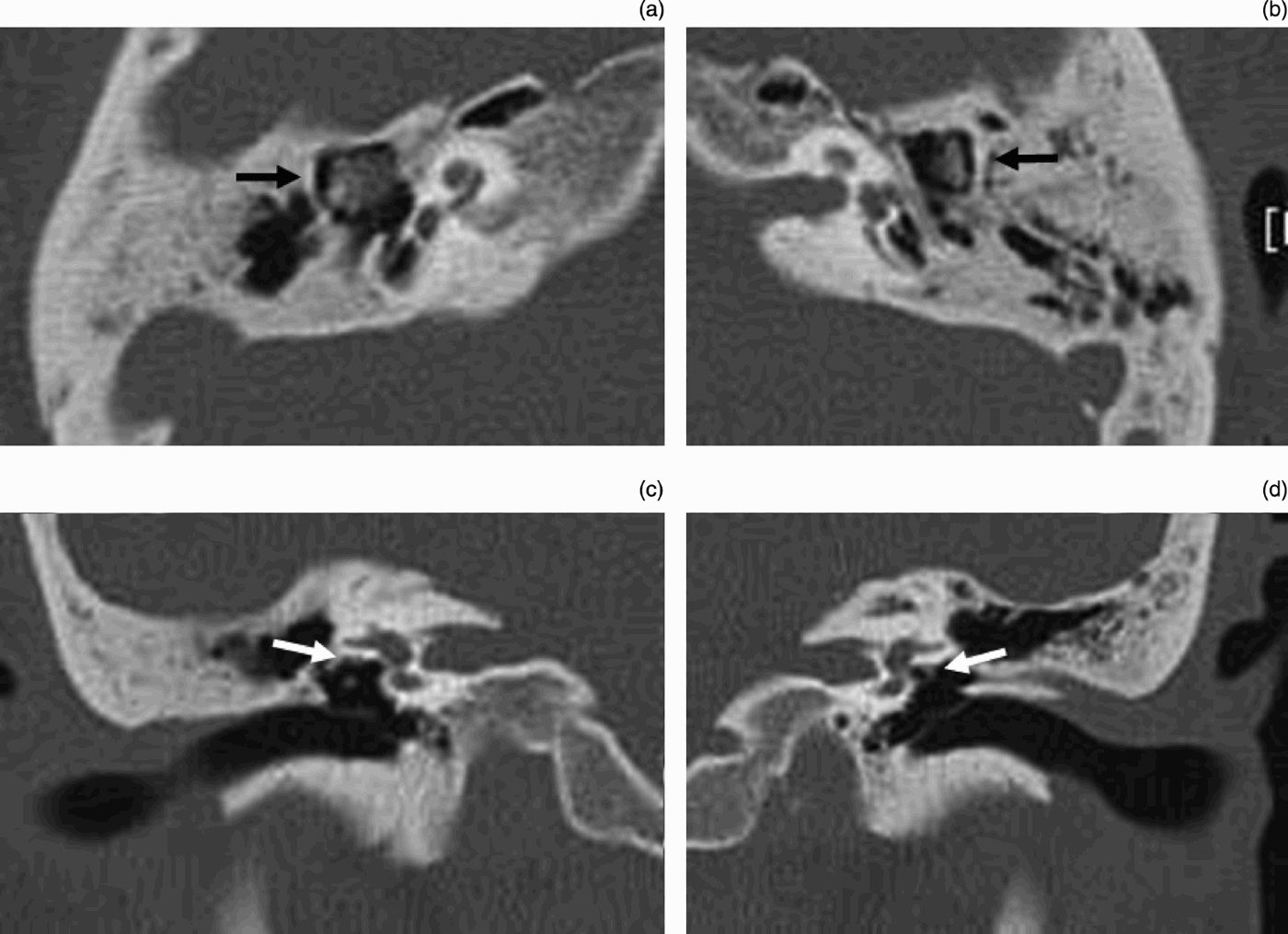
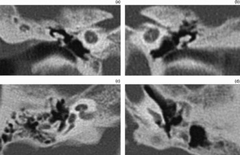
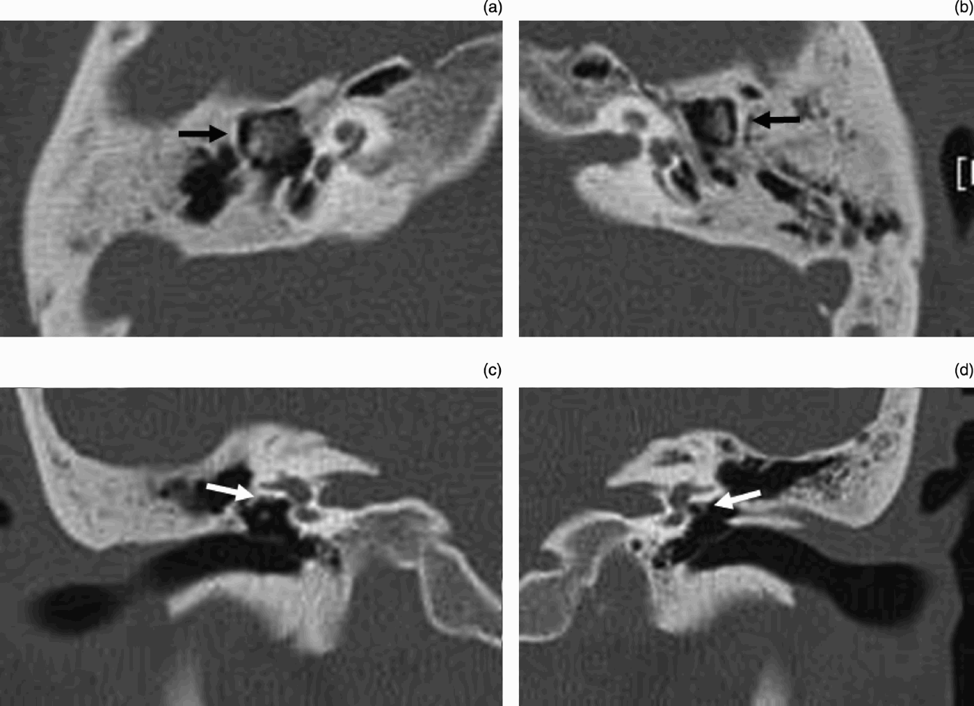
Auditory testing revealed bilateral conductive hearing loss (59 dB on the right and 65 dB on the left), and tympanometric measurements showed normal compliance in both ears. Pre-operative high resolution computed tomography (CT) of the temporal bone showed abnormal ossicular shadows in both middle ears. Coronal CT imaging showed that the malleus and incus appeared to be fused, deformed and lateralised bilaterally. In the axial plane, the body of the incus was large and low density, and the malleus and incus were shifted to the lateral attic wall bilaterally (Figures 1 and 2).
Computed tomography images of our patient's temporal bones: (a) right bone, coronal view; (b) left bone, coronal view; (c) right bone, axial view; and (d) left bone, axial view. The malleus and incus were narrowed and in contact with the lateral attic wall (black arrows). The location and shape of the incus were both abnormal (white arrows).

Coronal computed tomography scans of our patient's temporal bone. (a,b) Both incuses were located more inferiorly and were abnormally shaped (black arrows). (c,d) The hypotrophic tympanic segment of the facial nerves were seen near the oval window on both sides (white arrows).
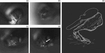
We performed exploratory tympanotomy on the left side, because the patient's hearing was worse on the left than on the right. At surgery, fixation of the malleus, incus and stapes was observed following tympanotomy. After dislocating the incudostapedial joint, the movement of the malleus and incus was essentially normal, while the stapes was fixed. After removing the suprastructure, a very narrow, fixed footplate and hanging facial nerve were seen (Figure 3). We could not perform stapedotomy because the hanging facial nerve blocked the operating field. The patient's hearing remained unchanged post-operatively.
Surgical findings. The patient's ossicles did not move when they were checked before manipulation (a). After dislocating the incudostapedial joint, the movements of the malleus and incus were almost normal (b), while the stapes was fixed (c). After removing the suprastructure, a very narrow, fixed footplate and hanging facial nerve were seen (d; white arrow). A schematic drawing of the middle ear (e) shows the abnormal ossicles and hanging facial nerve.

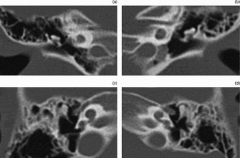
Review of the patient's family history indicated that both her mother and daughter also had hearing disturbances. Her 62-year-old mother used a hearing aid, and her five-year-old daughter used a hearing aid and was receiving speech therapy. The results of audiometry in all three cases were similar, as were findings for CT scanning of the temporal bone (Figures 4 and 5).
Computed tomography scans of the patient's daughter's temporal bones: (a) right bone, coronal view; (b) left bone, coronal view; (c) right bone, axial view; and (d) left bone, axial view.

Computed tomography scans of the patient's mother's temporal bones: (a) right bone, coronal view; (b) left bone, coronal view; (c) right bone, axial view; and (d) left bone, axial view.

Neither the mother nor the daughter agreed to surgical exploration of the middle ear.
Discussion
Familial congenital ossicular anomaly is rare and only a few cases have been reported worldwide. A review of the literature revealed several reports regarding familial ossicular anomaly without otosclerosis, familial expansile osteolysis, and ossicular problems in syndromic disease.
Wehrs reported three generations of one family suffering from bilateral conductive hearing loss due to congenital absence of the long process of the incus.Reference Wehrs1 In this family, the defect appeared to be an inherited trait due to either an autosomal dominant mutation or X-linked dominant inheritance. Higashi et al. reported two generations of congenital hypoplasia of the incus long process.Reference Higashi, Yamakawa, Itani and Togawa2 They suggested that this anomaly was inherited as an autosomal dominant trait.
Some authors have reported familial stapes superstructure fixation. In these cases, the head of the stapes was fixed to the pyramid by an ossified stapedius tendon.Reference Kinsella and Kerr3–Reference Doi, Nakazawa, Adachi, Kaneko, Munemoto and Komeda6 In some cases of this type, the defect appeared to be an inherited trait due to an autosomal dominant mutation, while in other cases it appeared to be due to an autosomal recessive mutation.
Another type of familial congenital ossicular anomaly which has been reported is stapedial fixation with a perilymph ‘gusher’ encountered during surgical intervention.Reference Carlson and Reeh7–Reference Cremers, Snik, Huygen, Joosten and Cremers9 The patient genealogy suggests that this anomaly is inherited as an X-linked recessive trait.
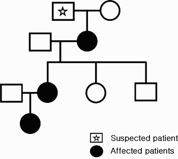
Here, we present three cases of bilateral congenital incus anomaly, stapes fixation and facial nerve anomaly in one family. The individuals involved were all female. However, not all female family members were affected, and the first generation male in the pedigree suffered from hearing disturbance. Unfortunately, he could not be evaluated as he was deceased. The other siblings were unaffected. Therefore, we postulate that this inherited trait is due to an autosomal dominant mutation, although it may also involve X-linked dominant inheritance (Figure 6).
Family tree showing the three generations affected. The male in the first generation had died several years prior, but had suffered impaired hearing since childhood.

• This paper reports three generations of one family suffering from bilateral conductive hearing loss due to a congenital anomaly of the incus and stapes fixation
• An abnormally shaped incus, fixed stapes and hanging tympanic portion of the facial nerve were seen at surgery
• The overhanging facial nerve prevented stapedotomy from being undertaken
Conclusion
Although several cases of familial ossicular anomaly have been reported, the current patients represent the first reported cases showing incus anomaly and stapes fixation combined with a facial nerve anomaly. These cases illustrate the fact that any congenital conductive or mixed hearing loss should be evaluated further by taking a family history.