INTRODUCTION
As the importance of microzooplankton as fundamental grazers in planktonic food webs became recognized, diverse techniques for determining the grazing impact of microzooplankters were developed (Kivi & Setälä, Reference Kivi and Setälä1995). The most widely used method to estimate in situ grazing rates is Landry & Hassett's dilution technique (Landry & Hassett, Reference Landry and Hassett1982; Calbet & Landry, Reference Calbet and Landry2004) which facilitates the estimation of grazing rates in barely manipulated grazer communities. Consequently, this method is now standard for assessments of in situ grazing rates of smaller microzooplankton (<200 µm).
As part of these experiments, mesozooplankton is removed and samples are checked for screening effectiveness with regard to the mesozooplankton (Fonda Umani et al., Reference Fonda Umani, Tirelli, Beran and Guardiani2005) or losses in the phytoplankton fraction. However, to our knowledge no published study has so far considered potential losses of microzooplankton during the set-up of these experiments (Suzuki et al., Reference Suzuki, Tsuda, Kiyosawa, Takeda, Nishioka, Saino, Takahashi and Wong2002; Paterson et al., Reference Paterson, Knott, Koslow and Waite2008; Pearce et al., Reference Pearce, Davidson, Wright and Van den Enden2008). This is especially problematic if the in situ abundance and biodiversity of microzooplankton grazers are the main targets of an experiment and results are transferred to the field.
Microzooplankton mainly consists of very delicate organisms (in particular ciliates and dinoflagellates: Gifford, Reference Gifford1985; Suzuki et al., Reference Suzuki, Tsuda, Kiyosawa, Takeda, Nishioka, Saino, Takahashi and Wong2002; Broglio et al., Reference Broglio, Jónasdóttir, Calbet, Jakobsen and Saiz2003), thus manipulation of water samples while setting up grazing experiments could significantly alter the grazer community through the loss of sensitive taxa, affecting estimates of grazing rates. This would defeat the goal of a grazing experiment aimed at the determination of the in situ grazing rate.
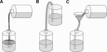
To avoid the loss of microzooplankters during the experimental set-up a widely used technique involves the siphoning off of water (Figure 1B) using silicone tubing and leaving the end of the tubing submerged in the water (Stelfox-Widdicombe et al., Reference Stelfox-Widdicombe, Archer, Burkill and Stefels2004; Strom et al., Reference Strom, Macri and Olson2007; Paterson et al., Reference Paterson, Knott, Koslow and Waite2008). This technique prevents destructive air bubbles that can occur in pouring processes (Figure 1A) and is thus believed to conserve fragile species.
Illustration of the different filling techniques: (A) a simple, more destructive pouring process producing a lot of air bubbles; (B) siphoning the water via a tube into a container without air bubbles; (C) our new, more gentle approach to fill incubation bottles: combination of a funnel and tube (FTT).

In preliminary experiments we found lower abundances of sensitive microzooplankton in siphoned samples when compared to the field, a pattern which was especially true for ciliates. Thus, we hypothesized that the set-up technique (here siphoning) might cause: (1) diminished microzooplankton abundances; (2) pronounced effects on ciliates compared to dinoflagellates; and (3) a selection in species composition in favour of non-sensitive species and thus an artificially modified grazer community. Consequently we compared two gentle filling techniques, siphoning (Figure 1B) versus a modified application, the alternative funnel-transfer technique (FTT) (Figure 1C) while setting up dilution experiments. Here, we report the effects of both filling techniques on microzooplankton abundance and Margalef diversity (dinoflagellates and ciliates) of North Sea samples. We concentrate on in situ grazing experiments, but the results are equally applicable to any situation where zooplankton communities containing physically fragile species are to be manipulated in the laboratory.
MATERIALS AND METHODS
Two different methods of filling experimental bottles for grazing experiments were tested in two separate experiments using water taken from the North Sea.
Sampling site
Helgoland is located in the German Bight (southern North Sea) approximately 50 km off the German coast. It is subject to both coastal influences from the shallow Wadden Sea as well as marine influences from the open North Sea. Since 1962 bucket water samples have been taken as part of a long term monitoring programme at the ‘Kabeltonne’ site at Helgoland Roads (54°11.3′N 7°54.0′E) (Wiltshire et al., Reference Wiltshire, Malzahn, Wirtz, Greve, Janisch, Mangelsdorf, Manly and Boersma2008). Water samples for the experiments were taken here.
Filling techniques
We compared two filling techniques: siphoning and FTT. Siphoning is believed to be conservative for sensitive species as it avoids bubbling when the flow-end of the tube is kept under the water surface (Stelfox-Widdicombe et al., Reference Stelfox-Widdicombe, Archer, Burkill and Stefels2004; Strom et al., Reference Strom, Macri and Olson2007; Paterson et al., Reference Paterson, Knott, Koslow and Waite2008). Water is transferred only via hydrostatic pressure through a tube from one container to another when both containers have different water levels relative to geodetic height (Figure 1B). The FTT consists of a funnel mounted on a tube. For the transfer to a container, water is filled gently into the funnel keeping the tube compressed completely until the funnel is half-full. Thereafter water flow to the container is adjusted via the internal diameter by compressing the flexible tube compartment (Figure 1C). This technique also avoids air bubbles that could harm sensitive species.
Experiment 1
Experiment 1 (July 2007) was conducted to compare the two filling techniques using the experimental set-up of dilution grazing experiments. During the experiment both techniques were tested with a 200 µm net for pre-screening. Pre-screening is routine in dilution experiments and excludes mesozooplankton grazers ensuring that only the microzooplankton grazing is measured (Liu & Dagg, Reference Liu and Dagg2003; Fonda Umani et al., Reference Fonda Umani, Tirelli, Beran and Guardiani2005; Sakka Hlaili et al., Reference Sakka Hlaili, Grami, Hadj Mabrouk, Gosselin and Hamel2007).
Containers, bottles, tubing and other material used for the experiments were acid washed (10% HCL) and rinsed with deionized water. Approximately 50 l of surface seawater were sampled at Helgoland Roads using a bucket and poured without bubbling into a wide-necked carboy. The samples were brought to the laboratory immediately.
The homogeneous distribution of the plankton in the initial seawater was ensured by gentle mixing. After homogenization, 10 l were gently transferred into a 10 l carboy (as usually used for the set-up of a dilution series) with the FTT (Figure 1C). Special care was taken during the filling process that no air bubbles were produced. When the carboy was filled, the flow was terminated by compressing the tube. In parallel to the FTT method 10 l of seawater were siphoned off with a silicone tube into a separate carboy (Figure 1B). 2.3 l narrow-necked polycarbonate incubation bottles were filled with water from each corresponding 10 l carboy using both methods in three replicates. Before each filling step gentle mixing was carried out. In both approaches a 200 µm mesh was fixed at the end of the tube. All incubation bottles were filled to the top, closed and stored cool in the dark until sampling for microzooplankton and chlorophyll-a.
Experiment 2
Experiment 2 (April 2008) was designed to investigate both filling techniques with and without the 200 µm pre-screening to record additional effects of the pre-screening process on the monitored parameters.
The experimental set-up in Experiment 2 was as described for Experiment 1 above. Siphoning and the FTT were also applied without pre-screening, resulting in a total of four treatments. Furthermore, a control with pure seawater served as a reference value: water for the determination of chlorophyll-a content and microzooplankton species composition was scooped out with a 1 l beaker from the initial seawater at the beginning of the experiment. During the set-up of both experiments special care was taken not to produce bubbles.
Determination of microzooplankton species
A 250 ml aliquot of each incubation bottle was subsampled into amber bottles and immediately fixed at a final concentration of 2% acid Lugol's iodine solution (Throndsen, Reference Throndsen and Sournia1978). Samples were stored cool and dark until further analysis. For species determination 50 ml samples were settled in Utermöhl sedimentation chambers (HYDRO-BIOS) for 24 hours (Utermöhl, Reference Utermöhl1958). To reduce a possible counting bias caused by patchy settlement, the whole surface of the sedimentation chamber was counted at 200-fold magnification under a Zeiss Axiovert 135 inverted microscope. The microzooplankton fractions were considered as two major groups: dinoflagellates and ciliates. Each group was identified to genus or species level or when this was impossible pooled into size-dependent groups or morphotypes. The identification of dinoflagellates was primarily based on Drebes (Reference Drebes1974), Dodge (Reference Dodge1982) and Tomas (Reference Tomas1996). Ciliates were determined based on Kahl (Reference Kahl and Dahl1932) and Montagnes (Reference Montagnes2003).
Chlorophyll-a analysis
Filtration of a subsample was carried out in a laboratory under dim light (<5 µmol PAR) to avoid the loss of pigments during the filtration procedure. We used the method of extraction and analysis as described by Wiltshire et al. (Reference Wiltshire, Harsdorf, Smidt, Blöcker, Reuter and Schroeder1998). Pigments were separated via high-performance liquid chromatography (HPLC) (Waters 2695 Separation Module), and detected with a Waters 996 Photodiode Array Detector (Wiltshire et al., Reference Wiltshire, Boersma, Möller and Buhtz2000).
Data analysis
The Margalef index ‘d’ was calculated for the most abundant ciliate and dinoflagellate species according to Equation (1).

This index was chosen as a simple measure for the relationship between total number of species (S) and total abundance of individuals (N) in a sample (Washington, Reference Washington1984). Therefore, it is a useful tool for the analysis of samples in the present study. If in two samples the total number of species is the same (as in our analysis), then the Margalef index gives direct information about the differences in the total abundance of individuals between the two samples. The index will be lower in the sample with the higher total abundance and therefore reflects changes due to the different filling methods.
Margalef indices, chlorophyll-a contents and abundances of dinoflagellates or ciliates between the treatments in Experiment 1 were tested for significant differences using t-tests. Differences between the four treatments and the control sample in Experiment 2 were in a first step analysed using t-tests. This was necessary because the control was not influenced by any treatment and could therefore not be included in an ANOVA. The effects of the two filling techniques and the pre-screening process on the above-mentioned parameters as well as the interaction between both were tested using a 2 × 2 factorial ANOVA.
Due to the normal count uncertainties of microzooplankton samples and, connected to that, a high variability in abundance of rare species, only dinoflagellate and ciliate species or groups that contained more than 200 cells l−1 in one of the two treatments (Experiment 1) or the control (Experiment 2) are shown here. It has been shown that many chloroplast-bearing dinoflagellate species are capable of mixotrophic nutrition via phagotrophy (Tillmann, Reference Tillmann2004). Therefore, all dinoflagellate species were considered as potential grazers and were included in the analysis. Statistical analyses were conducted with the software Statistica 7.1 (Stat Soft).
RESULTS
Experiment 1
MICROZOOPLANKTON
Microzooplankton was classified into 12 categories of ciliates and 15 categories of dinoflagellates. The microzooplankton community was numerically dominated by dinoflagellates (61%, FTT).
Ciliates
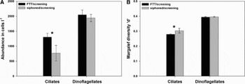
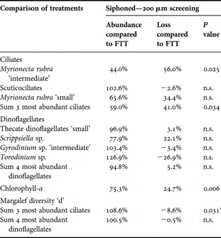
The three most abundant ciliate groups were: Myrionecta rubra ‘intermediate’ (35–50 μm length), scuticociliates and Myrionecta rubra ‘small’ (15–25 µm length). These three groups accounted for 84% of the total abundance of the FTT treatment and 77% of the siphoned treatment (mean values). They therefore dominated the ciliate community. We found a significant ciliate cell loss of 41% due to siphoning (t = 3.162, df = 4, P = 0.034, two-sided t-test) (Figure 2A; Table 1) for pooled values of the three most abundant groups leading to significant changes in diversity. The Margalef index ‘d’ (Figure 2B; Table 1) for the three most abundant ciliate groups was significantly lower for the FTT (t = –2.569, df = 4, P = 0.031, single-sided t-test) compared to siphoning. No difference in the Margalef index was observed between both treatments for the whole ciliate community (data not shown).
Experiment 1. (A) Mean abundance and (B) Margalef diversity ‘d’ of the most abundant ciliate and dinoflagellate species. Both treatments were done using a 200 µm pre-screened natural seawater sample. Significant difference marked with asterisk. Error bars correspond to one standard deviation.

Percentage of loss based on abundances of the most common ciliate and dinoflagellate groups, chlorophyll-a contents and Margalef indices ‘d’ during siphoning compared to the FTT for Experiment 1 (4 July 2007). P values derived from double-sided t-tests (N = 3). n.s., not significant (P > 0.05). *P value derived from single-sided t-test.

At species/group level the loss of cells was most pronounced in the category M. rubra ‘intermediate’. This size-class of M. rubra accounted for 54% of total ciliate cells (FTT) and displayed a significant loss of 56% in cell numbers in the siphoned treatments (t = 3.483, df = 4, P = 0.025, two-sided t-test). The category M. rubra ‘small’ showed a similar pattern to M. rubra ‘intermediate’ with a 34% lower mean cell number when siphoned. However, this effect was not statistically significant given the large count uncertainties. For the category ‘scuticociliates’ no significant difference between both treatments could be detected.
Although chlorophyll-a concentrations during our experiment were relatively low (<0.2 µg/l), we detected significant differences between both methods, with lower chlorophyll-a contents in the siphoned treatments (t = 5.366, df = 4, P = 0.006, two-sided t-test) (Table 1), possibly linked to the loss of M. rubra cells.
Dinoflagellates
Four groups, accounting for 85% of the total dinoflagellate abundance in both treatments, were used for statistical analysis: thecate dinoflagellates ‘small’ (20–25 µm length), Scrippsiella sp., Gyrodinium sp. ‘intermediate’ (30–50 µm length) and Torodinium sp. (Table 1). In contrast to observations for the most abundant ciliate categories, we found no differences between the two treatments for the four most abundant dinoflagellate groups pooled together or at species level (Figure 2A; Table 1). Margalef indices ‘d’ calculated for the four most abundant and all dinoflagellate categories also revealed no differences in abundance for both treatments (Figure 2B; Table 1).
Experiment 2
MICROZOOPLANKTON
In the second experiment 21 categories were established for ciliates and dinoflagellates corresponding to different species, group or size-classes. Ciliates numerically dominated the microzooplankton community (60%, control).
Ciliates
Ciliate categories that presented abundances above 200 cells l−1 were: Lohmanniella oviformis, Strombidium cf. tressum, Strombidium cf. epidemum, Balanion comatum and Myrionecta rubra ‘small’ (15–25 µm length). The five ciliate categories grouped together made up over 80% of the total ciliates' community in all treatments.
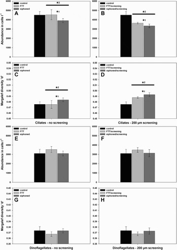
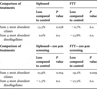
In a first step we pooled together the top five abundant ciliates and carried out a single-sided t-test (Table 2). It revealed a significant, 13% decline in abundance (t = 2.390, df = 4, P = 0.038) when siphoned without pre-screening compared to the control, while the FTT without pre-screening showed no difference. Both treatments with additional pre-screening exhibited significant losses in cell numbers compared to the control (siphon technique: mean loss of 26%, t = 4.820, df = 4, P = 0.004; FTT: mean loss 19%, t = 3.927, df = 4, P = 0.009, single-sided t-tests).
Experiment 2. (A, B) Mean abundance of the most abundant ciliate species, (E, F) mean abundance of the most abundant dinoflagellate species and (C, D, G, H) corresponding Margalef diversity ‘d’ in the control and using the FTT or the siphoning technique in experiments without (A, C, E, G) or with pre-screening with a 200 µm net (B, D, F, H). Control treatments were obtained by scooping samples from the initial seawater without pre-screening. Significant differences are marked with asterisks. Asterisk *1symbolizes significant differences between siphoning and the FTT; asterisk *2on the top of the horizontal bar symbolizes differences between ‘pre-screening’ and ‘no pre-screening’ independently from the filling technique used (therefore compare bars under the horizontal bar for each filling treatment in A and B, C and D). Error bars correspond to one standard deviation.

Percentage of loss in abundance of the most abundant ciliate and dinoflagellate groups due to the different treatments and treatment combinations compared to the control values during Experiment 2 (1 April 2008). P values derived from single-sided t-tests (N = 3). n.s., not significant (P > 0.05).

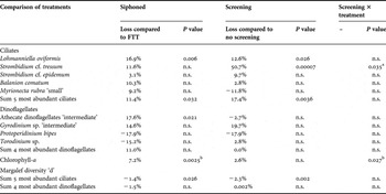
In the two-factorial ANOVA pooled abundances of the five most abundant ciliates showed significantly lower cell concentrations after siphoning (F1,8 = 6.685, P = 0.03) and pre-screening (F1,8 = 16.508, P = 0.004) as compared to the FTT and no screening, respectively (Figure 3A + B; Table 3), leading in both cases to significant changes in diversity. The Margalef index ‘d’ (Table 3; Figure 3 C + D) showed no difference at the community level (data not shown) but was significantly lower for the five most abundant ciliate species when ‘no pre-screening’ took place (F1,8 = 19.0, P = 0.002). Similar to Experiment 1, the Margalef index ‘d’ was also lower for the FTT (F1,8 = 7.4, P = 0.026; Table 3). No significant treatment interactions were observed for either abundances or Margalef index.
Percentage of loss in abundance of the most abundant ciliate and dinoflagellate species, chlorophyll-a contents and Margalef indices ‘d’ due to siphoning in comparison to the FTT and pre-screening in comparison to no pre-screening and effects of the combination between pre-screening and treatment during Experiment 2 (1 April 2008). P values are derived from ANOVA comparing all treatments (N = 6). n.s., not significant (P > 0.05); asignificantly lower abundance in the FTT with pre-screening treatments; bsignificantly higher content in the FTT with pre-screening treatments.

At the species level our results could be ascribed to the two dominant ciliates. Lohmanniella oviformis, showed a significant cell loss (17%, F1,8 = 14.173, P = 0.006) after siphoning as well as after pre-screening (loss: 13%, F1,8 = 7.471, P = 0.026). For Strombidium cf. tressum no significant difference between both filling treatments was found but pre-screening resulted in a significant loss of cells (up to 51% cell loss, F1,8 = 56.3325, P = 0.00007). The interaction of both filling methods and pre-screening showed a significant negative effect (F1,8 = 6.4228, P = 0.035) on S. cf. tressum with a stronger reduction in cell numbers when the combination of the FTT and pre-screening was used. The other three important ciliate species (Strombidium cf. epidemum, Balanion comatum and Myrionecta rubra ‘small’) were not significantly affected by any treatment. With a mean concentration of ~1.3 µg/l the chlorophyll-a content was approximately 10 fold higher in Experiment 2 as compared to Experiment 1. However, the differences between the treatments were not as clear as in Experiment 1.
Dinoflagellates
The four most abundant dinoflagellate categories were considered for analyses: athecate dinoflagellates ‘intermediate’ (25–40 µm length), Gyrodinium sp. ‘intermediate’ (30–50 µm length), Protoperidinium bipes and Torodinium sp. These four species contributed 82% to the dinoflagellate community. As in Experiment 1, no differences in abundance were found when the four dinoflagellate groups pooled were compared to the control (Table 2). For the pooled group an ANOVA proved insignificant (Figure 3E + F; Table 3) for the treatments as well as their interaction. At species level the athecate dinoflagellates ‘intermediate’ revealed significantly lower cell numbers in the siphoned treatment (F1,8 = 8.2718, P = 0.02; Table 3) as compared to the FTT. No impact of the pre-screening process or the combination of treatment and pre-screening on the other dinoflagellates could be found. As in Experiment 1, no differences were found for the Margalef index ‘d’ calculated for the whole community (data not shown) or when considering only the five most abundant groups (Table 3; Figure 3 G + H).
DISCUSSION
Transfer technique
The main aim of this study was to determine whether filling and screening techniques can influence abundance and diversity of microzooplankton during the set-up of grazing experiments. Although the FTT superficially seems to represent only a marginal modification of siphoning the change has a significant effect on microzooplankton diversity. Overall, siphoning significantly reduced the abundance of ciliates in North Sea water, while the dinoflagellate community seemed to be unaffected. The lower impact on the ciliate community using the FTT was reflected in the lower Margalef index ‘d’ for the most abundant ciliates in both experiments. The abundance and Margalef index ‘d’ of both microzooplankton groups were found to be similar to the control (Experiment 2) when the FTT was used, indicating that in contrast to siphoning no significant effect occurred using the FTT.
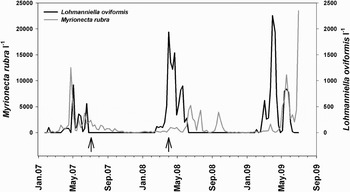
While the overall ciliate numbers appeared to be lower when siphoning was used, this was statistically significant only for two of the dominant ciliate species: Myrionecta rubra and Lohmanniella oviformis. The taxa Lohmanniella and M. rubra are widely distributed in coastal and open ocean environments (Smetacek, Reference Smetacek1984; Kivi & Setälä, Reference Kivi and Setälä1995; Myung et al., Reference Myung, Yih, Kim, Park and Cho2006; Park et al., Reference Park, Myung, Kim, Cho and Yih2007) and are also important members of the microzooplankton community in the North Sea around Helgoland (Figure 4). Both are known to feed on nanoplankton (Jonsson, Reference Jonsson1986; Christaki et al., Reference Christaki, Dolan, Pelegri and Rassoulzadegan1998; Aberle et al., Reference Aberle, Lengfellner and Sommer2007; Park et al., Reference Park, Myung, Kim, Cho and Yih2007) and bacteria (Myung et al., Reference Myung, Yih, Kim, Park and Cho2006).
Abundance of Myrionecta rubra and Lohmanniella oviformis during a 2.5 year period. Values from a weekly monitoring programme (N = 128). Arrows mark dates of the two experiments.

Both ciliates can be considered important grazers in temperate waters in general and dominated the communities during our experiments. Therefore, retaining natural abundances is important for accurate estimations of microzooplankton grazing.
In contrast to ciliates the sensitivity of dinoflagellates to the transfer method was more variable and species-dependent. Our results show that only the most abundant dinoflagellate group (athecate dinoflagellates ‘intermediate’) in Experiment 2 was significantly affected by siphoning. No effect was detected for all other investigated species. Heterotrophic dinoflagellates, including both thecate and athecate species are microzooplankton grazers of global importance (Tillmann, Reference Tillmann2004) frequently contributing more than 50% to the microzooplankton biomass (Sherr & Sherr, Reference Sherr and Sherr2007). As they often occur at high abundances during diatom blooms and are known to be more efficient grazers of bloom-forming diatoms than copepods and other mesozooplankters (Sherr & Sherr, Reference Sherr and Sherr2007) it is vital not to lose dinoflagellates during grazing experiment set-ups.
Our results also show that siphoning can affect chlorophyll-a concentration when setting up experiments. It has been reported that mixotrophic ciliates like Myrionecta rubra dominate plankton assemblages at certain times of the year (Stoecker et al., Reference Stoecker, Michaels and Davis1987). This was also the case during Experiment 1 (Figure 4). Our results show that the loss of these mixotrophic cells due to siphoning may induce an additional bias into dilution experiments through the concomitant loss of chlorophyll-a, a commonly used estimate for overall phytoplankton biomass.
The effect of siphoning on microzooplankton might be explained by hydro-mechanical disturbance of water when using this method. Mechanoreceptors are common in planktonic organisms (Singarajah, Reference Singarajah1969; Titelman & Kiørboe, Reference Titelman and Kiørboe2003; Robinson et al., Reference Robinson, Finelli and Buskey2007), including ciliates (Buskey & Stoecker, Reference Buskey and Stoecker1989; Jakobsen, Reference Jakobsen2001, Reference Jakobsen2002; Jakobsen et al., Reference Jakobsen, Everett and Strom2006) and dinoflagellates (Jakobsen et al., Reference Jakobsen, Everett and Strom2006). Different ciliates are reported to respond with long jumps to siphon-simulated feeding currents (Jakobsen, Reference Jakobsen2001; Fenchel & Hansen, Reference Fenchel and Hansen2006) and to orientate themselves against the current in a siphon flow (Fenchel & Hansen, Reference Fenchel and Hansen2006). They also respond with escape during the isolation processes with a pipette (tip: 1 mm diameter) during culture attempts (Löder, personal observation). Although the siphon-tube diameter (7 mm) we used was bigger than that used by Jakobsen (Reference Jakobsen2001) (0.25–0.48 mm), the flow velocity and therefore also the shear stress around the tube tip was in the same order of magnitude as that of Jakobsen (Reference Jakobsen2001). The ability of ciliates to detect suction at the top of a tube and respond with escape is reported by Jakobsen (Reference Jakobsen2002) and could explain their lower numbers in the incubation bottles when siphoning was used. Interestingly along with Lohmanniella oviformis only the larger Myrionecta rubra ‘intermediate’ cells were significantly affected by the siphon technique, whereas no significant difference was found for the category M. rubra ‘small’. This could be linked to the ability of the larger M. rubra to perform longer escape jumps than the smaller, which probably enables them to escape the siphon suction more successfully.
Similar to ciliates, mechanoreceptors that enable predator detection have also been described for dinoflagellates (Maldonado & Latz, Reference Maldonado and Latz2007). Jakobsen et al. (Reference Jakobsen, Everett and Strom2006) reported highly effective escape behaviour for two different dinoflagellates when being attacked by a predator. Although the differences were less obvious for dinoflagellates, siphoning had an effect on one naked dinoflagellate species in Experiment 2. These differences could also be due to the same predator detection and escape behaviour as described above for ciliates.
The high fragility of microzooplankton could also be an explanation for the effect of siphoning on some ciliate species (Gifford, Reference Gifford1985; Suzuki et al., Reference Suzuki, Tsuda, Kiyosawa, Takeda, Nishioka, Saino, Takahashi and Wong2002; Broglio et al., Reference Broglio, Jónasdóttir, Calbet, Jakobsen and Saiz2003). During the passage of the organism through the tube physical stress is likely to act on the organism and the period of time the organism is exposed to that should increase with the length of the tubing. Necessarily, siphoning requires a longer tube compared to FTT and could therefore also cause greater damage in fragile species.
Importantly, the error introduced by the siphoning technique in our experiments was not uniform across taxa. If experiments are replicated at different times of the year this could potentially lead to a different source of error in every single experiment depending on the community structure present leading to difficulties in comparison.
Pre-screening
Several authors recommend not using pre-screening for dilution experiments to avoid retention of phytoplankton chains or breakage of the cells. Our results also show that pre-screening had an effect on ciliates. This effect was stronger than the effect of siphoning as it led to a greater loss of ciliates at species level. Importantly, different ciliate species did not respond uniformly to pre-screening. A negative effect of pre-screening on fragile ciliates due to mechanical shear forces has been reported previously (Gifford, Reference Gifford1985) and could be shown here for Strombidium cf. tressum and more moderately for the strobilid Lohmanniella oviformis. The reduced abundance of ciliates resulted in a higher Margalef index (Table 3) compared to the unscreened treatments. The difference in response to the pre-screening process could indicate different degrees of fragility in ciliates, even in the same genus such as Strombidium cf. tressum and Strombidium cf. epidemum. Both ciliates are almost in the same size-range (25–40 µm maximal length) but the impact on S. cf. tressum was large while S. cf. epidemum was not significantly affected. The impact on S. cf. tressum (51% loss) was very strong taking into account that the mesh size was 200 µm and therefore at least five times the size of the biggest individuals of this species. Compared to the loss rates of oligotrichs due to 202 µm screening (23–37%) published by Gifford (Reference Gifford1985) our results for the pool of the most abundant ciliates are almost in the same size-range (17%). In contrast to the ciliates, no effect of the pre-screening process was detectable on dinoflagellates. To our knowledge no work has been published on whether dinoflagellates are sensitive to pre-screening. Despite the observed effect of pre-screening due to a high loss of particular ciliate species, the 200 µm pre-screening process remains unavoidable whenever mesozooplankton grazers are present in the water column in high numbers in order to prevent trophic cascade effects and to ensure correct estimates of microzooplankton grazing impact alone.
CONCLUSIONS
We have demonstrated that care is required when setting up grazing experiments particularly when sensitive species are present. Commonly used techniques such as siphoning can reduce microzooplankton abundance and diversity. Siphoning had a negative effect on ciliates compared to dinoflagellates resulting in shifts in species composition and producing an ‘artificial’ community that did not realistically reflect the in situ situation. Therefore, extrapolations to the field are difficult.
The FTT presented here was shown to be a good alternative to siphoning, conserving the natural microzooplankton abundance and diversity. Negative effects of pre-screening on natural microzooplankton communities were determined. With the methods currently available this cannot be avoided if mesozooplankters are to be excluded. We, therefore, highly recommend the use of a control sample taken from the initial seawater during experiments whenever a loss of species is expected. This enables the evaluation of possible biases in abundances and diversity, especially of ciliates, introduced by the pre-screening or filling techniques. We recommend the use of conservative techniques such as FTT during in situ grazing experiments when sensitive microzooplankton is present. This technique maintains the natural grazer community more closely and thus enables an accurate estimation of the grazing rate.
ACKNOWLEDGEMENTS
This study was part of a PhD thesis within the Food Web Project at the Alfred-Wegener-Institute for Polar and Marine Research and we are grateful for the funding. We want to thank David Montagnes for help with the identification of ciliates, the crew of the RV ‘Aade’, Kristine Carstens, Silvia Peters and last but not least the whole team of the AWI Food Web Project for helpful comments on this manuscript.